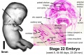
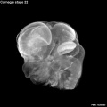
Carnegie stage 22
| Embryology - 24 Jul 2026 |
|---|
| Google Translate - select your language from the list shown below (this will open a new external page) |
|
العربية | català | 中文 | 中國傳統的 | français | Deutsche | עִברִית | हिंदी | bahasa Indonesia | italiano | 日本語 | 한국어 | မြန်မာ | Pilipino | Polskie | português | ਪੰਜਾਬੀ ਦੇ | Română | русский | Español | Swahili | Svensk | ไทย | Türkçe | اردو | ייִדיש | Tiếng Việt These external translations are automated and may not be accurate. (More? About Translations) |
Introduction

Facts
Week 8, 54 - 56 days, 23 - 28 mm
Gestational age GA week 10
Summary
- Ectoderm:
- Mesoderm: heart prominence, ossification continues
- Head: nose, eye, external acoustic meatus
- Body: straightening of trunk, heart, liver, umbilical cord
- Limb: upper limbs longer and bent at elbow, foot plate with webbed digits, wrist, hand plate with separated digits
See also Carnegie stage 22 Events
Features
- Identify: straightening of trunk, pigmented eye, eyelid, nose, external acoustic meatus, ear auricle, scalp vascular plexus, separated digits (fingers), thigh, ankle, umbilical cord
- hearing - cochlea continues spiral growth.
| Stage 22 Links: Week 8 | System Development | Lecture - Limb | Lecture - Head Development | Lecture - Sensory | Science Practical - Head | Science Practical - Sensory | Science Practical - Urogenital | Historic - Skull Development | Carnegie Embryos | Madrid Embryos | Category:Carnegie Stage 22 | Next Stage 23 |
| Week 8, GA week 10, 54 - 56 days, CRL 23 - 28 mm, Carnegie Embryos |
| Historic Papers: 1914 | 1954 Stage 19-23 |
| Week: | 1 | 2 | 3 | 4 | 5 | 6 | 7 | 8 |
| Carnegie stage: | 1 2 3 4 | 5 6 | 7 8 9 | 10 11 12 13 | 14 15 | 16 17 | 18 19 | 20 21 22 23 |
- Carnegie Stages: 1 | 2 | 3 | 4 | 5 | 6 | 7 | 8 | 9 | 10 | 11 | 12 | 13 | 14 | 15 | 16 | 17 | 18 | 19 | 20 | 21 | 22 | 23 | About Stages | Timeline
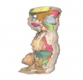
Kyoto Collection

Lateral view. Amniotic membrane removed.
Image source: The Kyoto Collection images are reproduced with the permission of Prof. Kohei Shiota and Prof. Shigehito Yamada, Anatomy and Developmental Biology, Kyoto University Graduate School of Medicine, Kyoto, Japan for educational purposes only and cannot be reproduced electronically or in writing without permission.
Carnegie Collection


| Carnegie Collection - Stage 22 | |||||||||||
|---|---|---|---|---|---|---|---|---|---|---|---|
| Serial No. | Size (mm) | Grade | Fixative | Embedding Medium | Plane | Thinness (µm) | Stain | Point Score | Sex | Year | Notes |
| 392 | E., 23 Ch., 45x45x25 | Poor | ... | P | Sagittal | 50 | Al. coch. | 42 | Female | 1907 | Brödel Collection. Injected |
| 405 | E., 26 | Good | Formalin | C | Sagittal | 40 | Carmine | 42.5 | Male | 1907 | |
| 464 | E.,26 Ch., 45x40x30 | Good | Formol? alc? | P | Sag. | 100 | Al. coch. | 44.5 | Male | 1910 | |
| 584A | E.,25 Ch., 50x42x40 | Poor | Formalin | P | Sagittal | 50 | Al. coch. | 41 | ? | 1913 | |
| 630 | E., 25 | Poor | Formalin | P | Transverse | 100 | Al. coch. | 46 | Male | 1913 | |
| 840 | E, 24.8 | Good | Formalin | P | Transverse | 50 | Al. coch. | 44.5 | Female | 1914 | |
| 875 | E, 27 Ch., 40x28x22 | Good | Formalin | P | Sagittal | 40 | Al. coch. | 45 | Male | 1914 | |
| 895 | E., 26 Ch., 67x62x54 | Good | Formalin | P | Transverse | 25 | Al. coch. | 46.5 | Female | 1914 | |
| 1315 | E.,25 | Good | Formalin | P | Sagittal | 50 | Al. coch. | 40.5 | Female | 1915 | Spina bifida and anencephaly |
| 1458 | E., 27.5 Ch, 45x45x30 | Exc. | Formalin | C | Sagittal | 50 | (Stain - Haematoxylin Eosin) aur, or. G. | 45.5 | Male | 1916 | |
| 1894 | E, 24.6 | Good | Formalin | c | Sagittal | 40,80 | (Stain - Haematoxylin Eosin) aur, or. G. | 41 | Female | 1917 | |
| 2206 | E, 27 Ch, 50x30x18 | Poor | Formalin | p | Transverse | 40 | (Stain - Haematoxylin Eosin) | 44.5 | Male | 1918 | |
| 3681 | E, 26.3 Ch, 36x36x34 | Good | Formalin | p | Transverse | 25 | Al. coch. | 44.5 | Male | 1921 | |
| 4304 | E,25 Ch, 66x45x45 | Good | Bouin | p | Transverse | 20 | (Stain - Haematoxylin Eosin) | 44.5 | Female | 1923 | Injected |
| 4339 | E, 24.5 | Good | Formalin | p | Transverse | 15 | Al. coch, Mallory | 46,5 | Female | 1923 | |
| 4476 | E., 26.2 | Good | Bouin | p | Transverse | 40 | (Stain - Haematoxylin Eosin) | 46 | Female | 1923 | Tubal |
| 4638 | E, 23.4 | Exc. | Bouin | p | Transverse | 15,20 | Al. coch, or. G. | 41.5 | Male | 1924 | |
| 6701 | E, 24 | Poor | Formalin | p | Coronal | 20 | (Stain - Haematoxylin Eosin) | 41 | Female | 1933 | |
| 6832 | E, 25.8 | Exc. | Bouin | C-P | Coronal | 20 | (Stain - Haematoxylin Eosin) | 42 | Female | 1934 | |
| 8394 | E, 25.3 Ch, 48x50x34 | Exc. | Bouin | C-P | Transverse | 20 | (Stain - Haematoxylin Eosin), Masson | 44.5 | Female | 1946 | |
| 8948 | E, 26.7 Ch, 61x51x50 | Poor | Formol-Zenker | p | Transverse | 15 | Ag | ? | ? | 1952 | |
Abbreviations
| |||||||||||
| iBook - Carnegie Embryos | |
|---|---|

|
|
Hinrichsen Collection

ME 31 Carnegie stage 22
Image source: The Hinrichsen Collection images are reproduced with the permission of Prof. Beate Brand-Saberi, Head, Department of Anatomy and Molecular Embryology, Ruhr-Universität Bochum. Images are for educational purposes only and cannot be reproduced electronically or in writing without permission.
Madrid Collection
| Madrid Collection Embryos | ||||||
|---|---|---|---|---|---|---|
| Carnegie Stage |
Embryo | Days | CRL (mm) | Section thickness |
Staining | Section plane |
| 22 | RX | 53 | 23.5 | 10 | (Stain - Haematoxylin Eosin) | transverse |
| 22 | H7 | 54 | 24 | 10 | (Stain - Haematoxylin Eosin) | transverse |
| 22 | R3 | 55 | 25 | 10 | (Stain - Haematoxylin Eosin) | frontal |
| 22 | Eo2 | 55 | 26 | 10 | (Stain - Haematoxylin Eosin)-trichrome | transverse |
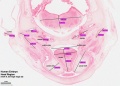
Stage 22 Movies
Movies - Embryo Carnegie stage 22 - These are rotating embryo animations based upon reconstruction of serial slice images.
|
|
|
|
|
|
These 3d movies were part of the UNSW Medical degree Independent Learning Project (ILP) prepared by Aashish Kumar (2006).
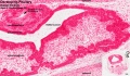
Stage 22 Serial Section Images
Virtual Slides
| These are high resolution histological sections (Stain - Haematoxylin Eosin) through regions of the Stage 22 human embryo from the original serial section set shown below. See also High resolution image extracts. |
|
|
|
| ||||||||||||
|
|
| |||||||||||||
|
|
|


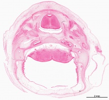
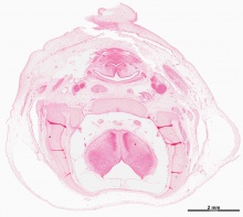
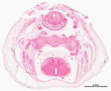

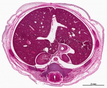
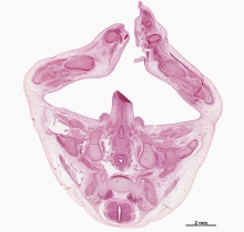
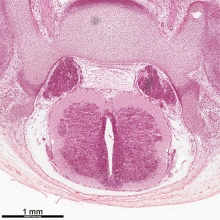
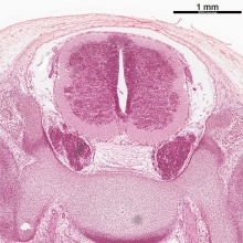
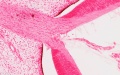
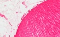
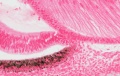
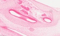
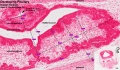
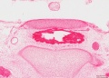
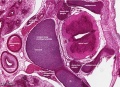
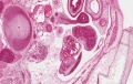
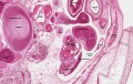
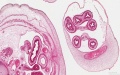
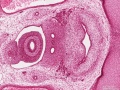
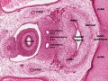
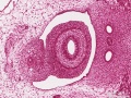
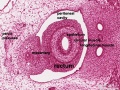
High Resolution Images
- Selected Human Embryo Histology - Stage 22 (week 8)
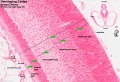
developing cortex labeled
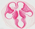
central nervous system and ventricles
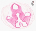
central nervous system and ventricles
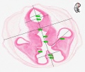
measured image
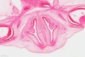
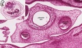
nose and eyes
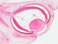
eye
optic nerve
lens
lens and pigmented epithelium

lens and pigmented epithelium
head region measured
developing cochlea
pituitary
pituitary measured
pituitary
vertebra and spinal cord
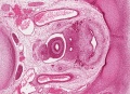
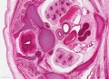
mesonephric ducts and bladder

developing testis
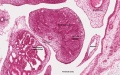
developing testis labeled
developing testis
developing testis labeled
midgut herniation
pelvic region - rectum and bladder
pelvic region - rectum and bladder labeled
pelvic region - rectum
pelvic region - rectum labeled
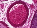
Aorta filled with red blood cells
Lungs and thymus
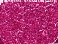
Dorsal aorta
Placental blood vessels


Serial Sections
These are the original serial images prepared from embryo sections for Embryology practical classes and transferred online in 1996.
- Links: Carnegie stage 22 - serial sections | Carnegie stage 13 - serial sections | Carnegie stage 13
Labeled

|

|

|

|

|

|

|
| A1L | A2L | A3L | A4L | A5L | A6L | A7L |

|

|

|

|

|

|

|
| B1L | B2L | B3L | B4L | B5L | B6L | B7L |

|

|

|

|

|

|

|
| C1L | C2L | C3L | C4L | C5L | C6L | C7L |

|

|

|

|

|

|

|
| D1L | D2L | D3L | D4L | D5L | D6L | D7L |

|

|

|

|

|

|

|
| E1L | E2L | E3L | E4L | E5L | E6L | E7L |

|

|

|

|

|

|

|
| F1L | F2L | F3L | F4L | F5L | F6L | F7L |

|

|

|

|

|

|

|
| G1L | G2L | G3L | G4L | G5L | G6L | G7L |
Unlabeled

|

|

|

|

|

|

|
| A1 | A2 | A3 | A4 | A5 | A6 | A7 |

|

|

|

|

|

|

|
| B1 | B2 | B3 | B4 | B5 | B6 | B7 |

|

|

|

|

|

|

|
| C1 | C2 | C3 | C4 | C5 | C6 | C7 |

|

|

|

|

|

|

|
| D1 | D2 | D3 | D4 | D5 | D6 | D7 |

|

|

|

|

|

|

|
| E1 | E2 | E3 | E4 | E5 | E6 | E7 |

|

|

|

|

|

|

|
| F1 | F2 | F3 | F4 | F5 | F6 | F7 |

|

|

|

|

|

|

|
| G1 | G2 | G3 | G4 | G5 | G6 | G7 |
Stage 22 Selected Serial Section Images
About Stage 22 Embryo Sections - The Carnegie stage 22 human embryo is 27 mm (CRL) in size and approximately equal to day 54 - 56 of development (week 8). These images have been selected to show some key features of late embryo development.
- Links: Carnegie stage 22 - selected serial sections | Carnegie stage 22 - serial sections | Carnegie stage 22 | Embryo Serial Sections
| Stage 22 serial unlabeled images | Stage 22 Serial labeled images |
Events
- vision - (stage 19 -22) the eyelid folds develop into the eyelids and cover more of the eye as the palpebral fissure takes shape. The upper and the lower eyelids meet at the outer canthus in Stage 19. [2]
- endocrine[3]
- parathyroid - Parathyroids 4 become detached from the pharyngeal endoderm (Jirfisek 1980).
- adrenal cortex - the C2 cells have changed and resemble fibrocytes.[4]
- meninges (spinal cord) - cavity formation in the meninx primitiva has progressed, and the rudimentary ventral dura is more distinct. Ventral to the medial edge of the ganglia, this dense dural rudiment is separated from the lateral extremities of the centra and intervertebral disks. Within this space a large longitudinal venous channel is developing. The dura can be identified around the spinal ganglia, which are now shifting into the intervertebral foramina, but becomes less distinct as it passes dorsad. The ventral dura can be followed throughout the cervical, thoracic, and lumbar segments, but in the sacral segments it can be identified only on the dorsal surfaces of the intervertebral disks.[5]
- genital[6]
- submandibular gland - Secondary branching of duct. Practically solid duct; suggestion of lumen in distal part. Definite lumen in oral part of duct.[1]
References
- ↑ 1.0 1.1 Streeter GL. Developmental Horizons In Human Embryos Description Or Age Groups XIX, XX, XXI, XXII, And XXIII, Being The Fifth Issue Of A Survey Of The Carnegie Collection. (1957) Carnegie Instn. Wash. Publ. 611, Contrib. Embryol., 36: 167-196.
- ↑ Pearson AA. The development of the eyelids. Part I. External features. (1980) J. Anat.: 130(1): 33-42. PMID 7364662 PDF
- ↑ O'Rahilly R. The timing and sequence of events in the development of the human endocrine system during the embryonic period proper. (1983) Anat. Embryol., 166: 439-451. PMID 6869855
- ↑ Crowder RE. The development of the adrenal gland in man, with special reference to origin and ultimate location of cell types and evidence in favor of the "cell migration" theory. (1957) Contrib. Embryol., Carnegie Inst. Wash. 36, 193-210.
- ↑ Sensenig EC. The early development of the meninges of the spinal cord in human embryos. (1951) Contrib. Embryol., Carnegie Inst. Wash. Publ. 611.
- ↑ O'Rahilly R. (1983). The timing and sequence of events in the development of the human reproductive system during the embryonic period proper. Anat. Embryol. , 166, 247-61. PMID: 6846859
- ↑ Gillman J. The development of the gonads in man, with a consideration of the role of fetal endocrines and the histogenesis of ovarian tumors. (1948) Contr Embryol Carneg Instn. 32: 81-131.
- ↑ Koff A. Development of the vagina in the human fetus. (1933) Contrib. Embryol., Carnegie Inst. Wash. Publ. 443, 24: 59-60.
Additional Images
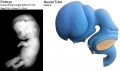
Neural tube model
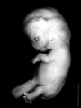
Stage 22 embryo rotated vertically
Stage 22 ear
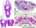
Stage 13 and 22 thyroid development
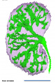
renal branching
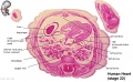
Human heart E3L

External ear Stages 14-23 and adult
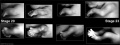
Stage 20-23 limbs
Stage 22 Optical Projection Tomography


- Carnegie Stages: 1 | 2 | 3 | 4 | 5 | 6 | 7 | 8 | 9 | 10 | 11 | 12 | 13 | 14 | 15 | 16 | 17 | 18 | 19 | 20 | 21 | 22 | 23 | About Stages | Timeline
Cite this page: Hill, M.A. (2026, July 24) Embryology Carnegie stage 22. Retrieved from https://embryology.med.unsw.edu.au/embryology/index.php/Carnegie_stage_22
- © Dr Mark Hill 2026, UNSW Embryology ISBN: 978 0 7334 2609 4 - UNSW CRICOS Provider Code No. 00098G