Placenta - Histology
Introduction
This page introduces the histology of the placenta, the placental cord and fetal membranes.
Various developmental stages of the placental villi are shown including maternal decidua.
There are images of placental cord and the placental vessels (vein and arteries).
| Histology Links: stains | fixatives | artifacts | menstrual histology | placenta histology | heart histology | liver histology | Pancreas | Gall Bladder | Colon | Renal | Respiratory Histology | Bone | Category:Histology | UNSW Histology |
| Historic Histology Textbooks: 1941 Histology] | 1944 Oral Histology |
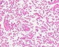
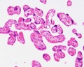
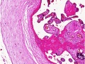
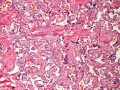
Hofbauer Cells
![Placental villi Hofbauer cells[1]](/embryology/index.php?title=File:Placenta_Hofbauer_cells_01.jpg)
- human villous macrophages
- highly vacuolated cells
- located the core of placental villi and cord
- macrophages with micropinocytotic activity and phagocytosis ability
- possible paracrine role for early stages of placental vasculogenesis
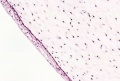
Villi Histology

Placenta anchoring villi and maternal decidua
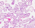
First trimester overview
First trimester villi
First trimester detail
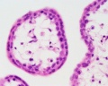
Term Overview
Term villi
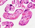
Term detail
Week 15 (GA)
Week 15 (GA)


- Villi Histology: First trimester (overview | villi | detail) | fetal and maternal RBCs | Term (overview | villi | detail) | Cord Histology | Placenta Histology | Placenta Development
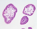
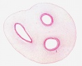
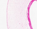
Cord Histology

Placental cord cross-section
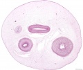
Placental cord cross-section

Placental vein
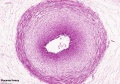
Placental artery

Placental artery

Placental allantois
Placental epithelium
Placental cord cross-section
Placental vein
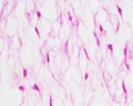
Whartons jelly


- Placental Cord Histology: Cord overview | Vein | Artery | Artery | Allantois | Epithelium | Cord overview 1 unlabeled | overview 2 unlabeled | unlabeled vein and connective tissue | unlabeled connective tissue | Villi histology | Placenta Histology
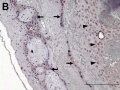
Abnormal Histology
Term placenta
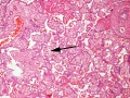
Term placenta chorangiosis


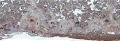
Other Species Placenta
Guinea Pig related Placenta

References
Reviews
Articles
Search PubMed
Search Pubmed: Placenta Histology
Glossary Links
- Glossary: A | B | C | D | E | F | G | H | I | J | K | L | M | N | O | P | Q | R | S | T | U | V | W | X | Y | Z | Numbers | Symbols | Term Link
Cite this page: Hill, M.A. (2024, June 26) Embryology Placenta - Histology. Retrieved from https://embryology.med.unsw.edu.au/embryology/index.php/Placenta_-_Histology
- © Dr Mark Hill 2024, UNSW Embryology ISBN: 978 0 7334 2609 4 - UNSW CRICOS Provider Code No. 00098G