Carnegie stage 17: Difference between revisions
mNo edit summary |
|||
| Line 142: | Line 142: | ||
==Other Specimens== | ==Other Specimens== | ||
{{Ref-Volcher1959}} | {{Ref-Volcher1959}} describes the peripheral nervous system. | ||
{{Ref-Hendrix1985}} | {{Ref-Hendrix1985}} | ||
Revision as of 11:40, 27 January 2017
| Embryology - 15 Jun 2024 |
|---|
| Google Translate - select your language from the list shown below (this will open a new external page) |
|
العربية | català | 中文 | 中國傳統的 | français | Deutsche | עִברִית | हिंदी | bahasa Indonesia | italiano | 日本語 | 한국어 | မြန်မာ | Pilipino | Polskie | português | ਪੰਜਾਬੀ ਦੇ | Română | русский | Español | Swahili | Svensk | ไทย | Türkçe | اردو | ייִדיש | Tiếng Việt These external translations are automated and may not be accurate. (More? About Translations) |
Introduction

Facts
Week 6, 42 - 44 days, 11 - 14 mm
Gestational Age GA week 8
Summary
- Ectoderm: sensory placodes, lens pit, otocyst,nasal pits moved ventrally, fourth ventricle of brain
- Mesoderm: heart prominence
- Head: 1st, 2nd and 3rd pharyngeal arch, forebrain, eye, auricular hillocks
- Body: heart, liver, umbilical cord, mesonephric ridge
- Limb: upper and lower limb buds, hand digital rays
See also Carnegie stage 17 Events
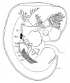
Features
- pigmented eye, nasal pit, nasolacrimal groove, external acoustic meatus, auricular hillock, heart, digital rays, liver pronminance, thigh, ankle, foot plate, umbilical cord
- Identify: pigmented eye, nasal pit, nasolacrimal groove, external acoustic meatus, auricular hillock, heart, digital rays, liver prominence, thigh, ankle, foot plate, umbilical cord
- Links: Week 6 | System Development | Head | Lecture - Limb | Lecture - Head Development | Lecture - Sensory | Science Practical - Head | Science Practical - Sensory | Science Practical - Urogenital | Category:Carnegie Stage 17 | Stage 18
| Week: | 1 | 2 | 3 | 4 | 5 | 6 | 7 | 8 |
| Carnegie stage: | 1 2 3 4 | 5 6 | 7 8 9 | 10 11 12 13 | 14 15 | 16 17 | 18 19 | 20 21 22 23 |
- Carnegie Stages: 1 | 2 | 3 | 4 | 5 | 6 | 7 | 8 | 9 | 10 | 11 | 12 | 13 | 14 | 15 | 16 | 17 | 18 | 19 | 20 | 21 | 22 | 23 | About Stages | Timeline
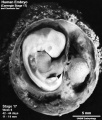
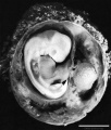
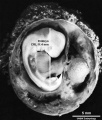
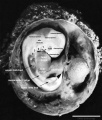
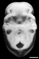
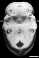
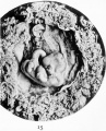
Kyoto Collection

View: This is a left lateral view of embryo. Amniotic membrane removed.

Ventral view of head region (1 mm scale).
- Human Embryo (Carnegie stage 17)
Embryo and membranes
Embryo unlabeled
Embryo CRL
Embryo features

Membranes
Membrane measurements
Oral cavity roof unlabeled
Oral cavity roof features


Right lateral view of embryo enclosed in chorionic sac. scale bar 5 mm.

Kyoto embryo (16834) showing detail of umbilicus Carnegie stage 17 (1 mm scale bar)
|
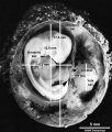
This is a MRI off-axis sagittal (not in at the exact anatomical plane) section through the week 6 embryo |
Image source: The Kyoto Collection images are reproduced with the permission of Prof. Kohei Shiota and Prof. Shigehito Yamada, Anatomy and Developmental Biology, Kyoto University Graduate School of Medicine, Kyoto, Japan for educational purposes only and cannot be reproduced electronically or in writing without permission.
Carnegie Collection


| iBook - Carnegie Embryos | |
|---|---|

|
|
Blechschmidt Collection
|
Model from serial section reconstruction.
|


Image source: The Blechschmidt Collection images are reproduced with the permission of Prof. Christoph Viebahn, director of the Institute of Anatomy and Embryology, , University Medical Center Göttingen. Images are for educational purposes only and cannot be reproduced electronically or in writing without permission.
Hill Collection
| Hill H58 | |
|---|---|

|

|
| left dorsolateral | left lateral |
| Hill H202 | |

|

|
Embryo Virtual Slide
|
|
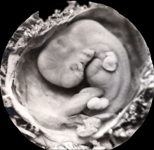
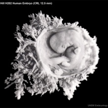
Image source: The images from the Hill Collection (part of the Embryological Collection) are reproduced with the permission of the Museum für Naturkunde, Leibniz Institute for Research on Evolution and Biodiversity. Images are for educational purposes only and must not be reproduced electronically or in writing without permission from the Museum für Naturkunde Berlin.
Hinrichsen Collection

|
Hinrichsen collection Human Embryo ME16 (stage17).
Note the developing mandible and maxilla in this ventrolateral view of the head. The developing maxilla in this ventral view of the nasal opening.
|

Image source: The Hinrichsen Collection images are reproduced with the permission of Prof. Beate Brand-Saberi, Head, Department of Anatomy and Molecular Embryology, Ruhr-Universität Bochum. Images are for educational purposes only and cannot be reproduced electronically or in writing without permission.
Scanning EM

|

|
| Ventral view of head showing upper lip, maxilla and nasal region. | Note that a ventral image of only half the head has been "mirrored" to generate this image.
Image Source: Prof Virginia Diewert |
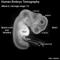
Tomography
Optical projection tomography movie of rotating stage 17 embryo. Note the detailed structural view of neural system development. |

Stage 17 Optical Projection Tomography (left) |

Stage 17 Optical Projection Tomography (right) |
Other Specimens
Volcher R. Le systeme nerveux pe'riphe'rique cninien d'un embryon humain de 12 mm. (1959) Arch. Biol. (Liege), 70:179-215. describes the peripheral nervous system.
Hendrix MJ Brailey JL and Shenker L. SEM-dissection of a human embryo derived from an ectopic pregnancy. (1985) Early Hum Dev. 11(1): 61-8. PMID 4006825
Events
- Neural
- Hearing - otic capsule now dense mesenchyme. Otic vesicle vestibular part wall thins prior to semicircular duct appearing. Geniculate ganglion forms. Auditory ossicles, tubotympanic recess and chorda tympani appear. First pharyngeal groove (cleft or hyomandibular groove) begins to form the concha and the external acoustic meatus. Six auricular hillocks present (1 tragus, 2 and 3 crus helicis, 4 and 5 helix, and 6 antitragus).
- Smell olfactory nerve fibres enter the brain[3]
- Vision - Retinal pigment is visible and the retinal fissure is largely closed. Eyelids grooves deepen, eyelid folds develop, first below, and then above, the eye.[4]
- Eyelid sulcus (groove) above and below eye deepen and eyelid folds develop (below first and then above)[4]
- Diaphragm - pleuroperitoneal fold (PPF) no longer separated from the diaphragm (CRL 14mm)[5]
- Abdominal Wall muscle cells now migrated approximately 50% of the distance to the ventral midline, inner and outer layers were not discernible yet.[6]
- Cardiovascular
- Coronary circulation acquires coronary sinus connection.[7]
- Endocrine[8]
- Hypophysis - juxtacerebral wall of the craniopharyngeal pouch is the thicker. The lateral lobes (future infundibular, or tuberal, part) and the anterior chamber (Vorraum) are clearly visible (O'Rahilly 1973 a). The infundibular recess displays a characteristically folded wall, namely the neurohypophysis (O'Rahilly 1973 a).
- Thymus - connection of the thymus with the pharynx has been severed (Weller 1933). The thymus is intimately approximated to the cervical duct (ibid.) According to Norris (1937), both third and fourth pouches make contact with the ectoderm, although only the third "receives an increment from the ectoderm".
- Parathyroids -parathyroid 4 is attached to the lateral surface of what Weller (1933) termed the "lateral thyroid component"
- Thyroid. The lobes of the thyroid curve around the carotid arteries and are connected by a delicate isthmus. Lacunae "should not be confused with lumina of follicles" (Weller 1933).
- Adrenal Cortex - dorsal part of the whole suprarenal primordium is disorganized by the invasion of sympathetic nerves and cells, while the band of C2 cells and the coelomic epithelium remain intact (Crowder 1957).
- Adrenal Medulla - first neural migration is at its height. Growth of the para-aortic complex is extensive. The plexiform complex is derived from paravertebral sympathetic ganglia T6-12 and usually L 1. Included in it are the primordia of the suprarenal medulla and of the celiac, superior mesenteric, and renal plexuses. Nerve fibres and "paraganglion" (M3) cells enter.
- Pancreas - ventral pancreas has now fused with dorsal (Streeter 1948). Perhaps the ventral and dorsal ducts have begun to blend (Russu and Vaida 1959).
References
- ↑ <pubmed>2802187</pubmed>
- ↑ <pubmed>15478101</pubmed>
- ↑ <pubmed>15604533</pubmed>
- ↑ 4.0 4.1 <pubmed>7364662</pubmed>
- ↑ <pubmed>19711422</pubmed>
- ↑ <pubmed>22976993</pubmed>
- ↑ <pubmed>3286038</pubmed>
- ↑ O'Rahilly R. The timing and sequence of events in the development of the human endocrine system during the embryonic period proper. (1983) Anat. Embryol., 166: 439-451. PMID 6869855
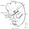
Additional Images

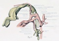
External ear Stages 14-23 and adult
Streeter 1921 Plate 3
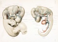
Embryo 940 cardiovascular lateral view
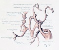
Embryo 940 cardiovascular ventrolateral view
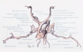
Embryo 940 cardiovascular ventral view
His 1897 Fig. 15
Sabin 1909 Fig. 4
Keith 1921 Fig. 45


- Carnegie Stages: 1 | 2 | 3 | 4 | 5 | 6 | 7 | 8 | 9 | 10 | 11 | 12 | 13 | 14 | 15 | 16 | 17 | 18 | 19 | 20 | 21 | 22 | 23 | About Stages | Timeline
Cite this page: Hill, M.A. (2024, June 15) Embryology Carnegie stage 17. Retrieved from https://embryology.med.unsw.edu.au/embryology/index.php/Carnegie_stage_17
- © Dr Mark Hill 2024, UNSW Embryology ISBN: 978 0 7334 2609 4 - UNSW CRICOS Provider Code No. 00098G