File:Stage12 sem3a.jpg: Difference between revisions
(== Human Embryo Carnegie Stage 12== Carnegie Stage 12 Facts: Week 4, 26 days, 5 mm, Somite Number 25 View: Dorsolatera view, day 26, 25 somites, amniotic membrane removed Features: caudal (dorsal) neuropore region [[:File:Stage12 bf2.jpg|Bright fiel) |
mNo edit summary |
||
| (3 intermediate revisions by 2 users not shown) | |||
| Line 1: | Line 1: | ||
== Human Embryo Carnegie | ==Human Embryo Caudal Neuropore (Carnegie stage 12)== | ||
Dorsolateral view, day 26, 25 somites, amniotic membrane removed. | |||
Carnegie | This is an SEM image of the human embryo ([[Carnegie stage 12]], [[week 4]]) caudal end. There is a [[:File:Stage12 bf2.jpg|Bright field image version]] also available. | ||
The neural tube is now shown closed at the caudal neuropore (posterior neuropore). | |||
Image version links: [[:File:Stage12 sem3.jpg|Large 1200px]] | [[:File:Stage12 sem3a.jpg|1000px]] | [[:File:Stage12 sem3b.jpg|Medium 600px]] | [[:File:Stage12 sem3c.jpg|Small 400px]] | |||
===Neural=== | |||
{{Neural Links}} | |||
===SEM Images=== | |||
{{Stage 12 SEM images}} | |||
===Stage 12=== | |||
{{Carnegie_stages}} | |||
{{Carnegie_stage_table_1}} | |||
{{Carnegie stage 12 links}} | |||
===Reference=== | |||
{{SEM}} | |||
[[Category:Week 4]] [[Category:Ectoderm]] [[Category:Neural]] | |||
[[Category:Carnegie Stage]] [[Category:Carnegie Stage 12]] | |||
[[Category: | |||
[[Category: | |||
Latest revision as of 14:02, 19 May 2017
Human Embryo Caudal Neuropore (Carnegie stage 12)
Dorsolateral view, day 26, 25 somites, amniotic membrane removed.
This is an SEM image of the human embryo (Carnegie stage 12, week 4) caudal end. There is a Bright field image version also available.
The neural tube is now shown closed at the caudal neuropore (posterior neuropore).
Image version links: Large 1200px | 1000px | Medium 600px | Small 400px
Neural
SEM Images
- Stage 12 SEM Images: Bright Field 1 | Bright Field 3 | Bright Field 3 | SEM1 | SEM2 | SEM3 | SEM4 dorsolateral head and arches | SEM5 lateral head and arches | SEM6 ventrolateral head and arches | SEM7 lateral | SEM8 ventrolateral | SEM9 cloacal membrane | SEM9 labeled | Carnegie stage 12
Stage 12
- Carnegie Stages: 1 | 2 | 3 | 4 | 5 | 6 | 7 | 8 | 9 | 10 | 11 | 12 | 13 | 14 | 15 | 16 | 17 | 18 | 19 | 20 | 21 | 22 | 23 | About Stages | Timeline
| Week: | 1 | 2 | 3 | 4 | 5 | 6 | 7 | 8 |
| Carnegie stage: | 1 2 3 4 | 5 6 | 7 8 9 | 10 11 12 13 | 14 15 | 16 17 | 18 19 | 20 21 22 23 |
Reference
Image Source: Scanning electron micrographs of the Carnegie stages of the early human embryos are reproduced with the permission of Prof Kathy Sulik, from embryos collected by Dr. Vekemans and Tania Attié-Bitach. Images are for educational purposes only and cannot be reproduced electronically or in writing without permission.
File history
Click on a date/time to view the file as it appeared at that time.
| Date/Time | Thumbnail | Dimensions | User | Comment | |
|---|---|---|---|---|---|
| current | 14:39, 14 May 2011 | 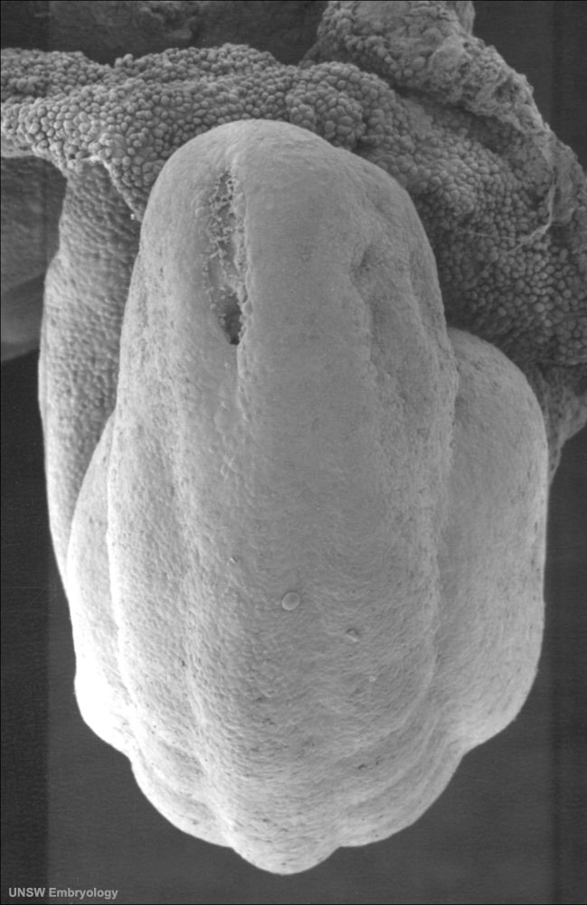 | 649 × 1,000 (95 KB) | S8600021 (talk | contribs) | |
| 13:14, 14 May 2011 |  | 649 × 1,000 (94 KB) | S8600021 (talk | contribs) | == Human Embryo Carnegie Stage 12== Carnegie Stage 12 Facts: Week 4, 26 days, 5 mm, Somite Number 25 View: Dorsolatera view, day 26, 25 somites, amniotic membrane removed Features: caudal (dorsal) neuropore region [[:File:Stage12 bf2.jpg|Bright fiel |
You cannot overwrite this file.
File usage
The following page uses this file: