Renal System Development: Difference between revisions
mNo edit summary |
mNo edit summary |
||
| (70 intermediate revisions by the same user not shown) | |||
| Line 1: | Line 1: | ||
{{Header}} | |||
== Introduction == | |||
[[File:Gray1127.jpg|right|200px]] | [[File:Gray1127.jpg|right|200px]] | ||
The paired adult kidneys consist of a functional unit called the "nephron", that filters blood, excretes waste, reabsorbs water (and other compounds) and has endocrine functions. Each adult human kidney typically contains about 750,000 nephrons, though the total number can vary significantly from as few as 250,000 to as many as 2,000,000.{{#pmid:1546799|PMID1546799}}{{#pmid:17495859|PMID17495859}} | |||
In the embryo, nephron development, '''nephrogenesis''', occurs through several stages involving classical epithelial/mesenchyme type of interactions. Nephrogenesis continues into the late fetal period ({{GA}} week 34–35) and while the fetal kidney does produce urine, not until after birth does the glomerular filtration rate (GFR) increases rapidly due to a postnatal drop in kidney vascular resistance and an increase in renal blood flow. | |||
The urinary system is developmentally and anatomically associated with genital development, often described as the "urogenital system". (More? {{Genital}}) | |||
{{Renal Links}} | {{Renal Links}} | ||
==Some Recent Findings== | ==Some Recent Findings== | ||
[[File:Cloacal septation model.jpg|thumb|Cloacal septation model | [[File:Human embryonic renal branching 1.jpg|thumb|alt=Human renal branching development between week 5 to 8|Human renal branching development between week 5 to 8.{{#pmid:30192900|PMID30192900}}]] | ||
[[File:Cloacal septation model.jpg|thumb|Cloacal septation model{{#pmid:22253716|PMID22253716}}]] | |||
{| | {| | ||
|-bgcolor="F5FAFF" | |-bgcolor="F5FAFF" | ||
| | | | ||
* ''' | |||
* ''' | * '''A practical guide to the stereological assessment of glomerular number, size, and cellular composition'''{{#pmid:31960613|PMID31960613}} "The evaluation of a range of measures in the kidneys, such as developmental stage, rate and success, injury, and disease processes, relies on obtaining information on the three-dimensional structure of the renal corpuscles, and in particular the glomerular capillary tufts. To do this in the most accurate, comprehensive, and unbiased manner depends on a knowledge of stereological methods. In this article, we provide a practical guide for researchers on how to quantitate a number of structures in the kidneys, including the estimation of total glomerular number, glomerular capillary length and filtration surface area, and the cellular composition of individual glomeruli. Guidance is also provided on how to apply these methods to kidneys at different sizes and levels of maturity." | ||
* ''' | |||
* '''Branching morphogenesis of the urinary collecting system in the human embryonic metanephros'''{{#pmid:30192900|PMID30192900}} "An elaborate system of ducts collects urine from all nephrons, and this structure is known as the urinary collecting system (UCS). This study focused on how the UCS is formed during human embryogenesis. Fifty human embryos between the Carnegie stage {{CS14}} and {{CS23}} were selected from the [[Kyoto Collection]] at the Congenital Anomaly Research Center of Kyoto University, Japan. Metanephroses, including the UCS, were segmented on serial digital virtual histological sections. Three-dimensional images were computationally reconstructed for morphological and quantitative analyses. A CS timeline was plotted. It consisted of the 3-D structural morphogenesis of UCS and quantification of the total amount of end-branching, average and maximum numbers of generations, deviation in the metanephros, differentiation of the urothelial epithelium in the renal pelvis, and timing of the rapid expansion of the renal pelvis. The first UCS branching generation occurred by {{CS16}}. The average branching generation reached a maximum of 8.74 ± 1.60 and was already the twelfth in {{CS23}}. ...Differentiation may have continued up until the tenth generation to allow for renal pelvis expansion. The branching speed was not uniform. There were significantly more branching generations in the polar- than in the interpolar regions (P < 0.05). Branching speed reflects the growth orientation required to form the metanephros. Further study will be necessary to understand the renal pelvis expansion mechanism in {{CS23}}." | |||
* '''Spatiotemporal heterogeneity and patterning of developing renal blood vessels'''{{#pmid:29627966|PMID29627966}} "Here, we examine the developing kidney vasculature to assess its 3-dimensional structure and transcriptional heterogeneity. First, we observe that endothelial cells (ECs) grow coordinately with the kidney bud as early as {{ME10.5}}, and begin to show signs of specification by {{ME13.5}} when the first arteries can be identified. We then focus on how ECs pattern and remodel with respect to the developing nephron and collecting duct epithelia. ECs circumscribe nephron progenitor populations at the distal tips of the ureteric bud (UB) tree and form stereotyped cruciform structures around each tip. Beginning at the renal vesicle (RV) stage, ECs form a continuous plexus around developing nephrons. The endothelial plexus envelops and elaborates with the maturing nephron, becoming preferentially enriched along the early distal tubule." | |||
|} | |} | ||
{| class="wikitable collapsible collapsed" | {| class="wikitable mw-collapsible mw-collapsed" | ||
! More recent papers | ! More recent papers | ||
|- | |- | ||
| [[File:Mark_Hill.jpg|90px|left]] {{Most_Recent_Refs}} | | [[File:Mark_Hill.jpg|90px|left]] {{Most_Recent_Refs}} | ||
Search term: [http://www.ncbi.nlm.nih.gov/pubmed/?term=Renal+Embryology ''Renal Embryology''] | Search term: [http://www.ncbi.nlm.nih.gov/pubmed/?term=Renal+Embryology ''Renal Embryology''] | [http://www.ncbi.nlm.nih.gov/pubmed/?term=Renal+Development ''Renal Development''] | [http://www.ncbi.nlm.nih.gov/pubmed/?term=Nephrogenesis ''Nephrogenesis''] | [http://www.ncbi.nlm.nih.gov/pubmed/?term=Intermediate+Mesoderm ''Intermediate Mesoderm''] | [http://www.ncbi.nlm.nih.gov/pubmed/?term=Ureter+Development ''Ureter Development''] | [http://www.ncbi.nlm.nih.gov/pubmed/?term=Urethera+Development ''Urethera Development''] | [http://www.ncbi.nlm.nih.gov/pubmed/?term=Renal+Pelvis+Development ''Renal Pelvis Development''] | [http://www.ncbi.nlm.nih.gov/pubmed/?term=Urinary+Bladder+Development ''Urinary Bladder Development''] | ||
|} | |||
{| class="wikitable mw-collapsible mw-collapsed" | |||
! Older papers | |||
|- | |||
| {{Older papers}} | |||
* '''Conserved and Divergent Features of Human and Mouse Kidney Organogenesis'''{{#pmid:29449453|PMID29449453}} "Human kidney function is underpinned by approximately 1,000,000 nephrons, although the number varies substantially, and low nephron number is linked to disease. Human kidney development initiates around 4 weeks of gestation and ends around 34-37 weeks of gestation. Over this period, a reiterative inductive process establishes the nephron complement. Studies have provided insightful anatomic descriptions of human kidney development, but the limited histologic views are not readily accessible to a broad audience. In this first paper in a series providing comprehensive insight into human kidney formation, we examined human kidney development in 135 anonymously donated human kidney specimens. We documented kidney development at a macroscopic and cellular level through histologic analysis, RNA in situ hybridization, immunofluorescence studies, and transcriptional profiling, contrasting human development (4-23 weeks) with {{mouse}} development at selected stages (embryonic day {{ME15.5}} and postnatal day 2). The high-resolution histologic interactive atlas of human kidney organogenesis generated can be viewed at the GUDMAP database (www.gudmap.org) together with three-dimensional reconstructions of key components of the data herein. At the anatomic level, human and mouse kidney development differ in timing, scale, and global features such as lobe formation and progenitor niche organization. The data also highlight differences in molecular and cellular features, including the expression and cellular distribution of anchor gene markers used to identify key cell types in mouse kidney studies." {{mouse}} | |||
* '''Reciprocal Spatiotemporally Controlled {{Apoptosis}} Regulates Wolffian Duct Cloaca Fusion'''{{#pmid:29326158|PMID29326158}} "The epithelial Wolffian duct (WD) inserts into the cloaca (primitive bladder) before metanephric kidney development, thereby establishing the initial plumbing for eventual joining of the ureters and bladder. Defects in this process cause common anomalies in the spectrum of congenital anomalies of the kidney and urinary tract (CAKUT). However, developmental, cellular, and molecular mechanisms of WD-cloaca fusion are poorly understood. Through systematic analysis of early WD tip development in mice, we discovered that a novel process of spatiotemporally regulated apoptosis in WD and cloaca was necessary for WD-cloaca fusion." | |||
* '''{{Zebrafish}} Pronephros Development'''{{#pmid:28409341|PMID28409341}} "The pronephros is the first kidney type to form in vertebrate embryos. The first step of pronephrogenesis in the zebrafish is the formation of the intermediate mesoderm during gastrulation, which occurs in response to secreted morphogens such as BMPs and Nodals. Patterning of the intermediate mesoderm into proximal and distal cell fates is induced by retinoic acid signaling with downstream transcription factors including wt1a, pax2a, pax8, hnf1b, sim1a, mecom, and irx3b. In the anterior intermediate mesoderm, progenitors of the glomerular blood filter migrate and fuse at the midline and recruit a blood supply. More posteriorly localized tubule progenitors undergo epithelialization and fuse with the cloaca. The Notch signaling pathway regulates the formation of multi-ciliated cells in the tubules and these cells help propel the filtrate to the cloaca. The lumenal sheer stress caused by flow down the tubule activates anterior collective migration of the proximal tubules and induces stretching and proliferation of the more distal segments. Ultimately these processes create a simple two-nephron kidney that is capable of reabsorbing and secreting solutes and expelling excess water-processes that are critical to the homeostasis of the body fluids. The zebrafish pronephric kidney provides a simple, yet powerful, model system to better understand the conserved molecular and cellular progresses that drive nephron formation, structure, and function." {{Zebrafish}} | |||
* '''Histone deacetylase 1 and 2 regulate Wnt and p53 pathways in the ureteric bud epithelium'''{{#pmid:25758227|PMID25758227}} "Histone deacetylases (HDACs) regulate a broad range of biological processes through removal of acetyl groups from histones as well as non-histone proteins. Our previous studies showed that Hdac1 and Hdac2 are bound to promoters of key renal developmental regulators and that HDAC activity is required for embryonic kidney gene expression. However, the existence of many HDAC isoforms in embryonic kidneys raises questions concerning the possible specificity or redundancy of their functions. We report here that targeted deletion of both the Hdac1 and Hdac2 genes from the ureteric bud (UB) cell lineage of mice causes bilateral renal hypodysplasia. One copy of either Hdac1 or Hdac2 is sufficient to sustain normal renal development." | |||
* '''{{Bmp}}7 functions via a polarity mechanism to promote cloacal septation'''{{#pmid:22253716|PMID22253716}} "During normal development in human and other placental mammals, the embryonic cloacal cavity separates along the axial longitudinal plane to give rise to the urethral system, ventrally, and the rectum, dorsally. Defects in cloacal development are very common and present clinically as a rectourethral fistula in about 1 in 5,000 live human births. Yet, the cellular mechanisms of cloacal septation remain poorly understood. ...Our results strongly indicate that Bmp7/JNK signaling regulates remodeling of the cloacal endoderm resulting in a topological separation of the urinary and digestive systems. Our study points to the importance of Bmp and JNK signaling in cloacal development and rectourethral malformations." | |||
* '''Size and location of the kidneys during the {{fetal}} period'''{{#pmid:21110022|PMID21110022}} "The level of the left kidney was higher than the level of the right kidney in the {{fetal}} period. The posterior surface relations to the ribs showed certain ascendance during gestation, corresponding to vertebral levels. However, fetal kidneys do not reach the same level as adults at full term. The kidneys move farther apart from the midline of the body during the fetal period. The dimensions, weight, and volume of the kidneys increased with gestational age during the fetal period. The ratio between kidney weights and fetal body weights were determined, and we observed that the ratio decreased during the fetal period. There were no sex or laterality differences in any parameter." (See also [[Fetal Development]]) | |||
* '''Characterization of Mesonephric Development and Regeneration Using Transgenic Zebrafish.'''{{#pmid:20810610|PMID20810610}} "The majority of previous studies have focused on the pronephros of zebrafish, which consists of only two nephrons and is structurally simpler than the mesonephros of adult fish and the metanephros of mammals. To evaluate the zebrafish system for more complex studies of kidney development and regeneration, we investigated the development and post-injury regeneration of the mesonephros in adult zebrafish." (See also [[Zebrafish Development]]) | |||
|} | |} | ||
== Objectives == | == Objectives == | ||
[[File: | [[File:Nephron histology.jpg|thumb|Renal Nephron]] | ||
* Understand the 3 main stages of kidney development. | * Understand the 3 main stages of kidney development. | ||
* Understand development of the nephron and renal papilla. | * Understand development of the nephron and renal papilla. | ||
| Line 35: | Line 60: | ||
== Textbook References == | == Textbook References == | ||
[[File:Stage 13 kidney sections.jpg|right]] | |||
* '''The Developing Human: Clinically Oriented Embryology''' (8th Edition) by Keith L. Moore and T.V.N Persaud - Moore & Persaud Chapter 13 p303-346 | * '''The Developing Human: Clinically Oriented Embryology''' (8th Edition) by Keith L. Moore and T.V.N Persaud - Moore & Persaud Chapter 13 p303-346 | ||
* '''Larsen’s Human Embryology''' by GC. Schoenwolf, SB. Bleyl, PR. Brauer and PH. Francis-West - Chapter 10 p261-306 | * '''Larsen’s Human Embryology''' by GC. Schoenwolf, SB. Bleyl, PR. Brauer and PH. Francis-West - Chapter 10 p261-306 | ||
| Line 43: | Line 69: | ||
== Background== | == Background== | ||
* Mesoderm then intermediate mesoderm | * Mesoderm then intermediate mesoderm | ||
* Vascular Development | * Vascular Development | ||
| Line 52: | Line 77: | ||
==Kidney Anatomy== | ==Kidney Anatomy== | ||
[[File:Gray1128.jpg|thumb|Adult nephron structure]] | [[File:Gray1128.jpg|thumb|Adult nephron structure]] | ||
* Nephron - Functional unit of kidney | * Nephron - Functional unit of kidney | ||
* Humans up to 1 million | * Humans up to 1 million | ||
| Line 77: | Line 101: | ||
==Intermediate Mesoderm== | ==Intermediate Mesoderm== | ||
{| | |||
| [[File:Stage7_intermediate-mesoderm.jpg|200px]] | |||
Week 3 - [[Carnegie stage 7|Stage 7]] dorsal view<br> | |||
intermediate mesoderm (orange lateral strips) | |||
| [[File:Mesoderm-cartoon4.jpg|250px]] | |||
Cross-section showing mesoderm regions | |||
| | |||
* development occurs laterally symmetrical (left right) | * development occurs laterally symmetrical (left right) | ||
* intermediate mesoderm lying beside the '''dorsal aorta''' | * intermediate mesoderm lying beside the '''dorsal aorta''' | ||
| Line 84: | Line 116: | ||
* the mesonephric duct then extends within the mesoderm, rostro-caudally | * the mesonephric duct then extends within the mesoderm, rostro-caudally | ||
* eventually making contact with the '''cloaca''' | * eventually making contact with the '''cloaca''' | ||
|} | |||
==Mesonephric Duct== | ==Mesonephric Duct== | ||
Later in development, both the mesonephric duct and the cloaca both continue to differentiate and undergo extensive remodelling (and renaming) | Later in development, both the mesonephric duct and the cloaca both continue to differentiate and undergo extensive remodelling (and renaming) | ||
=== | ===Ureteric Bud=== | ||
[[File:Mouse-kidney in vitro.jpg|thumb|Mouse E12.5 kidney in vitro]] | [[File:Mouse-kidney in vitro.jpg|thumb|Mouse E12.5 kidney in vitro]] | ||
* arise near the cloacal connection of the mesonephric duct | * arise near the cloacal connection of the mesonephric duct | ||
* branch from the mesonephric duct laterally into the intermediate mesoderm | * branch from the mesonephric duct laterally into the intermediate mesoderm | ||
* induce the surrounding mesoderm to differentiate - metanephric blastema | * induce the surrounding mesoderm to differentiate - metanephric blastema | ||
** this mesoderm will in turn signal back to differentiate the | ** this mesoderm will in turn signal back to differentiate the ureteric bud | ||
'''Epithelial - mesenchymal interaction''' | '''Epithelial - mesenchymal interaction''' | ||
Ureteric Bud forms - ureter, pelvis, calyces, collecting ducts | |||
===Metanephric Blastema=== | ===Metanephric Blastema=== | ||
| Line 118: | Line 150: | ||
===Mesonephros=== | ===Mesonephros=== | ||
The mesonephros, historically called the Wolffian body, is the embryonic stage of renal development that is entirely lost during development of the later fetal /adult metanepros. The only developmental structure not lost is the {{mesonephric duct}}. | |||
{| | |||
| Human E24, Mouse {{ME9.5}} caudal to pronephros | |||
* forms by induction from pronephros | * forms by induction from pronephros | ||
* pronephric duct now becomes mesonephric duct (also called Wolffian Duct) | * pronephric duct now becomes mesonephric duct (also called Wolffian Duct) | ||
* extends downwards in intermediate mesoderm towards cloaca, later urogenital sinus | |||
| [[File:Stage 13 kidney sections 2.jpg|200px]] | |||
Week 5 - Stage {{CS13}} mesonephros<br>(extending the length of the body) | |||
| [[File:Stage22 mesonephros.jpg|200px]] | |||
Week 8 - {{CS22}} mesonephros<br>(now degenerating) | |||
|} | |||
====Wolffian body==== | |||
Historic name for the developing combined renal (mesonephros) and genital (paramesonephrotic blastema) structures. This term is no longer used in describing development. | |||
{{Male Vignette}} | |||
===Metanephros=== | ===Metanephros=== | ||
* Human E35-37, Mouse | [[File:Fetal_10wk_urogenital_1.jpg|thumb|200px|Early fetal kidney (week 10)]] | ||
* Human E35-37, Mouse {{ME11}} epithelia bud at end of mesonephric duct ureteric bud and associated metanephric mesenchyme | |||
Ureteric Bud | |||
* induced by metanephric mesenchyme to differentiate | * induced by metanephric mesenchyme to differentiate | ||
* forms collecting tubules, renal pelvis, ureter | * forms collecting tubules, renal pelvis, ureter | ||
* metanephric mesenchyme induced by | * metanephric mesenchyme induced by ureteric to differentiate forms nephron | ||
== Nephron == | == Nephron == | ||
{| | |||
| In humans, nephrogenesis only occurs before birth, though nephron maturation continues postnatally. Mean glomerular number shown to level at 36 weeks, increasing from about 15,000 at 15 weeks to 740,000 at 40 weeks. | |||
In humans, nephrogenesis only occurs before birth, though nephron maturation continues postnatally. Mean glomerular number shown to level at 36 weeks, increasing from about 15,000 at 15 weeks to 740,000 at 40 weeks. | | [[File:Gray1128.jpg|200px]] | ||
Adult nephron structure | |||
| [[File:Nephron histology.jpg|200px]] | |||
Nephron histology | |||
| {{Nephron movie}} | |||
|} | |||
Nephron development has four identifiable developmental stages: | |||
# Vesicle (V) stage (13-19 weeks, second trimester) | # Vesicle (V) stage (13-19 weeks, second trimester) | ||
# S-shaped body (S) stage ( 20-24 weeks, second trimester) | # S-shaped body (S) stage ( 20-24 weeks, second trimester) | ||
# Capillary loop (C) stage (25-29 weeks, third trimester) | # Capillary loop (C) stage (25-29 weeks, third trimester) | ||
# Maturation (M) stage (infants aged 1-6 months, neonatal and postnatal) | # Maturation (M) stage (infants aged 1-6 months, neonatal and postnatal) | ||
{| | |||
| | |||
* disorganised mesenchymal cells become a highly organised epithelial tubule | * disorganised mesenchymal cells become a highly organised epithelial tubule | ||
* Condensation - groups of about 100 cells condense tightly together to form a distinct mass | * Condensation - groups of about 100 cells condense tightly together to form a distinct mass | ||
* Epithelialisation - condensed cells lose their mesenchymal character and gain epithelial | * Epithelialisation - condensed cells lose their mesenchymal character and gain epithelial | ||
* At end of this period formed a small epithelial cyst complete with a basement membrane, cell-cell junctions and a defined cellular apico-basal polarity. | * At end of this period formed a small epithelial cyst complete with a basement membrane, cell-cell junctions and a defined cellular apico-basal polarity. | ||
| [[File:Nephron development 01.jpg|600px]] | |||
Nephron development{{#pmid:23338209|PMID23338209}} | |||
* '''a''' - condensed aggregate forms | |||
* '''b''' - renal vesicle forms | |||
* '''c''' - S-shaped body (red), renal progenitors are localised, at this stage podocyte-committed progenitors (red + blue) as well as tubular-committed progenitors (orange) can already be seen. | |||
* '''d''' - mature nephron, renal progenitors (red), podocyte-committed progenitors (red + blue), as well as tubular-committed progenitors (orange) are distributed along the nephron. | |||
* '''e''' - glomerulus, renal progenitors (red) are localized at the urinary pole of the Bowman capsule. Podocyte-committed progenitors (red + blue) localize along the Bowman capsule. | |||
|} | |||
* cyst invaginates twice to form a comma | * cyst invaginates twice to form a comma | ||
* then a S-shaped body one invagination site later becomes the glomerular cleft | * then a S-shaped body one invagination site later becomes the glomerular cleft | ||
* At about this time blood vessel progenitors invade cleft to begin construction of vascular component of glomerulus | * At about this time blood vessel progenitors invade cleft to begin construction of vascular component of glomerulus | ||
* Tubule maturation specialised transporting segments of nephron differentiate complex of convoluted tubules is created | * Tubule maturation specialised transporting segments of nephron differentiate complex of convoluted tubules is created | ||
{| | |||
| valign=top| | |||
# ureteric bud (gray) extends from the mesonephric (Wolffian) duct | |||
# upon contact with the metanephric mesenchyme (red), reciprocal signaling induces both bud bifurcation and condensation of the mesenchyme to generate the cap mesenchyme. | |||
# blastemal mesenchyme then undergo a MET to generate the renal vesicle | |||
# renal vesicle continues to form the comma-shaped and S-shaped body | |||
# distal end fuses with the ureteric bud (which forms the collecting duct) | |||
# proximal end joins to form the glomerulus, generating the mature nephron (dark orange). | |||
| [[File:Renal development cartoon01.jpg|600px]] | |||
Renal Development Interactions{{#pmid:25737276|PMID25737276}} | |||
|} | |||
{| | |||
! colspan=2|Bowmans Capsule | |||
|- | |||
| Bowmans Capsule forms two layers with a space between these layers. | |||
* '''Outer later''' - (parietal) single layer of '''simple squamous epithelium'''. Does not function in filtration. | |||
* '''Space''' - (Bowman's space, "urinary space", "capsular space") space filled by fluid (filtrate) passing through podocyte filtration slits | |||
* '''Inner layer''' - (visceral) formed by podocytes on thickened glomerular basement membrane covering glomerular capillaries. | |||
| [[File:Nephron EM01.jpg|400px]] | |||
Within the glomerulus a high magnification view of a podocyte showing the interdigitated foot processes (pedicels) that are wrapped around the exterior of glomerular capillaries.{{#pmid:25918223|PMID25918223}} | |||
|- | |||
| colspan=2|[[File:Nephron EM02.jpg|600px]] | |||
|} | |||
==Embryonic Kidney== | |||
Embryonic stage descriptions based on [[Carnegie Collection]] embryos.<ref name=O'RahillyMüller1987>{{Ref-O'RahillyMüller1987}}</ref> | |||
===Week 4=== | |||
* [[Carnegie stage 12]] - 29 somite embryo mesonephros tubules begin at the level of somite 8 and are distinct as far caudally as somite 20, whence they extend as a continuous nephrogenic cord to t he level of somite 24. Opposite each somite there are two or more tubules. Thus they are not metameric, any more than the mesonephric duct is metameric, or the umbilical vein. The number of nephric vesicles is being increased by progressive differentiation caudally from the nephrogenic cord.<ref name=Torrey1954>{{Ref-Torrey1954}}</ref> The mesonephric duct at first ends blindly immediately short of the cloaca, but soon becomes attached to the cloaca (i.e., to the terminal part of the hindgut) and acquires a lumen. | |||
* [[Carnegie stage 13]] - Mesonephros glomeruli begin to develop, and nephric tubules become S-shaped.<ref name=Shikinami1926>{{Ref-Shikinami1926}}</ref> A ureteric bud may possibly be present in some specimens<ref name=Wells1954>{{Ref-Wells1954}}</ref> fig. 4), although further confirmation is needed. The mesonephric duct, which becomes separated from the surface ectoderm except in its caudal portion, is fused to the cloaca, into which it may open. A urorectal cleavage line is apparent. | |||
===Week 5=== | |||
* [[Carnegie stage 14]] - In embryos of this stage the mesonephros is well along in its organogenesis. The steps in this process are made easier to follow by the fact that the development occurs progressively in a rostrocaudal direction. Here again is an organ in which the epithelial elements constitute its primary tissue and seem largely to determine its form. The non-epithelial mesonephric elements, though necessary complements for epithelial-mesenchymal interaction, give the appearance of being subsidiary. It has already been seen that the coelomic surface cells possess various inherent potentialities. The surface of the coelom can be mapped in definite areas in accordance with the distribution of these various kinds of surface cells. Running along each side of the median plane is a narrow strip of coelom where, by the proliferation and delamination of its surface cells, there is produced a longitudinal series of epithelial tubules that constitute the units of the mesonephros. This follows the manner in which nephric elements were formed in previous stages, and it is now about to be repeated, with certain modifications, in the development of the metanephros, which is still in the primordial state of a budding ureter with its nephrogenic capsule (fig. 14-6). One can go a step further in regard to the inherent constitution of these coelomic epithelial tubules. Not only do they become tubules, but from the beginning they show regional differentiation. The proximal end promptly blends with and opens into the mesonephric duct, and this part of the tubule persists as a collecting duct. The distal free end at the same time begins its expansion into a highly specialized part of the tubule, namely the mesonephric corpuscle. The intervening central segment of the tubule becomes the convoluted secretory portion. Embryos in this stage are especially favorable for the study of the process of formation of the mesonephric corpuscle. The proliferation of the tubular epithelium at the free end results in its maximum expansion. This occurs in such a way as to produce an indented flattened vesicle, known as a glomerular capsule. As seen in section, it has an arched floor-plate several cells thick and a thin, single-layered roof membrane. The two are continuous with each other but are very different in their potentialities. The roof membrane becomes attenuated as an impermeable membrane. The floor plate continues active proliferation and many of its cells were believed by Streeter to delaminate and apparently become angioblasts, participating in the formation of the vascular glomerulus and its supporting tissues. It is now maintained, however, that the glomerular capillaries (in the metanephros) come from adjacent vessels and never develop in situ from epithelial cells (Potter, 1965). Further details of renal development have been provided by several authors (e.g., Potter, 1972). The residual cells facing the capsular lumen in the mesonephros at stage 14 are reduced in the more advanced phases to a single layer, covering and conforming everywhere to the tabulations of the underlying capillary tufts. Angiogenesis around the secretory part of the tubule is not far advanced. Angiogenic strands connect with the caudal cardinal vein, and throughout the mesonephros there are isolated clumps of angioblasts, particularly around the capsules. These show the typical difference in complexity of the three parts of the tubule: (1) collecting duct, (2) secretory segment, and (3) glomerular capsule. | |||
* [[Carnegie stage 15]] - The ureteric bud is longer, and its tip is expanded as the pelvis of the ureter (fig. 15-10). The primary urogenital sinus is distinguishable. | |||
===Week 6=== | |||
* [[Carnegie stage 16]] - The metanephros, which is now reniform, is still sacral in level. The ureter is elongating and, in more advanced embryos, the pelvis of the ureter divides into rostral and caudal poles. The urorectal septum, the formation of which is disputed, is well marked. | |||
* [[Carnegie stage 17]] - The mesonephros shows epithelial plaques in the visceral layer of the glomerular capsule and hence can produce urine (Silverman, 1969){{#pmid:5374935|PMID5374935}}. The pelvis of the ureter usually shows three main divisions, and calices appear. The urogenital sinus presents a pelvic part (vesico-urethral canal) and a phallic part (definitive urogenital sinus). | |||
===Week 7=== | |||
* Carnegie stage {{CS18}} - Collecting tubules develop from the calices at stages [[Carnegie stage 17|17]] and [[Carnegie stage 18|18]]. They are surrounded by sharply outlined condensed primordia in the process of forming secretory tubules. Renal corpuscules are not yet present. By stage 18 the mesonephric duct and the ureter open almost independently into the vesico-urethral canal<ref name=Shikinami1926>{{Ref-Shikinami1926}}</ref>: i.e., the common excretory duct is disappearing. The cloacal membrane is ready to rupture. | |||
* Carnegie stage {{CS19}} - Lack of orientation in metanephrogenic tissue. Beginning formation of renal vesicles.<ref name=Streeter1957>{{Ref-Streeter1957}}</ref> | |||
===Week 8=== | |||
* Carnegie stage {{CS20}} - The external surface of the metanephros is said to be slightly lobulated. A reconstruction of the urinary system has been published by Shikinami<ref name=Shikinami1926>{{Ref-Shikinami1926}}</ref> (fig. 5). S-shaped lumen in renal vesicles. Spoon-shaped capsules (Bowman’s).<ref name=Streeter1957>{{Ref-Streeter1957}}</ref> | |||
* Carnegie stage {{CS21}} - Metanephros is spoon-shaped glomerular capsules are developing, no large glomeruli.<ref name=Streeter1957>{{Ref-Streeter1957}}</ref> | |||
* Carnegie stage {{CS22}} - Metanephros few large glomeruli.<ref name=Streeter1957>{{Ref-Streeter1957}}</ref> | |||
* Carnegie stage {{CS23}} - Short secretory tubules. Numerous large glomeruli. Long secretory tubules. High epithelium in some tubules. Increased number and convolutions of tubules.<ref name=Streeter1957>{{Ref-Streeter1957}}</ref> Comparison between the mesonephros and the metanephros in staged embryos is lacking. In metanephros, the kidneys have ascended from a sacral level at stages 13–15 to a lumbar level at stages 17–23. At stage 23 they are generally at the level of lumbar vertebrae 1–3.. | |||
==Fetal Kidney== | |||
{| | |||
| [[File:Fetal kidney MRI 01.jpg|400px]] | |||
| valign=top|MRI appearance of normal fetal kidney.{{#pmid:25685519|PMID25685519}} Sagittal T2- SSFSE of a fetal abdomen at {{GA}} 25 week. Adequate volume of the amniotic fluid and the developing lungs indicate good renal function. | |||
* two arrowheads - note the size and the signal appearance of the normal kidney. | |||
* white arrow - the fluid-filled urinary bladder | |||
* black arrow - the developing lung. | |||
Note that the urinary bladder can occupy a considerable portion of the abdomen as a normal finding. | |||
:'''Links:''' [[Magnetic Resonance Imaging]] | |||
|} | |||
{| | |||
! Fetal nephron development{{#pmid:23338209|PMID23338209}} | |||
|- | |||
| [[File:Fetal nephron development 01.jpg|600px]] | |||
After nephron development has completed and concomitant with the development of the renal papilla in the newborn, the thin ascending limb of Henle’s loops is generated as an outgrowth from the S3 segment of the proximal tubule and from the distal tubule anlage of the nephron. | |||
==Endocrine Kidney== | ==Endocrine Kidney== | ||
| Line 178: | Line 315: | ||
|} | |} | ||
===Common | ===Common Urogenital Sinus=== | ||
* superior end continuous with '''allantois''' | * superior end continuous with '''allantois''' | ||
* common urogenital sinus and mesonephric duct fuse (connect) | * common urogenital sinus and mesonephric duct fuse (connect) | ||
| Line 222: | Line 359: | ||
{{Trigone movie}} | {{Trigone movie}} | ||
===Urethra Development=== | |||
The entire human male and female urethra is {{endoderm}}al in origin based on the presence of FOXA1, KRT 7, uroplakin, and the absence of KRT10 staining.{{#pmid:30287094|PMID30287094}} A recent study of male penile urethra describes a theory of a "two-step process of urethral plate canalization and urethral fold fusion to form the human penile urethra. Canalization ("opening zipper") opens the solid urethral plate into a groove, and fusion ("closing zipper")."{{#pmid:27397682|PMID27397682}}{{#pmid:29155192|PMID29155192}} | |||
==Kidney Ascent== | ==Kidney Ascent== | ||
| Line 310: | Line 450: | ||
===Renal Cysts=== | ===Renal Cysts=== | ||
The Bosniak classification system (Category I - IV) was designed to separate identified cystic renal masses by analysis of computed tomography (CT) features into surgical and nonsurgical categories. | The Bosniak classification system (Category I - IV) was designed to separate identified cystic renal masses by analysis of computed tomography (CT) features into surgical and nonsurgical categories.{{#pmid:16040900|PMID16040900}} Named after Morton Bosniak, Yale University School of Medicine, the developer of this classification system. | ||
==Molecular== | |||
{| | |||
|-bgcolor="CEDFF2" | |||
! Abbreviation | |||
! Growth Factor | |||
! Renal Development | |||
! Expression Location | |||
|- | |||
| BMP4 | |||
| Bone Morphogenetic Protein 4 | |||
| prevents ectopic ureteric bud outgrowth and extra ureteric bud divisions | |||
| mesenchymal cells surrounding mesonephric duct and stromal mesenchyme surrounding steric bud stalks | |||
|-bgcolor="F5FAFF" | |||
| BMP7 | |||
| Bone Morphogenetic Protein 7 | |||
| survival of metanephric mesenchyme | |||
| metanephric mesenchyme | |||
|- | |||
| Fgf8 | |||
| Fibroblast Growth Factor 8 | |||
| transition of the induced cap mesenchyme into RVs | |||
| cap mesenchyme | |||
|-bgcolor="F5FAFF" | |||
| GDNF | |||
| Glial-cell derived neurotrophic factor | |||
| induces steric bud outgrowth from mesonephric duct, interacts with Ret | |||
| metanephric mesenchyme | |||
|- | |||
| VEGF | |||
| Vascular endothelial growth factor | |||
| promotes endothelial cell proliferation, differentiation | |||
| s-shaped body | |||
|-bgcolor="F5FAFF" | |||
| Wnt4 | |||
| Wingless-Type MMTV Integration Site Family, Member 4 | |||
| mesenchymal-to-epithelial transition | |||
| cap metanephric mesenchyme, pre-tubular aggregate, nephron progenitors | |||
|- | |||
| Wnt5a | |||
| Wingless-Type MMTV Integration Site Family, Member 5a | |||
| nephrogenesis induction, ectopic bud formation | |||
| steric bud, metanephric mesenchyme | |||
|-bgcolor="F5FAFF" | |||
| Wnt9b | |||
| Wingless-type MMTV integration site family, Member 9B | |||
| renewal and differentiation of nephron progenitors and normal ureteric bud branching, mesenchymal-to-epithelial transition | |||
| steric bud stalk epithelial cells | |||
|} | |||
* '''Foxd1''' - (Brain Factor-2) transcription factor that is a renal stroma specific gene. | |||
:'''Links:''' [[Renal System - Molecular]] | [http://omim.org/entry/601091 OMIM Foxd1] | |||
== References == | == References == | ||
<references/> | <references/> | ||
===Reviews=== | |||
{{#pmid:32765290}} | |||
===Textbooks=== | ===Textbooks=== | ||
* '''The Developing Human: Clinically Oriented Embryology''' (8th Edition) by Keith L. Moore and T.V.N Persaud - Moore & Persaud Chapter 13 p303-346 | * '''The Developing Human: Clinically Oriented Embryology''' (8th Edition) by Keith L. Moore and T.V.N Persaud - Moore & Persaud Chapter 13 p303-346 | ||
| Line 328: | Line 529: | ||
===Reviews=== | ===Reviews=== | ||
{{#pmid:25918223}} | |||
{{#pmid:25737276}} | |||
{{#pmid:25128732}} | |||
{{#pmid:25088264}} | |||
{{#pmid:25080023}} | |||
{{Ref-JacobYusufJacob2012}} | |||
{{#pmid:20691850}} | |||
{{#pmid:19906853}} | |||
{{#pmid:19828308}} | |||
{{#pmid:19615554}} | |||
{{#pmid:18184729}} | |||
{{#pmid:17442697}} | |||
{{#pmid:16916378}} | |||
[http://www.nature.com/ng/meetings/nephrogenetics/index.html Forefronts Symposium on Nephrogenetics: from development to physiology March 8-11, 2007 Danvers, MA] A meeting to synthesize an integrated view of the normal development and function of the kidney from the genetic standpoint. | [http://www.nature.com/ng/meetings/nephrogenetics/index.html Forefronts Symposium on Nephrogenetics: from development to physiology March 8-11, 2007 Danvers, MA] A meeting to synthesize an integrated view of the normal development and function of the kidney from the genetic standpoint. | ||
===Articles=== | ===Articles=== | ||
{{#pmid:31960613}} | |||
{{#pmid:29158444}} | |||
{{#pmid:26335195}} | |||
{{#pmid:24154527}} | |||
{{#pmid:18846389}} | |||
===Search PubMed=== | ===Search PubMed=== | ||
'''Search Pubmed:''' [http://www.ncbi.nlm.nih.gov/sites/entrez?db=pubmed&cmd=search&term=renal+system+development Renal System Development] | [http://www.ncbi.nlm.nih.gov/sites/entrez?db=pubmed&cmd=search&term=renal+development Renal Development] | [http://www.ncbi.nlm.nih.gov/sites/entrez?db=pubmed&cmd=search&term=intermediate%20mesoderm intermediate mesoderm] | [http://www.ncbi.nlm.nih.gov/sites/entrez?db=pubmed&cmd=search&term=kidney%20development kidney development] | [http://www.ncbi.nlm.nih.gov/sites/gquery?itool=toolbar&cmd=search&term=renal_development renal development] | [http://www.ncbi.nlm.nih.gov/sites/entrez?db=pubmed&cmd=search&term=ureteric%20bud ureteric bud] | [http://www.ncbi.nlm.nih.gov/sites/entrez?db=pubmed&cmd=search&term=nephron%20development nephron development] | [http://www.ncbi.nlm.nih.gov/sites/entrez?db=pubmed&cmd=search&term=bladder%20development bladder development] | '''Search Pubmed:''' [http://www.ncbi.nlm.nih.gov/sites/entrez?db=pubmed&cmd=search&term=renal+system+development Renal System Development] | [http://www.ncbi.nlm.nih.gov/sites/entrez?db=pubmed&cmd=search&term=renal+development Renal Development] | [http://www.ncbi.nlm.nih.gov/sites/entrez?db=pubmed&cmd=search&term=intermediate%20mesoderm intermediate mesoderm] | [http://www.ncbi.nlm.nih.gov/sites/entrez?db=pubmed&cmd=search&term=kidney%20development kidney development] | [http://www.ncbi.nlm.nih.gov/sites/gquery?itool=toolbar&cmd=search&term=renal_development renal development] | [http://www.ncbi.nlm.nih.gov/sites/entrez?db=pubmed&cmd=search&term=ureteric%20bud ureteric bud] | [http://www.ncbi.nlm.nih.gov/sites/entrez?db=pubmed&cmd=search&term=nephron%20development nephron development] | [http://www.ncbi.nlm.nih.gov/sites/entrez?db=pubmed&cmd=search&term=bladder%20development bladder development] | ||
| Line 354: | Line 577: | ||
<gallery> | <gallery> | ||
File:Nephron physiology.jpg|Nephron physiology | File:Nephron physiology.jpg|Nephron physiology | ||
File:Nephrons-cortical_and_juxtamedullary.jpg|Nephrons - cortical and juxtamedullary | File:Nephrons-cortical_and_juxtamedullary.jpg|Nephrons - cortical and juxtamedullary | ||
File:Endoderm cartoon.jpg|Endoderm cartoon | File:Endoderm cartoon.jpg|Endoderm cartoon | ||
File:Fetal 10wk urogenital 1.jpg|Fetal urogenital region most lateral right | File:Fetal 10wk urogenital 1.jpg|Fetal urogenital region most lateral right | ||
| Line 368: | Line 586: | ||
File:Fetal_10wk_urogenital_1.jpg|Fetal kidney (10 weeks) | File:Fetal_10wk_urogenital_1.jpg|Fetal kidney (10 weeks) | ||
File:Bladder_histology.jpg|Bladder histology | File:Bladder_histology.jpg|Bladder histology | ||
File:Australian_abnormalities_pie_urogen.png | File:Australian_abnormalities_pie_urogen.png|Australian abnormalities | ||
File:Drug-clearance-rates.png|Drug clearance rates | |||
File:Horseshoe kidney.jpg|Horseshoe kidney | File:Horseshoe kidney.jpg|Horseshoe kidney | ||
File:Hydronephrosis.jpg|Hydronephrosis | File:Hydronephrosis.jpg|Hydronephrosis | ||
| Line 375: | Line 594: | ||
File:Renal_outflow_obstruction.jpg|Renal outflow obstruction | File:Renal_outflow_obstruction.jpg|Renal outflow obstruction | ||
File:Bladder Exstrophy.jpg|Bladder Exstrophy | File:Bladder Exstrophy.jpg|Bladder Exstrophy | ||
File:Mouse-kidney in vitro.jpg|Mouse | File:Mouse-kidney in vitro.jpg|Mouse {{ME12.5}} kidney in vitro | ||
</gallery> | </gallery> | ||
===Historic=== | |||
{{Historic Disclaimer}} | |||
<gallery> | |||
File:Stage 11 historic-Atwell1930-3b.jpg|Stage 11 historic Atwell (1930) | |||
File:Stage_11_historic-Heuser1930-1c.jpg|Stage 11 historic Heuser (1930) | |||
File:Gray1128.jpg|Nephron structure | |||
File:Gray1127.jpg|Kidney and adrenal gland (adult) | |||
File:Gray1126.png|retroperitoneal | |||
</gallery> | |||
== Terms == | == Terms == | ||
Open table below to see list of renal terms. | |||
{{Renal terms}} | |||
==External Links== | ==External Links== | ||
{{External Links}} | {{External Links}} | ||
* Australia - Network for Genes & Environment in Development [http://www. | * Australia - Network for Genes & Environment in Development [http://www.monash.edu.au/research/people/profiles/profile.html?sid=2307&pid=3191 Professor John F. Bertram] | ||
* '''GenitoUrinary Development Molecular Anatomy Project''' (GUDMAP) [http://www.gudmap.org/About/Tutorial/DevMUS.html Renal Development Tutorial] | [http://www.gudmap.org/About/Tutorial/DevMRS.html Genital Development Tutorial] | * '''GenitoUrinary Development Molecular Anatomy Project''' (GUDMAP) [http://www.gudmap.org/About/Tutorial/DevMUS.html Renal Development Tutorial] | [http://www.gudmap.org/About/Tutorial/DevMRS.html Genital Development Tutorial] | ||
* [http://www.urinemetabolome.ca Urine Metabolome database] is a freely available electronic database containing detailed information about ~3100 small molecule metabolites found in human urine along with ~3900 concentration values. | |||
{{Systems}} | {{Systems}} | ||
Latest revision as of 09:42, 11 August 2020
| Embryology - 10 Jun 2024 |
|---|
| Google Translate - select your language from the list shown below (this will open a new external page) |
|
العربية | català | 中文 | 中國傳統的 | français | Deutsche | עִברִית | हिंदी | bahasa Indonesia | italiano | 日本語 | 한국어 | မြန်မာ | Pilipino | Polskie | português | ਪੰਜਾਬੀ ਦੇ | Română | русский | Español | Swahili | Svensk | ไทย | Türkçe | اردو | ייִדיש | Tiếng Việt These external translations are automated and may not be accurate. (More? About Translations) |
Introduction

The paired adult kidneys consist of a functional unit called the "nephron", that filters blood, excretes waste, reabsorbs water (and other compounds) and has endocrine functions. Each adult human kidney typically contains about 750,000 nephrons, though the total number can vary significantly from as few as 250,000 to as many as 2,000,000.[1][2]
In the embryo, nephron development, nephrogenesis, occurs through several stages involving classical epithelial/mesenchyme type of interactions. Nephrogenesis continues into the late fetal period (GA week 34–35) and while the fetal kidney does produce urine, not until after birth does the glomerular filtration rate (GFR) increases rapidly due to a postnatal drop in kidney vascular resistance and an increase in renal blood flow.
The urinary system is developmentally and anatomically associated with genital development, often described as the "urogenital system". (More? genital)
Some Recent Findings


|
| More recent papers |
|---|
 This table allows an automated computer search of the external PubMed database using the listed "Search term" text link.
More? References | Discussion Page | Journal Searches | 2019 References | 2020 References Search term: Renal Embryology | Renal Development | Nephrogenesis | Intermediate Mesoderm | Ureter Development | Urethera Development | Renal Pelvis Development | Urinary Bladder Development |
| Older papers |
|---|
| These papers originally appeared in the Some Recent Findings table, but as that list grew in length have now been shuffled down to this collapsible table.
See also the Discussion Page for other references listed by year and References on this current page.
|
Objectives
- Understand the 3 main stages of kidney development.
- Understand development of the nephron and renal papilla.
- Brief understanding of the mechanisms of nephron development.
- Understand the development of the cloaca, ureter and bladder.
- Brief understanding of abnormalities of the urinary system.
Textbook References

- The Developing Human: Clinically Oriented Embryology (8th Edition) by Keith L. Moore and T.V.N Persaud - Moore & Persaud Chapter 13 p303-346
- Larsen’s Human Embryology by GC. Schoenwolf, SB. Bleyl, PR. Brauer and PH. Francis-West - Chapter 10 p261-306
Renal Movies
|
|
|
| ||||||||||||
|
|
|
Background
- Mesoderm then intermediate mesoderm
- Vascular Development
- Gastrointestional
- Cloacal development
- Endocrine - covered in future lecture/lab
Kidney Anatomy
- Nephron - Functional unit of kidney
- Humans up to 1 million
- Filtration of waste from blood
- Endocrine
- Blood pressure regulation
The key structure of the adult nephron is the glomerulus (renal corpuscle), which represents the initial vascular/renal interface.
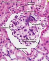
Glomerulus structure
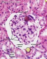
Vascular and renal poles


Related Images: Nephron histology overview | glomerulus structure | vascular and renal poles
Ureter
- Bladder - Urine storage
- Endoderm allantois
Mesoderm
- Intermediate mesoderm - Lies between somites and lateral plate
Intermediate Mesoderm

Week 3 - Stage 7 dorsal view |

Cross-section showing mesoderm regions |
|
Mesonephric Duct
Later in development, both the mesonephric duct and the cloaca both continue to differentiate and undergo extensive remodelling (and renaming)
Ureteric Bud

- arise near the cloacal connection of the mesonephric duct
- branch from the mesonephric duct laterally into the intermediate mesoderm
- induce the surrounding mesoderm to differentiate - metanephric blastema
- this mesoderm will in turn signal back to differentiate the ureteric bud
Epithelial - mesenchymal interaction
Ureteric Bud forms - ureter, pelvis, calyces, collecting ducts
Metanephric Blastema
- forms glomeruli, capsule, nephron tubules
- this development continues through fetal period
Nephros Development
Three pairs appearing in sequence within intermediate mesoderm during development.
- pronephros
- mesonephros
- metanephros
Pronephros
- week 4 few cells in cervical region fish
- Human E18, Mouse E7.5pronephric duct forms first with associated nephrogenic mesenchyme
- grows rostro caudally cervical -> cloaca
- E22 nephrogenic mesenchyme differentiates to form pronephroi not functional in mammals degenerates rapidly
Mesonephros
The mesonephros, historically called the Wolffian body, is the embryonic stage of renal development that is entirely lost during development of the later fetal /adult metanepros. The only developmental structure not lost is the mesonephric duct.
Human E24, Mouse E9.5 caudal to pronephros
|

Week 5 - Stage 13 mesonephros |

Week 8 - 22 mesonephros |
Wolffian body
Historic name for the developing combined renal (mesonephros) and genital (paramesonephrotic blastema) structures. This term is no longer used in describing development.
| Historic Embryology |
  Caspar Friedrich Wolff (1734-1794) was a German embryologist and anatomist best known today for identifying the Wolffian duct (mesonephric duct; ductus deferens, epididymis), Wolffian body (mesonephros) and Wolffian cyst (mesonephric origin uterine broad ligament cyst) that bear his name. Thought also to be a founder of the germ layer theory. His doctorate dissertation Theoria generationis (1774) discarded the developmental theory of preformation. Later in his career, his teaching in Berlin was opposed by the professors of the Medical-Surgical College, who had guild privileges to teach medicine. |
Metanephros

- Human E35-37, Mouse E11 epithelia bud at end of mesonephric duct ureteric bud and associated metanephric mesenchyme
Ureteric Bud
- induced by metanephric mesenchyme to differentiate
- forms collecting tubules, renal pelvis, ureter
- metanephric mesenchyme induced by ureteric to differentiate forms nephron
Nephron
| In humans, nephrogenesis only occurs before birth, though nephron maturation continues postnatally. Mean glomerular number shown to level at 36 weeks, increasing from about 15,000 at 15 weeks to 740,000 at 40 weeks. | 
Adult nephron structure |

Nephron histology |
|
Nephron development has four identifiable developmental stages:
- Vesicle (V) stage (13-19 weeks, second trimester)
- S-shaped body (S) stage ( 20-24 weeks, second trimester)
- Capillary loop (C) stage (25-29 weeks, third trimester)
- Maturation (M) stage (infants aged 1-6 months, neonatal and postnatal)
|

Nephron development[13]
|
- cyst invaginates twice to form a comma
- then a S-shaped body one invagination site later becomes the glomerular cleft
- At about this time blood vessel progenitors invade cleft to begin construction of vascular component of glomerulus
- Tubule maturation specialised transporting segments of nephron differentiate complex of convoluted tubules is created
|

Renal Development Interactions[14] |
| Bowmans Capsule | |
|---|---|
Bowmans Capsule forms two layers with a space between these layers.
|

Within the glomerulus a high magnification view of a podocyte showing the interdigitated foot processes (pedicels) that are wrapped around the exterior of glomerular capillaries.[15] |

| |
Embryonic Kidney
Embryonic stage descriptions based on Carnegie Collection embryos.[16]
Week 4
- Carnegie stage 12 - 29 somite embryo mesonephros tubules begin at the level of somite 8 and are distinct as far caudally as somite 20, whence they extend as a continuous nephrogenic cord to t he level of somite 24. Opposite each somite there are two or more tubules. Thus they are not metameric, any more than the mesonephric duct is metameric, or the umbilical vein. The number of nephric vesicles is being increased by progressive differentiation caudally from the nephrogenic cord.[17] The mesonephric duct at first ends blindly immediately short of the cloaca, but soon becomes attached to the cloaca (i.e., to the terminal part of the hindgut) and acquires a lumen.
- Carnegie stage 13 - Mesonephros glomeruli begin to develop, and nephric tubules become S-shaped.[18] A ureteric bud may possibly be present in some specimens[19] fig. 4), although further confirmation is needed. The mesonephric duct, which becomes separated from the surface ectoderm except in its caudal portion, is fused to the cloaca, into which it may open. A urorectal cleavage line is apparent.
Week 5
- Carnegie stage 14 - In embryos of this stage the mesonephros is well along in its organogenesis. The steps in this process are made easier to follow by the fact that the development occurs progressively in a rostrocaudal direction. Here again is an organ in which the epithelial elements constitute its primary tissue and seem largely to determine its form. The non-epithelial mesonephric elements, though necessary complements for epithelial-mesenchymal interaction, give the appearance of being subsidiary. It has already been seen that the coelomic surface cells possess various inherent potentialities. The surface of the coelom can be mapped in definite areas in accordance with the distribution of these various kinds of surface cells. Running along each side of the median plane is a narrow strip of coelom where, by the proliferation and delamination of its surface cells, there is produced a longitudinal series of epithelial tubules that constitute the units of the mesonephros. This follows the manner in which nephric elements were formed in previous stages, and it is now about to be repeated, with certain modifications, in the development of the metanephros, which is still in the primordial state of a budding ureter with its nephrogenic capsule (fig. 14-6). One can go a step further in regard to the inherent constitution of these coelomic epithelial tubules. Not only do they become tubules, but from the beginning they show regional differentiation. The proximal end promptly blends with and opens into the mesonephric duct, and this part of the tubule persists as a collecting duct. The distal free end at the same time begins its expansion into a highly specialized part of the tubule, namely the mesonephric corpuscle. The intervening central segment of the tubule becomes the convoluted secretory portion. Embryos in this stage are especially favorable for the study of the process of formation of the mesonephric corpuscle. The proliferation of the tubular epithelium at the free end results in its maximum expansion. This occurs in such a way as to produce an indented flattened vesicle, known as a glomerular capsule. As seen in section, it has an arched floor-plate several cells thick and a thin, single-layered roof membrane. The two are continuous with each other but are very different in their potentialities. The roof membrane becomes attenuated as an impermeable membrane. The floor plate continues active proliferation and many of its cells were believed by Streeter to delaminate and apparently become angioblasts, participating in the formation of the vascular glomerulus and its supporting tissues. It is now maintained, however, that the glomerular capillaries (in the metanephros) come from adjacent vessels and never develop in situ from epithelial cells (Potter, 1965). Further details of renal development have been provided by several authors (e.g., Potter, 1972). The residual cells facing the capsular lumen in the mesonephros at stage 14 are reduced in the more advanced phases to a single layer, covering and conforming everywhere to the tabulations of the underlying capillary tufts. Angiogenesis around the secretory part of the tubule is not far advanced. Angiogenic strands connect with the caudal cardinal vein, and throughout the mesonephros there are isolated clumps of angioblasts, particularly around the capsules. These show the typical difference in complexity of the three parts of the tubule: (1) collecting duct, (2) secretory segment, and (3) glomerular capsule.
- Carnegie stage 15 - The ureteric bud is longer, and its tip is expanded as the pelvis of the ureter (fig. 15-10). The primary urogenital sinus is distinguishable.
Week 6
- Carnegie stage 16 - The metanephros, which is now reniform, is still sacral in level. The ureter is elongating and, in more advanced embryos, the pelvis of the ureter divides into rostral and caudal poles. The urorectal septum, the formation of which is disputed, is well marked.
- Carnegie stage 17 - The mesonephros shows epithelial plaques in the visceral layer of the glomerular capsule and hence can produce urine (Silverman, 1969)[20]. The pelvis of the ureter usually shows three main divisions, and calices appear. The urogenital sinus presents a pelvic part (vesico-urethral canal) and a phallic part (definitive urogenital sinus).
Week 7
- Carnegie stage 18 - Collecting tubules develop from the calices at stages 17 and 18. They are surrounded by sharply outlined condensed primordia in the process of forming secretory tubules. Renal corpuscules are not yet present. By stage 18 the mesonephric duct and the ureter open almost independently into the vesico-urethral canal[18]: i.e., the common excretory duct is disappearing. The cloacal membrane is ready to rupture.
- Carnegie stage 19 - Lack of orientation in metanephrogenic tissue. Beginning formation of renal vesicles.[21]
Week 8
- Carnegie stage 20 - The external surface of the metanephros is said to be slightly lobulated. A reconstruction of the urinary system has been published by Shikinami[18] (fig. 5). S-shaped lumen in renal vesicles. Spoon-shaped capsules (Bowman’s).[21]
- Carnegie stage 21 - Metanephros is spoon-shaped glomerular capsules are developing, no large glomeruli.[21]
- Carnegie stage 22 - Metanephros few large glomeruli.[21]
- Carnegie stage 23 - Short secretory tubules. Numerous large glomeruli. Long secretory tubules. High epithelium in some tubules. Increased number and convolutions of tubules.[21] Comparison between the mesonephros and the metanephros in staged embryos is lacking. In metanephros, the kidneys have ascended from a sacral level at stages 13–15 to a lumbar level at stages 17–23. At stage 23 they are generally at the level of lumbar vertebrae 1–3..
Fetal Kidney

|
MRI appearance of normal fetal kidney.[22] Sagittal T2- SSFSE of a fetal abdomen at GA 25 week. Adequate volume of the amniotic fluid and the developing lungs indicate good renal function.
Note that the urinary bladder can occupy a considerable portion of the abdomen as a normal finding.
|
| Fetal nephron development[13] | ||||||||||||||||||||||||||||||||||||||||||||||||||||||||||||||
|---|---|---|---|---|---|---|---|---|---|---|---|---|---|---|---|---|---|---|---|---|---|---|---|---|---|---|---|---|---|---|---|---|---|---|---|---|---|---|---|---|---|---|---|---|---|---|---|---|---|---|---|---|---|---|---|---|---|---|---|---|---|---|

After nephron development has completed and concomitant with the development of the renal papilla in the newborn, the thin ascending limb of Henle’s loops is generated as an outgrowth from the S3 segment of the proximal tubule and from the distal tubule anlage of the nephron. Endocrine KidneyCovered also in Endocrine Development lecture
Cloaca
Common Urogenital Sinus
Urinary Bladder
Bladder StructureCan be described anatomically by its 4 layers from outside inward:
Detrusor Muscle
Ureter Development
Trigone Development
Urethra DevelopmentThe entire human male and female urethra is endodermal in origin based on the presence of FOXA1, KRT 7, uroplakin, and the absence of KRT10 staining.[23] A recent study of male penile urethra describes a theory of a "two-step process of urethral plate canalization and urethral fold fusion to form the human penile urethra. Canalization ("opening zipper") opens the solid urethral plate into a groove, and fusion ("closing zipper")."[24][25] Kidney Ascent
Renal Arteries
Note: Frequently a second renal artery (inferior renal) from abdominal aorta at a lower level, supplies lower portion of kidney AbnormalitiesThere are many different forms of renal development abnormalities associated with kidney, ureters, bladder and urethra. There are many genetic disorders associated with failure or abnormal renal development. Prenatal diagnosis of obstructive and renal agenesis/dysgenesis disorders are also important for early reproductive decisions by the parents. For example, with bilateral renal agenesis, failure of both kidneys to development, is not compatible with fetal/neonatal survival. Because of their close developmental association, often described as the urogenital system, there can be an associated genital abnormalities.
Horseshoe Kidney
Urorectal Septum Malformation
Bladder
Bladder Exstrophy
Ureter and Urethra
Polycystic Kidney Disease
Wilms' Tumor
Prune Belly Syndrome
Renal CystsThe Bosniak classification system (Category I - IV) was designed to separate identified cystic renal masses by analysis of computed tomography (CT) features into surgical and nonsurgical categories.[26] Named after Morton Bosniak, Yale University School of Medicine, the developer of this classification system. Molecular
References
ReviewsLumbers ER, Kandasamy Y, Delforce SJ, Boyce AC, Gibson KJ & Pringle KG. (2020). Programming of Renal Development and Chronic Disease in Adult Life. Front Physiol , 11, 757. PMID: 32765290 DOI.
Textbooks
Online TextbooksSearch Bookshelf intermediate mesoderm | kidney development | renal development | ureteric bud | nephron development | bladder development
ReviewsScott RP & Quaggin SE. (2015). Review series: The cell biology of renal filtration. J. Cell Biol. , 209, 199-210. PMID: 25918223 DOI. Hohenstein P, Pritchard-Jones K & Charlton J. (2015). The yin and yang of kidney development and Wilms' tumors. Genes Dev. , 29, 467-82. PMID: 25737276 DOI. Herzlinger D & Hurtado R. (2014). Patterning the renal vascular bed. Semin. Cell Dev. Biol. , 36, 50-6. PMID: 25128732 DOI. Upadhyay KK & Silverstein DM. (2014). Renal development: a complex process dependent on inductive interaction. Curr Pediatr Rev , 10, 107-14. PMID: 25088264 Blake J & Rosenblum ND. (2014). Renal branching morphogenesis: morphogenetic and signaling mechanisms. Semin. Cell Dev. Biol. , 36, 2-12. PMID: 25080023 DOI. Jacob M. Yusuf F. and Jacob HJ. Development, Differentiation and Derivatives of the Wolffian and Müllerian Ducts. (2012) The Human Embryo, Dr. Shigehito Yamada (Ed.), ISBN: 978-953-51-0124-6, InTech, Available from: https://www.intechopen.com/books/the-human-embryo/development-differentiation-and-derivatives-of-the-wolffian-and-m-llerian-ducts Little M, Georgas K, Pennisi D & Wilkinson L. (2010). Kidney development: two tales of tubulogenesis. Curr. Top. Dev. Biol. , 90, 193-229. PMID: 20691850 DOI. Dressler GR. (2009). Advances in early kidney specification, development and patterning. Development , 136, 3863-74. PMID: 19906853 DOI. Michos O. (2009). Kidney development: from ureteric bud formation to branching morphogenesis. Curr. Opin. Genet. Dev. , 19, 484-90. PMID: 19828308 DOI. Reidy KJ & Rosenblum ND. (2009). Cell and molecular biology of kidney development. Semin. Nephrol. , 29, 321-37. PMID: 19615554 DOI. Quaggin SE & Kreidberg JA. (2008). Development of the renal glomerulus: good neighbors and good fences. Development , 135, 609-20. PMID: 18184729 DOI. Brenner-Anantharam A, Cebrian C, Guillaume R, Hurtado R, Sun TT & Herzlinger D. (2007). Tailbud-derived mesenchyme promotes urinary tract segmentation via BMP4 signaling. Development , 134, 1967-75. PMID: 17442697 DOI. Costantini F. (2006). Renal branching morphogenesis: concepts, questions, and recent advances. Differentiation , 74, 402-21. PMID: 16916378 DOI. Forefronts Symposium on Nephrogenetics: from development to physiology March 8-11, 2007 Danvers, MA A meeting to synthesize an integrated view of the normal development and function of the kidney from the genetic standpoint.
ArticlesSutherland MR, Vojisavljevic D & Black MJ. (2020). A practical guide to the stereological assessment of glomerular number, size, and cellular composition. Anat Rec (Hoboken) , , . PMID: 31960613 DOI. Desgrange A, Heliot C, Skovorodkin I, Akram SU, Heikkilä J, Ronkainen VP, Miinalainen I, Vainio SJ & Cereghini S. (2017). HNF1B controls epithelial organization and cell polarity during ureteric bud branching and collecting duct morphogenesis. Development , 144, 4704-4719. PMID: 29158444 DOI. Hinata N, Suzuki R, Ishizawa A, Miyake H, Rodriguez-Vazquez JF, Murakami G & Fujisawa M. (2015). Fetal development of the mesonephric artery in humans with reference to replacement by the adrenal and renal arteries. Ann. Anat. , 202, 8-17. PMID: 26335195 DOI. Grinstein M, Yelin R, Herzlinger D & Schultheiss TM. (2013). Generation of the podocyte and tubular components of an amniote kidney: timing of specification and a role for Wnt signaling. Development , 140, 4565-73. PMID: 24154527 DOI. Rhodin MM, Anderson BJ, Peters AM, Coulthard MG, Wilkins B, Cole M, Chatelut E, Grubb A, Veal GJ, Keir MJ & Holford NH. (2009). Human renal function maturation: a quantitative description using weight and postmenstrual age. Pediatr. Nephrol. , 24, 67-76. PMID: 18846389 DOI.
Search PubMedSearch Pubmed: Renal System Development | Renal Development | intermediate mesoderm | kidney development | renal development | ureteric bud | nephron development | bladder development Additional Images
Historic
TermsOpen table below to see list of renal terms.
External LinksExternal Links Notice - The dynamic nature of the internet may mean that some of these listed links may no longer function. If the link no longer works search the web with the link text or name. Links to any external commercial sites are provided for information purposes only and should never be considered an endorsement. UNSW Embryology is provided as an educational resource with no clinical information or commercial affiliation.
Glossary Links
Cite this page: Hill, M.A. (2024, June 10) Embryology Renal System Development. Retrieved from https://embryology.med.unsw.edu.au/embryology/index.php/Renal_System_Development
|


