Mouse Heart
| Embryology - 30 Jun 2026 |
|---|
| Google Translate - select your language from the list shown below (this will open a new external page) |
|
العربية | català | 中文 | 中國傳統的 | français | Deutsche | עִברִית | हिंदी | bahasa Indonesia | italiano | 日本語 | 한국어 | မြန်မာ | Pilipino | Polskie | português | ਪੰਜਾਬੀ ਦੇ | Română | русский | Español | Swahili | Svensk | ไทย | Türkçe | اردو | ייִדיש | Tiếng Việt These external translations are automated and may not be accurate. (More? About Translations) |
Introduction

This page is organised to show day by day heart (cardiac) development features and approximate timing of key events.
| Mouse Links: Introduction | Mouse Stages | Mouse Timeline | Mouse Timeline Detailed | Mouse Estrous Cycle | Mouse Heart | Mouse Knockout | Movie - Cephalic Plexus | Movie - Blastocyst Cdx2 | ANAT2341 Project 2009 | Category:Mouse | |||||||||||||||||||||||||||||||||||||||||||||||||||||||||||||||||||||||||||||||||||||||||||||||||||||||||||||||||||||||||||
|
| ||||||||||||||||||||||||||||||||||||||||||||||||||||||||||||||||||||||||||||||||||||||||||||||||||||||||||||||||||||||||||
- Mouse Stages: E1 | E2.5 | E3.0 | E3.5 | E4.5 | E5.0 | E5.5 | E6.0 | E7.0 | E7.5 | E8.0 | E8.5 | E9.0 | E9.5 | E10 | E10.5 | E11 | E11.5 | E12 | E12.5 | E13 | E13.5 | E14 | E14.5 | E15 | E15.5 | E16 | E16.5 | E17 | E17.5 | E18 | E18.5 | E19 | E20 | Timeline | About timed pregnancy
| Carnegie | Stage | |||||||||||||||||||||||
| Human | Days | 1 | 2-3 | 4-5 | 5-6 | 7-12 | 13-15 | 15-17 | 17-19 | 20 | 22 | 24 | 28 | 30 | 33 | 36 | 40 | 42 | 44 | 48 | 52 | 54 | 55 | 58 |
| Mouse | Days | 1 | 2 | 3 | E4.5 | E5.0 | E6.0 | E7.0 | E8.0 | E9.0 | E9.5 | E10 | E10.5 | E11 | E11.5 | E12 | E12.5 | E13 | E13.5 | E14 | E14.5 | E15 | E15.5 | E16 |
| Rat | Days | 1 | 3.5 | 4-5 | 5 | 6 | 7.5 | 8.5 | 9 | 10.5 | 11 | 11.5 | 12 | 12.5 | 13 | 13.5 | 14 | 14.5 | 15 | 15.5 | 16 | 16.5 | 17 | 17.5 |
| Note these Carnegie stages are only approximate day timings for average of embryos. Links: Carnegie Stage Comparison | ||||||||||||||||||||||||
| ||||||||||||||||||||||||
| Timeline Links: human timeline | mouse timeline | mouse detailed timeline | chicken timeline | rat timeline | Medaka | Category:Timeline |
Some Recent Findings
|
| More recent papers |
|---|
 This table allows an automated computer search of the external PubMed database using the listed "Search term" text link.
More? References | Discussion Page | Journal Searches | 2019 References | 2020 References Search term: Mouse Heart Development <pubmed limit=5>Mouse Heart Development</pubmed> <pubmed limit=5>Mouse Cardiac Development</pubmed> |
Normal Images
The following images of normal heart development are arranged in timed sequence and are from a recent review.[2]
- Mouse E8
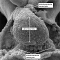
fig 1 Mouse E8 linear heart tube (SEM)
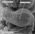
fig 2 Mouse E8 heart tube ventricular loop (SEM)
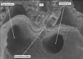
fig 5 Mouse E8.5 heart (SEM)


- Mouse E10.5
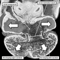
fig 3 Mouse E10.5 heart
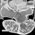
fig 4 Mouse E10.5 heart (EFIC)
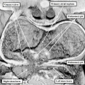
fig 6 Mouse E10.5 heart (EFIC)
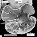
fig 8a Mouse E10.5 heart atrioventricular canal (EFIC)
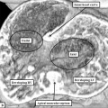
fig 8b Mouse E10.5 heart outflow tract (EFIC)
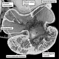
fig 13a Mouse E10.5 heart septum primum (EFIC)


- Mouse E11.5
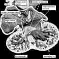
fig 12a Mouse E11.5 heart (EFIC)
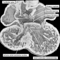
fig 24a Mouse E11.5 heart
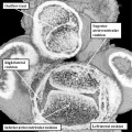
fig 25a Mouse E11.5 Heart atrioventricular junction
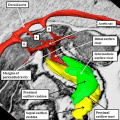
fig 38 Mouse E11.5 heart (EFIC)
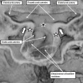
fig 39a Mouse E11.5 Heart aortic sac
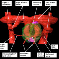
fig 39b Mouse E11.5 Heart arch arteries
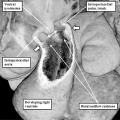
fig 40a Mouse E11.5 heart

fig 40b Mouse E11.5 heart (later)
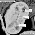
fig 42a Mouse E11.5 heart arterial roots and outflow tract


- Mouse E12.5
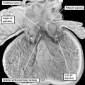
fig 24b Mouse E12.5 heart
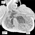
fig 25b Mouse E12.5 Heart atrioventricular junction
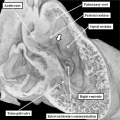
fig 31 Mouse E12.5 heart outflow tract
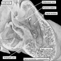
fig 46a Mouse E12.5 heart
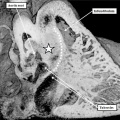
fig 33a Mouse E12.5 heart
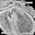
fig 33b Mouse E12.5 heart
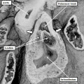
fig 42b Mouse E12.5 and E13.5 pulmonary valve
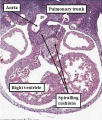
fig 50a Mouse E12.5 heart
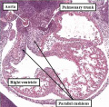
fig 50b Mouse E12.5 abnormal heart


- Mouse E13.5
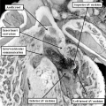
fig 26a Mouse E13.5 heart aortic root
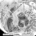
fig 26b Mouse E13.5 heart aortic root


- Mouse E14.5
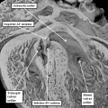
fig 27a Mouse E14.5 heart aortic root

fig 27b Mouse E14.5 heart aortic root
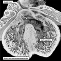
fig 32a Mouse E14.5 heart interventricular septum
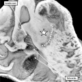
fig 32b Mouse E14.5 heart interventricular membranous septum


- Mouse E15.5
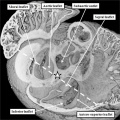
fig 28a Mouse E15.5 heart septation complete
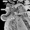
fig 28b Mouse E15.5 heart septation complete
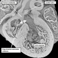
fig 34a Mouse E15.5 heart perimembranous defect


- Mouse E18.5
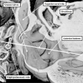
fig 17a Mouse E18.5 heart oval fossa (EFIC)
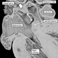
fig 17b Mouse E18.5 heart oval fossa (EFIC)
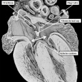
fig 29a Mouse E18.5 heart atrioventricular septal defect
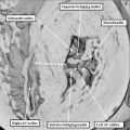
fig 29b Mouse E18.5 heart atrioventricular septal defect


References
- ↑ Chowdhury B, Xiang B, Liu M, Hemming R, Dolinsky VW & Triggs-Raine B. (2017). Hyaluronidase 2 Deficiency Causes Increased Mesenchymal Cells, Congenital Heart Defects, and Heart Failure. Circ Cardiovasc Genet , 10, . PMID: 28196902 DOI.
- ↑ Anderson RH. Teratogenecity in the setting of cardiac development and maldevelopment. (2016)
External Links
External Links Notice - The dynamic nature of the internet may mean that some of these listed links may no longer function. If the link no longer works search the web with the link text or name. Links to any external commercial sites are provided for information purposes only and should never be considered an endorsement. UNSW Embryology is provided as an educational resource with no clinical information or commercial affiliation.
Glossary Links
- Glossary: A | B | C | D | E | F | G | H | I | J | K | L | M | N | O | P | Q | R | S | T | U | V | W | X | Y | Z | Numbers | Symbols | Term Link
Cite this page: Hill, M.A. (2026, Haziran 30) Embryology Mouse Heart. Retrieved from https://embryology.med.unsw.edu.au/embryology/index.php/Mouse_Heart
- © Dr Mark Hill 2026, UNSW Embryology ISBN: 978 0 7334 2609 4 - UNSW CRICOS Provider Code No. 00098G