Domenech-Mateu Collection
| Embryology - 27 Apr 2024 |
|---|
| Google Translate - select your language from the list shown below (this will open a new external page) |
|
العربية | català | 中文 | 中國傳統的 | français | Deutsche | עִברִית | हिंदी | bahasa Indonesia | italiano | 日本語 | 한국어 | မြန်မာ | Pilipino | Polskie | português | ਪੰਜਾਬੀ ਦੇ | Română | русский | Español | Swahili | Svensk | ไทย | Türkçe | اردو | ייִדיש | Tiếng Việt These external translations are automated and may not be accurate. (More? About Translations) |
Introduction
(Anatomy and Embryology Unit, Morphological Science Department, Universitat Autònoma de Barcelona)

|

|
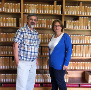
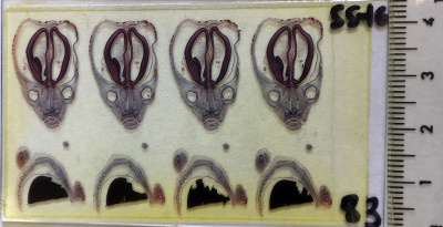
| Dr Mark Hill and Prof. Rosa Mirapeix in front of the human embryo collection. | Example of collection slide. |
- Carnegie Stages: 1 | 2 | 3 | 4 | 5 | 6 | 7 | 8 | 9 | 10 | 11 | 12 | 13 | 14 | 15 | 16 | 17 | 18 | 19 | 20 | 21 | 22 | 23 | About Stages | Timeline
Embryos
Stage 23
E109
Human Embryo (E109) 30 mm Abdomen, thorax
| Domenech-Mateu Collection Embryo - E109 | ||||||
|---|---|---|---|---|---|---|
| Carnegie Stage |
Embryo | Original | CRL (mm) | Section thickness |
Staining | Section plane |
| 23 | E109 | Bi-1 | 30 | 10 | ?? | Sagittal |
| Links: Carnegie Stage 23 | Domenech-Mateu Collection | DEC - Domenech-Mateu Collection | ||||||
Articles
Development of the arterial pattern in the upper limb of staged human embryos: normal development and anatomic variations
Arterial pattern in the human upper limb[1] "A total of 112 human embryos (224 upper limbs) between stages 12 and 23 of development were examined. It was observed that formation of the arterial system in the upper limb takes place as a dual process. An initial capillary plexus appears from the dorsal aorta during stage 12 and develops at the same rate as the limb. At stage 13, the capillary plexus begins a maturation process involving the enlargement and differentiation of selected parts. This remodelling process starts in the aorta and continues in a proximal to distal sequence. By stage 15 the differentiation has reached the subclavian and axillary arteries, by stage 17 it has reached the brachial artery as far as the elbow, by stage 18 it has reached the forearm arteries except for the distal part of the radial, and finally by stage 21 the whole arterial pattern is present in its definitive morphology. This differentiation process parallels the development of the skeletal system chronologically. A number of arterial variations were observed, and classified as follows: superficial brachial (7.7%), accessory brachial (0.6%). brachioradial (14%), superficial brachioulnar (4.7%), superficial brachioulnoradial (0.7%), palmar pattern of the median (18.7%) and superficial brachiomedian (0.7%) arteries. They were observed in embryos belonging to stages 17-23 and were not related to a specific stage of development. Statistical comparison with the rates of variations reported in adults did not show significant differences. It is suggested that the variations arise through the persistence, enlargement and differentiation of parts of the initial network which would normally remain as capillaries or even regress."
- Human embryos (224 upper limbs) belonging to the Bellaterra Collection (Prof. J. M. Domenech. Unit of Anatomy and Embryology, School of Medicine, Autonomous University of Barcelona, Spain) and the Boyd Collection (Department of Anatomy, University of Cambridge, UK) were studied.
References
Articles
Domènech-Mateu JM, Arnó Palau A & Martínez Pozo A. (1993). [The development of the atrioventricular node and bundle of His in the human embryonic period]. Rev Esp Cardiol , 46, 421-30. PMID: 8341829
Arango-Toro O & Domenech-Mateu JM. (1993). Development of the pelvic plexus in human embryos and fetuses and its relationship with the pelvic viscera. Eur J Morphol , 31, 193-208. PMID: 8217469
Domènech-Mateu JM, Martínez-Pozo A & Arnó-Palau A. (1994). Development of the tendon of Todaro during the human embryonic and fetal periods. Anat. Rec. , 238, 374-82. PMID: 8179219 DOI.
Orts Llorca F, Domenech Mateu JM & Puerta Fonolla J. (1983). [Typical complete transposition of the great arteries in a 19mm human embryo. A new theory of its embryogenesis]. Rev Esp Cardiol , 36, 81-8. PMID: 6878817
Orts Llorca F, Domenech Mateu JM & Puerta Fonolla J. (1979). Innervation of the sinu-atrial node and neighbouring regions in two human embryos. J. Anat. , 128, 365-75. PMID: 438095
Doménech-Mateu JM. (1988). Development and arterial supply of the supraventricular crest during the human embryonic and fetal periods. Acta Anat (Basel) , 132, 143-9. PMID: 3414360
Doménech-Mateu JM & Gonzalez-Compta X. (1988). Horseshoe kidney: a new theory on its embryogenesis based on the study of a 16-mm human embryo. Anat. Rec. , 222, 408-17. PMID: 3228209 DOI.
Nebot-Cegarra J & Domenech-Mateu JM. (1989). Association of tracheoesophageal anomalies with visceral and parietal malformations in a human embryo (Carnegie stage 21). Teratology , 39, 11-7. PMID: 2718136 DOI.
Search PubMed
External Links
External Links Notice - The dynamic nature of the internet may mean that some of these listed links may no longer function. If the link no longer works search the web with the link text or name. Links to any external commercial sites are provided for information purposes only and should never be considered an endorsement. UNSW Embryology is provided as an educational resource with no clinical information or commercial affiliation.
Glossary Links
- Glossary: A | B | C | D | E | F | G | H | I | J | K | L | M | N | O | P | Q | R | S | T | U | V | W | X | Y | Z | Numbers | Symbols | Term Link
Cite this page: Hill, M.A. (2024, April 27) Embryology Domenech-Mateu Collection. Retrieved from https://embryology.med.unsw.edu.au/embryology/index.php/Domenech-Mateu_Collection
- © Dr Mark Hill 2024, UNSW Embryology ISBN: 978 0 7334 2609 4 - UNSW CRICOS Provider Code No. 00098G
