Cardiovascular System - Atrial Septal Defects
| Embryology - 27 Apr 2024 |
|---|
| Google Translate - select your language from the list shown below (this will open a new external page) |
|
العربية | català | 中文 | 中國傳統的 | français | Deutsche | עִברִית | हिंदी | bahasa Indonesia | italiano | 日本語 | 한국어 | မြန်မာ | Pilipino | Polskie | português | ਪੰਜਾਬੀ ਦੇ | Română | русский | Español | Swahili | Svensk | ไทย | Türkçe | اردو | ייִדיש | Tiếng Việt These external translations are automated and may not be accurate. (More? About Translations) |
LB20 Congenital Anomaly of Atrioventricular Valves or Septum
| ICD-11 |
|---|
LB20 Congenital Anomaly of Atrioventricular Valves or Septum
|
| ICD-11 Structural developmental anomalies of the circulatory system (draft) |
|---|
| ICD-11 Beta Draft - NOT FINAL, updated on a daily basis, It is not approved by WHO, NOT TO BE USED for CODING except for agreed FIELD TRIALS.
20 Developmental Anomalies - Structural Developmental Anomalies Beta coding and tree structure for "structural developmental anomalies" within this section are shown in the table below. |
| Structural developmental anomalies of the circulatory system |
|
| CD-11 Beta Draft - NOT FINAL, updated on a daily basis, It is not approved by WHO, NOT TO BE USED for CODING except for agreed FIELD TRIALS.
|
Introduction
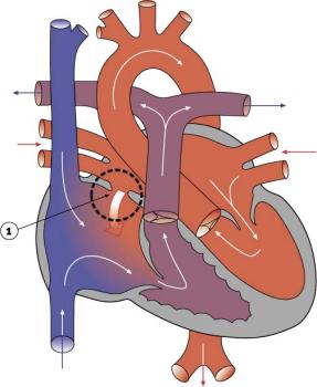
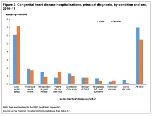
The atrial septal defects (ASD) are a group of common (1% of cardiac) congenital anomolies defects occuring in a number of different forms and more often in females. Patients have been reported living to old age with forms of this cardiac abnormality[1], that is also common in Trisomy 21.
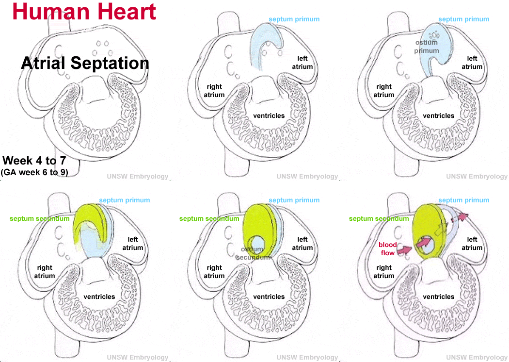
- patent foramen ovale - allows a continuation of the atrial shunting of blood, in 25% of people a probe patent foramen ovale (allowing a probe to bepassed from one atria to the other) exists.
- ostium secundum defect
- endocardial cushion defect involving ostium primum
- sinus venosus defect - contributes about 10% of all ASDs and occurs mainly in a common and less common form.
- Common ("usual type") - in upper atrial septum which is contiguous with the superior vena cava.
- Less common - at junction of the right atrium and inferior vena cava.
- common atrium
Some Recent Findings
|
| More recent papers |
|---|
|
This table allows an automated computer search of the external PubMed database using the listed "Search term" text link.
More? References | Discussion Page | Journal Searches | 2019 References | 2020 References Search term: Atrial Septal Defect | Congenital Anomaly of Atrioventricular Valves |
Movies
|
|
Oval Fossa Defect
|
|
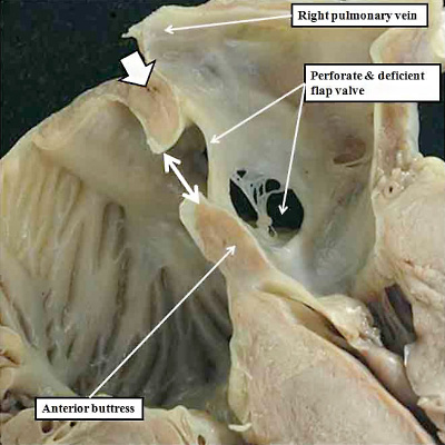
| The cross-section of a human heart shows defects (patent foramen ovale) in the floor of the oval fossa (fossa ovalis, annulus ovalis) shown by double headed white arrow due to deficiency and perforation of the flap valve derived from the primary atrial septum. The white arrow with black borders shows the intact infolded cranial rim of the fossa. |
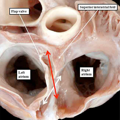
Human heart has been sectioned across the short axis of the atrial chambers, and photographed from above, to show a persistently patent oval foramen (double headed red arrow). The double headed white arrow shows the normal rims of the oval fossa, which are overlapped by the flap valve, but in absence of fusion with the left atrial aspect of the rims. |
| Heart specimen images produced by Diane Spicer. | |
Sinus Venosus Atrial Septal Defect
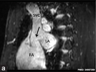
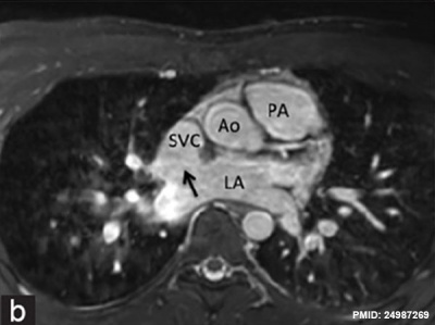
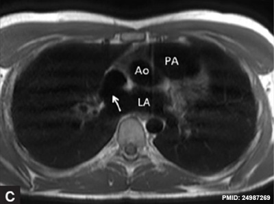
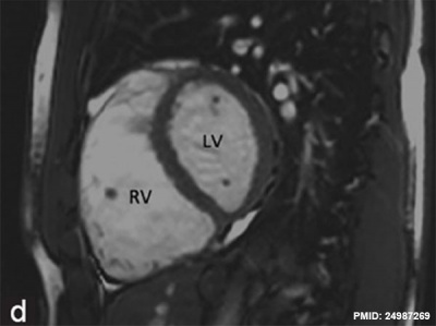
Sinus venosus atrial septal defects (SV-ASDs) are inter-atrial abnormality caused by a deficiency of the common wall between the superior or inferior vena cava and the right-sided pulmonary veins. The images below are MRI from a 25-year-old asymptomatic male[4] SSFP - Breath-held fat suppressed three-dimensional steady-state free precession.
|
|
| MRI SSFP sagittal view | SSFP axial view |
|
|
| Turbo spin-echo axial plane | SSFP dilated right ventricle |
- Links: Magnetic Resonance Imaging
History
1941
ATRIAL SEPTAL DEFECT.[5]
- "Patent foramen ovale and atrial (or auricular) septal defect (A.S.D.), though both characterised by an aperture in the atrial septum, are embryologically and pathologically different conditions."
Treatment
The surgical repair requires a cardiopulmonary bypass and is recommended in most cases of ostium secundum ASD, even though there is a significant risk involved. Ostium primum defects tend to present earlier and are often associated with endocardial cushion defects and defective mitral or tricuspid valves. In such cases, valve replacement may be necessary and the extended operation has a considerable chance of mortality.
- Increasingly closure by a transcatheter device closure has been applied.
- Repair of atrial septal defects on the perfused beating heart (atrial septal defect size 2 cm - 4.5 cm)[6]
Cardiovascular Abnormalities
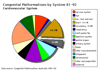
Heart defects and preterm birth are the most common causes of neonatal and infant death. The long-term development of the heart combined with extensive remodelling and post-natal changes in circulation lead to an abundance of abnormalities associated with this system.
A UK study literature showed that preterm infants have more than twice as many cardiovascular malformations (5.1 / 1000 term infants and 12.5 / 1000 preterm infants) as do infants born at term and that 16% of all infants with cardiovascular malformations are preterm. (0.4% of live births occur at greater than 28 weeks of gestation, 0.9% at 28 to 31 weeks, and 6% at 32 to 36 weeks. Overall, 7.3% of live-born infants are preterm)[7]
"Baltimore-Washington Infant Study data on live-born cases and controls (1981-1989) was reanalyzed for potential environmental and genetic risk-factor associations in complete atrioventricular septal defects AVSD (n = 213), with separate comparisons to the atrial (n = 75) and the ventricular (n = 32) forms of partial AVSD. ...Maternal diabetes constituted a potentially preventable risk factor for the most severe, complete form of AVSD."[8]
In addition, there are in several congenital abnormalities that exist in adults (bicuspid aortic valve, mitral valve prolapse, and partial anomalous pulmonary venous connection) which may not be clinically recognized.
References
- ↑ Matsumoto T, Tamiya E, Kanoh T, Takabe T, Kuremoto KI, Kamiyama T, Yamamoto S & Daida H. (2019). Atrial Septal Defect of the Ostium Secundum Type in A 101-Year-Old Patient. Int Heart J , , . PMID: 30799379 DOI.
- ↑ Australian Institute of Health and Welfare 2019. Congenital heart disease in Australia. Cat. no. CDK 14. Canberra: AIHW.
- ↑ Gervasi L & Basu S. (2009). Atrial septal defect devices used in the cardiac catheterization laboratory. Prog Cardiovasc Nurs , 24, 86-9. PMID: 19737165 DOI.
- ↑ Ganigara M, Tanous D, Celermajer D & Puranik R. (2014). The role of cardiac MRI in the diagnosis and management of sinus venosus atrial septal defect. Ann Pediatr Cardiol , 7, 160-2. PMID: 24987269 DOI.
- ↑ Bedford DE, Papp C & Parkinson J. (1941). ATRIAL SEPTAL DEFECT. Br Heart J , 3, 37-68. PMID: 18609869
- ↑ Pendse N, Gupta S, Geelani MA, Minhas HS, Agarwal S, Tomar A & Banerjee A. (2009). Repair of atrial septal defects on the perfused beating heart. Tex Heart Inst J , 36, 425-7. PMID: 19876418
- ↑ Tanner K, Sabrine N & Wren C. (2005). Cardiovascular malformations among preterm infants. Pediatrics , 116, e833-8. PMID: 16322141 DOI.
- ↑ Loffredo CA, Hirata J, Wilson PD, Ferencz C & Lurie IW. (2001). Atrioventricular septal defects: possible etiologic differences between complete and partial defects. Teratology , 63, 87-93. PMID: 11241431 <87::AID-TERA1014>3.0.CO;2-5 DOI.
Articles
Ng WP & Yip WL. (2012). Successful maternal-foetal outcome using nitric oxide and sildenafil in pulmonary hypertension with atrial septal defect and HIV infection. Singapore Med J , 53, e3-5. PMID: 22252195
Ma ZS, Dong MF, Yin QY, Feng ZY & Wang LX. (2012). Totally thoracoscopic closure for atrial septal defect on perfused beating hearts. Eur J Cardiothorac Surg , 41, 1316-9. PMID: 22219470 DOI.
Scheuerle A. (2011). Clinical differentiation of patent foramen ovale and secundum atrial septal defect: a survey of pediatric cardiologists in Dallas, Texas, USA. J Registry Manag , 38, 4-8. PMID: 22097699
Ağaç MT, Akyüz AR, Acar Z, Akdemir R, Korkmaz L, Kırış A, Erkuş E, Erkan H & Celik S. (2012). Evaluation of right ventricular function in early period following transcatheter closure of atrial septal defect. Echocardiography , 29, 358-62. PMID: 22066780 DOI.
Rodriguez FH, Moodie DS, Parekh DR, Franklin WJ, Morales DL, Zafar F, Graves DE, Friedman RA & Rossano JW. (2011). Outcomes of hospitalization in adults in the United States with atrial septal defect, ventricular septal defect, and atrioventricular septal defect. Am. J. Cardiol. , 108, 290-3. PMID: 21545985 DOI.
Search Pubmed
Search Pubmed: Atrial Septal Defect
External Links
External Links Notice - The dynamic nature of the internet may mean that some of these listed links may no longer function. If the link no longer works search the web with the link text or name. Links to any external commercial sites are provided for information purposes only and should never be considered an endorsement. UNSW Embryology is provided as an educational resource with no clinical information or commercial affiliation.
- OMIM Atrial Septal Defect
- Medline Plus ASD Repair Video
Glossary Links
- Glossary: A | B | C | D | E | F | G | H | I | J | K | L | M | N | O | P | Q | R | S | T | U | V | W | X | Y | Z | Numbers | Symbols | Term Link
Cite this page: Hill, M.A. (2024, April 27) Embryology Cardiovascular System - Atrial Septal Defects. Retrieved from https://embryology.med.unsw.edu.au/embryology/index.php/Cardiovascular_System_-_Atrial_Septal_Defects
- © Dr Mark Hill 2024, UNSW Embryology ISBN: 978 0 7334 2609 4 - UNSW CRICOS Provider Code No. 00098G
