File:Mall1912-fig02.jpg

Original file (797 × 730 pixels, file size: 180 KB, MIME type: image/jpeg)
Fig. 2. From the reconstruction of a heart of an embryo 3.5 mm long
Embryo (No. 164). X 66. View from the left side.
In the embryo 3.5 mm. long (No. 164) the completed heart tube is seen, which is S-shaped and twisted upon itself so that the arterial and venous ends are brought close together. At the venous end the muscle wall is slightly dilated which marks the atrium; this is lined closely with endothelium which encircles the cavity within. No delicate fibrils are here seen between the muscle wall and its endothelial lining. Then follows an upper bend to the heart after which there is a dilatation projecting towards the left side, the former marking the atrial: canal and the latter the left ventricle. The lower connecting piece unites the left ventricle with the bulb which later on gives rise to the right ventricle. In the atrial canal (Haller’s auricular canal) the endothclial tube is seen as a solid strand of cells suspended freely in the muscle wall by the delicate fibrils already mentioned. In the left ventricle the tube shows a distinct cavity, while throughout the rest of the heart tube the cavity is irregular but not pronounced. The form of the endothelial tube is shown in fig. 2 and again in a semi-diagrammatic figure of a transverse section through the atrium, ventricle and bulb in fig. 3. The delicate fibrils, which no doubt belong to the endothelial cells are present in large number throughout the whole heart tube, excepting in that which forms the atrium. In another embryo (No. 384, 2 mm. long), considerably smaller than the one just described and probably pathological, the degree of development of the heart is practically identical with the one 3.5 mm. long (No. 164).
| Mall Figure Legend | ||
|---|---|---|
|
|
|
| Historic Disclaimer - information about historic embryology pages |
|---|
 |
- Links: Fig 1 | Fig 2 | Fig 3 | Fig 4 | Fig 5 | Fig 6 | Fig 7 | Fig 8 | Fig 9 | Fig 10 | Fig 11 | Fig 12 | Fig 13 | Fig 14 | Fig 15 | Fig 16 | Fig 17 | Fig 18 | Fig 19 | Fig 20 | Fig 21 | Fig 22 | Fig 23 | Fig 24-25 | Fig 26 | Fig 27 | Fig 28 | Fig 31-33 | Mall 1912 | Historic Papers | Carnegie Collection | Franklin Mall | Cardiovascular Development

Reference
Mall FP. On the development of the human heart. (1912) Amer. J Anat. 13: 249-298.
Cite this page: Hill, M.A. (2024, April 27) Embryology Mall1912-fig02.jpg. Retrieved from https://embryology.med.unsw.edu.au/embryology/index.php/File:Mall1912-fig02.jpg
- © Dr Mark Hill 2024, UNSW Embryology ISBN: 978 0 7334 2609 4 - UNSW CRICOS Provider Code No. 00098G
File history
Click on a date/time to view the file as it appeared at that time.
| Date/Time | Thumbnail | Dimensions | User | Comment | |
|---|---|---|---|---|---|
| current | 00:36, 9 September 2015 | | 797 × 730 (180 KB) | Z8600021 (talk | contribs) | |
| 00:31, 9 September 2015 | 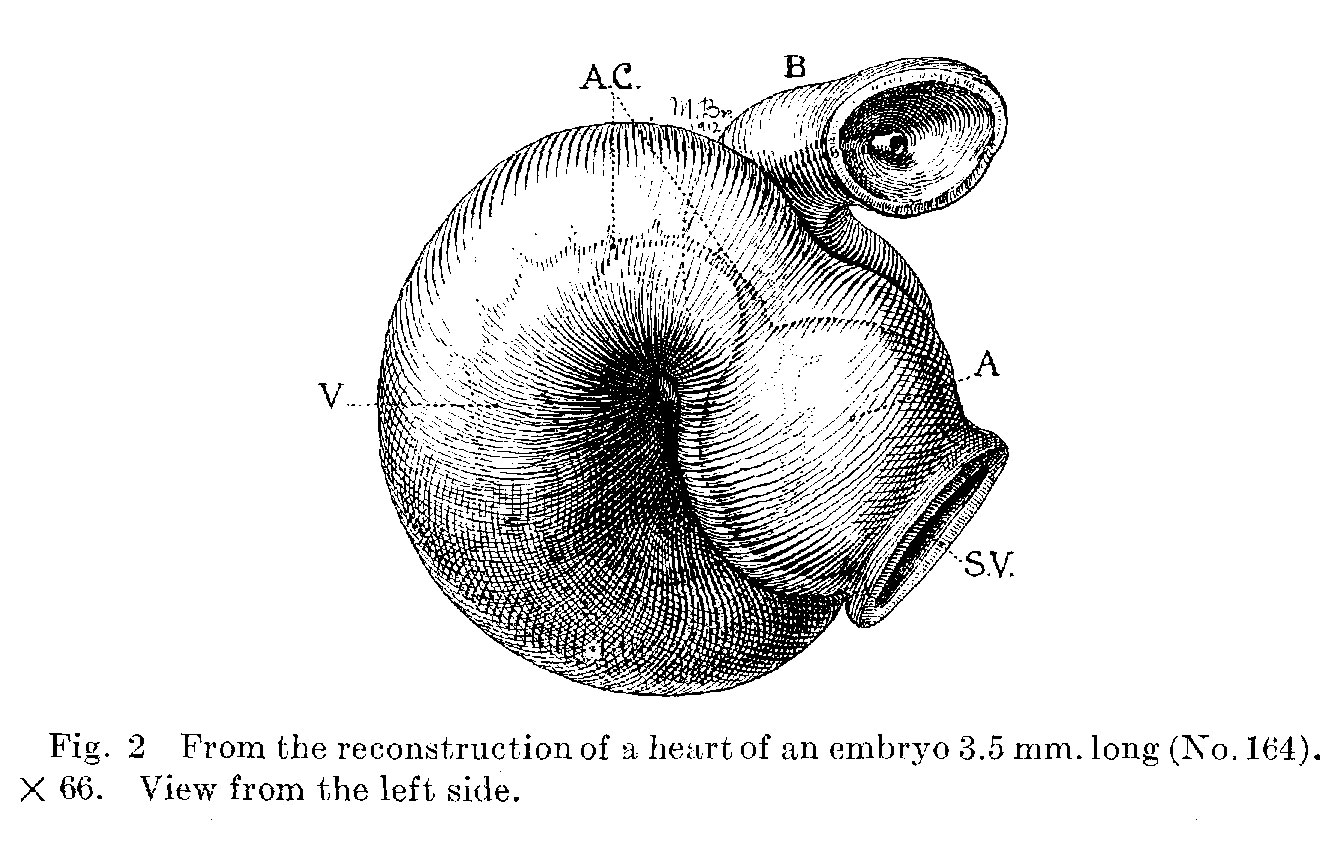 | 1,328 × 848 (235 KB) | Z8600021 (talk | contribs) | {{Mall1912 figures}} |
You cannot overwrite this file.
File usage
The following page uses this file: