File:Heart outflow tract stage 14 03.jpg
Original file (989 × 996 pixels, file size: 134 KB, MIME type: image/jpeg)
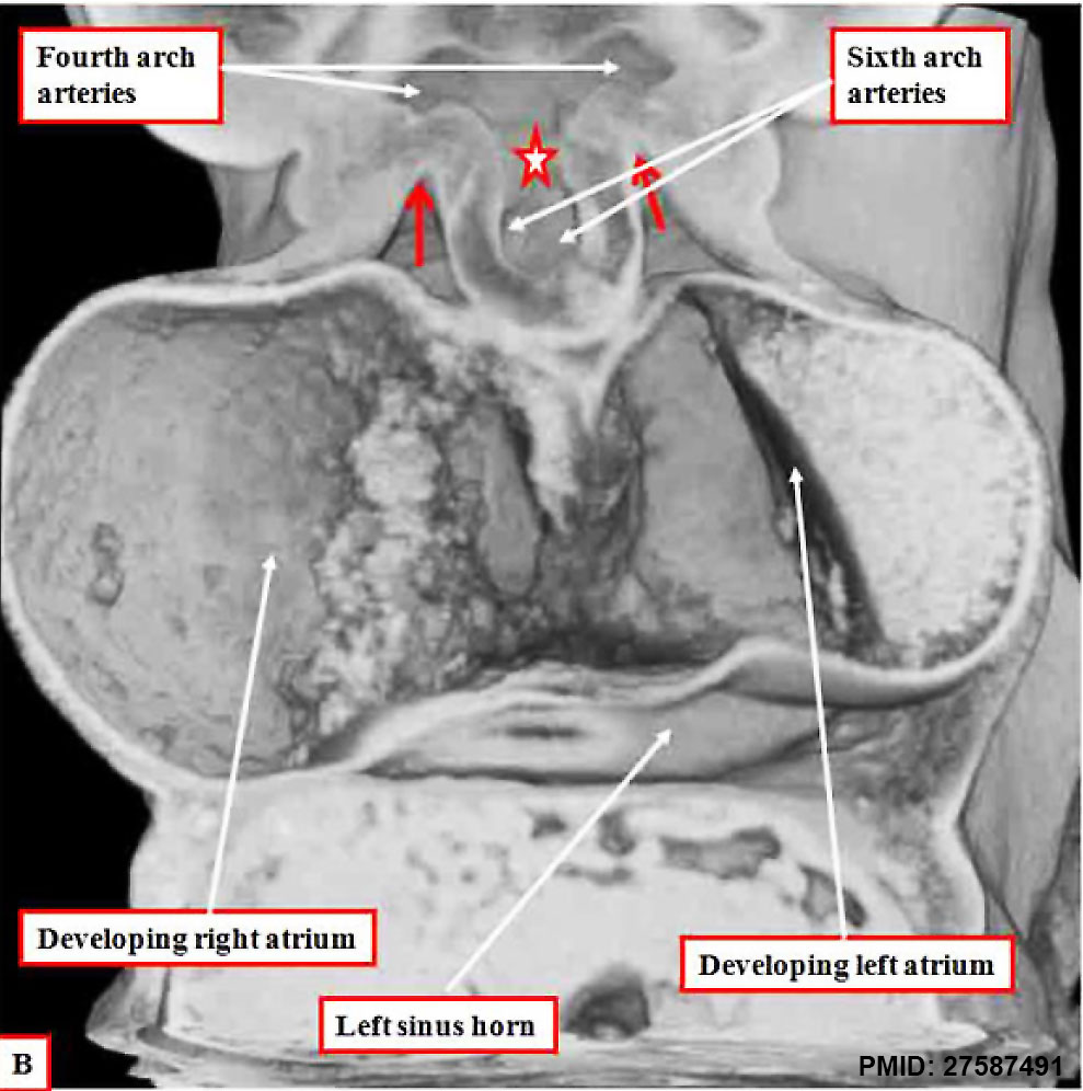
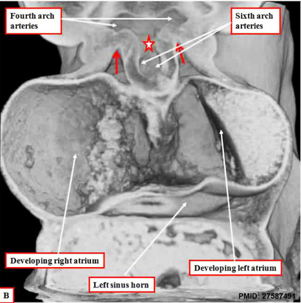
Heart Outflow Tract (Carnegie Stage 14) EFIC
EFIC images are from an episcopic data set prepared from a human embryo at Carnegie stage 14, representing the end of the fifth week of development.
B cut in frontal fashion at the junction of the outflow tract with the pharyngeal mesenchyme, shows the origins of the pharyngeal arch arteries from the aortic sac. The back wall of the sac, shown by the star, is the putative aortopulmonary septum. The two single-headed arrows show the extent of the pericardial cavity.
Sixth Arch Artery
- right arch - proximal part persists as right pulmonary artery, distal part degenerates
- left arch - left pulmonary artery and ductus arteriosus
- See Pharyngeal arches
| Pharyngeal Arch Derivatives | |||||
|---|---|---|---|---|---|
| Pharyngeal Arch | Nerve | Artery | Neural Crest (Skeletal Structures) |
Muscles | Ligaments |
| 1 (maxillary/mandibular) |
trigeminal (V) | maxillary artery (terminal branches) | mandible, maxilla, malleus, incus | muscles of mastication, mylohyoid, tensor tympanic, ant. belly digastric | ant lig of malleus, sphenomandibular ligament |
| 2 (hyoid) |
facial (VII) | stapedial (embryonic) corticotympanic (adult) |
stapes, styloid process, lesser cornu of hyoid, upper part of body of hyoid bone | muscles of facial expression, stapedius, stylohyoid, post. belly digastric | stylohyoid ligament |
| 3 | glossopharyngeal (IX) | common carotid, internal carotid arteries | greater cornu of hyoid, lower part of body of hyoid bone | stylopharyngeus | |
| 4 | vagus (X) superior laryngeal branch | part of aortic arch (left), part right subclavian artery (right) | thyroid, cricoid, arytenoid, corniculate and cuneform cartilages | crycothyroid, soft palate levator veli palatini (not tensor veli palatini) | |
| 6 | vagus (X) recurrent laryngeal branch | part of left pulmonary artery (left), part of right pulmonary artery (right) | thyroid, cricoid, arytenoid, corniculate and cuneform cartilages | larynx intrinsic muscles (not cricothyroid muscle) | |
- Links: Image EFIC outflow tract frontal | Image EFIC - OFT RA RV | Image EFIC - OFT RA LA | Carnegie stage 14 | Week 5 | EFIC
Reference
<pubmed>27587491</pubmed>
https://www.ncbi.nlm.nih.gov/pmc/articles/PMC5011314/
http://journals.sagepub.com/doi/abs/10.1177/2150135116651114
PMID 27587491
Copyright
© The Author(s) 2016
https://creativecommons.org/licenses/by/3.0/
Figure 2. cropped and resized.
Cite this page: Hill, M.A. (2024, April 27) Embryology Heart outflow tract stage 14 03.jpg. Retrieved from https://embryology.med.unsw.edu.au/embryology/index.php/File:Heart_outflow_tract_stage_14_03.jpg
- © Dr Mark Hill 2024, UNSW Embryology ISBN: 978 0 7334 2609 4 - UNSW CRICOS Provider Code No. 00098G
File history
Click on a date/time to view the file as it appeared at that time.
| Date/Time | Thumbnail | Dimensions | User | Comment | |
|---|---|---|---|---|---|
| current | 12:00, 29 January 2017 | | 989 × 996 (134 KB) | Z8600021 (talk | contribs) | |
| 12:00, 29 January 2017 | 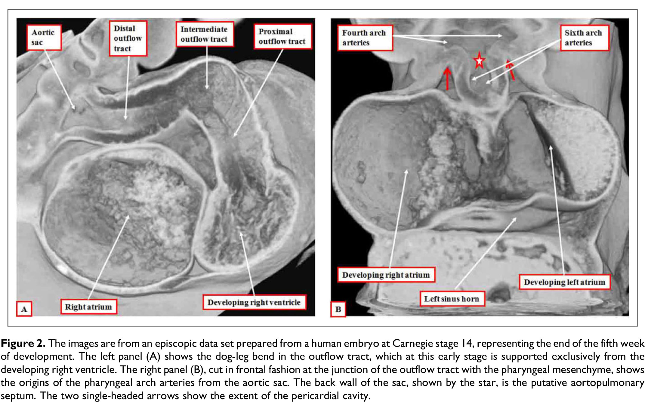 | 2,144 × 1,353 (421 KB) | Z8600021 (talk | contribs) |
You cannot overwrite this file.
File usage
The following page uses this file: