2009 Lecture 21
Redirect to:
Late Cardiovascular Development
Introduction
In lecture 7 - Early Vascular Development, there was an introduction to the origins of the cardiovascular system. This second lecture will now focus on the extensive remodeling that occurs in both the heart and vascular system during later development. In addition, there will be discussion on the major cardiovascular abnormalities. The laboratory this week will also give you the opportunity to work through some of these concepts using a new online teaching module.
This material is also presented in an online Cardiac Embryology tutorial.
- Lecture Audio Lecture Date: 12-10-2009 Lecture Time: 12:00 Venue: CLB 5 Speaker: Mark Hill Late Cardiovascular
Lecture Objectives
- Describe the development of primary and secondary atrial septa and the ventricular septum.
- Explain the changes occurring in the bulbis cordis and truncus arteriosus in its transformation from a single to a double tube.
- Describe the development of the aortic arches on the right and left sides from the fetus to the adult.
- Describe the development of arteries and veins.
- Describe the major cardiovascular developmental abnormalities.
Textbook references
- Human Embryology (3rd ed.) Larson Chapter 7 p151-188 Heart, Chapter 8 p189-228 Vasculature
- The Developing Human: Clinically Oriented Embryology (6th ed.) Chapter 14: p304-349
Other textbooks
- Before we Are Born (5th ed.) Moore and Persaud Chapter 12; p241-254
- Essentials of Human Embryology Larson Chapter 7 p97-122 Heart, Chapter 8 p123-146 Vasculature
- Human Embryology Fitzgerald and Fitzgerald Chapter 13-17: p77-111
Recent reviews
- Yutzey KE, Kirby ML. Wherefore heart thou? Embryonic origins of cardiogenic mesoderm. Dev Dyn. 2002 Mar;223(3):307-20. Review. PMID: 11891982
- Three-dimensional reconstruction of gene expression patterns during cardiac development. Soufan AT, Ruijter JM, van den Hoff MJ, de Boer PA, Hagoort J, Moorman AF. Physiol Genomics. 2003 May 13;13(3):187-95. Review. PMID: 12746463
- Moorman A, Webb S, Brown NA, Lamers W, Anderson RH. Development of the heart: (1) formation of the cardiac chambers and arterial trunks. Heart. 2003 Jul;89(7):806-14. PMID: 12807866
- Bruneau BG. Transcriptional regulation of vertebrate cardiac morphogenesis. Circ Res. 2002 Mar 22;90(5):509-19. Review. PMID: 11909814
Heart Development
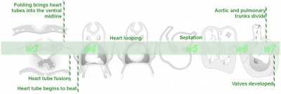
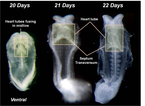
- endocardial tube in pericardial cavity
- dorsal mesentry (mesocardial) attachment lost
- attached at cranial (arterial) and caudal (venous) ends
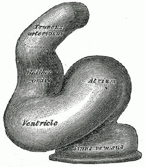
- tube elongation - bending and series of expansions
Bulbus Cordis - 3 parts
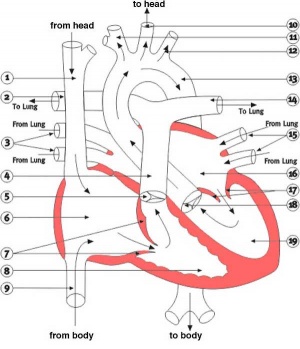
- distal - forms truncus arteriosus (aorta and pulmonary artery)
- middle - conus cordis ventricular outflow tract
- proximal - right ventricle trabeculate part
endocardial cushions
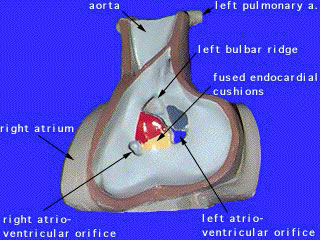
- form initial division of atria and ventricles
- fusion forma a left and right atrioventricular canals
Heart Layers
- pericardium - covers the heart, formed by 3 layers consisting of a fibrous pericardium and a double layered serous pericardium (parietal layer and visceral epicardium layer).
- myocardium - muscular wall of the heart, thickest layer formed by spirally arranged cardiac muscle cells.
- endocardium - lines the heart, epithelial (endothelial) tissue lining the inner surface of heart chambers and valves.
Embryonic Heart Rate
- Stage 9-10 2 mm embryo (gestational sac diameter of 20 mm) EHR at least 75 beats / minute
- Stage 11-12 5 mm embryo (gestational sac diameter of 30 mm) EHR at least 100 beats / minute
- Stage 16 10 mm embryo EHR at least 120 beats / minute
- Stage 18 15 mm embryo EHR at least 130 beats / minute
Week 12 fetal heart rate doppler Week 17 fetal heart rate audio
Atrial Septation
Through all development blood shunts from right to left atrium (bypass lungs)
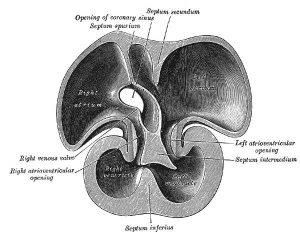
Septum Primum
Stage 13 Septum Primum Stage 22 Septum Primum
- dorso-cranial wall, membranous extension
- grows downward towards endocardial cushions
- opening is foramen primum (ostium primum)
- septum primum fuses with endocardial cushion, but cranially had begun to degenerate forming foramen secundum (ostium secundum)
Septum Secundum
- ventro-cranial wall
- grows as septum primum downward, does not fuse with endocardial cushion, opening is foramen ovale
Right Atrium
- right sinus horn incorporates into dorsal wall sinus venerum
- sinoatrial opening - has 2 flaps, left fuses with septum secundum, right forms valve to inferior vena cava and coronary sinus. Stage 13 - right venous valve Stage 22 - right venous valve
Left Atrium
- posterior wall - outgrowth forms single pulmonary vein, divided into 4 branches
- incorporation into the wall leads to 4 openings in posterior wall
- later moves to the right aligns with atrioventricular canal.
Ventricular Septation
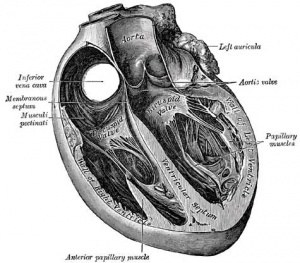
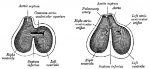
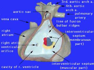
- 2 separate components - superior membranous, inferior muscular
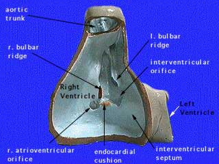
Muscular Septum
- growth of inferior wall
- fusion of 3 components - right and left bulbar ridges and dorsal endocardial cushion
Membranous Septum
- above the muscular septum, fusion continuous with septation of the outflow tract
Outflow Tract Septation
- truncus arteriousus - 2 growths from wall in spiral pattern, inferior upwards Stage 13 truncus arteriousus
- mesenchyme and neural crest contribute to this septation process
- fusion of outgrowths separate aortic and pulmonary outflow
| Normal Heart | Abnormal Heart | Abnormal Heart 2 |
Vascular Remodeling
Arterial System
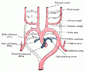
- Aortic sac - remodels forming 2 horns, R forms brachiocephalic artery, L forms common carotid artery
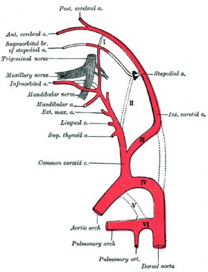
Pharyngeal arch arteries
- pharyngeal arch artery 1 and 2 regress
- pharyngeal arch artery 3 and the associated dorsal aorta form the paired internal carotid arteries, these in turn generate the external carotids
- Left pharyngeal arch artery 4 – forms the aortic arch
- paraxial mesoderm forms paired dorsal aorta
- head mesenchyme forms aortic arches
- connecting stalk contains unbilical (placental) arteries
- dorsal aortas give rise to
- vitelline arteries which connect to capillaries on yolk sac
- intersegmental arteries between somites
Venous System
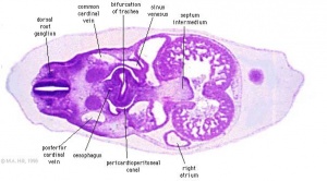
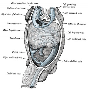
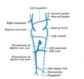
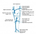
Cardinal veins contribute nearly all systemic venous system.
common cardinal veins - ducts of Cuvier
hepatic veins - drain de-oxygenated blood from the liver into the inferior vena cava.
internal iliac vein - hypogastric vein
Birth Changes
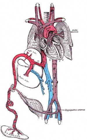
?| Fetal Structure | Adult Structure |
|---|---|
| foramen ovale | fossa ovalis |
| umbilical vein (intra-abdominal part) | ligamentum teres |
| ductus venosus | ligamentum venosum |
| umbilical arteries and abdominal ligaments | medial umbilical ligaments, superior vesicular artery (supplies bladder) |
| ductus arteriosum | ligamentum arteriosum |
Abnormalities
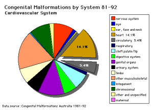
There are many different cardiac abnormalities, some more common than others, and only a few will be described in this lecture.
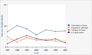
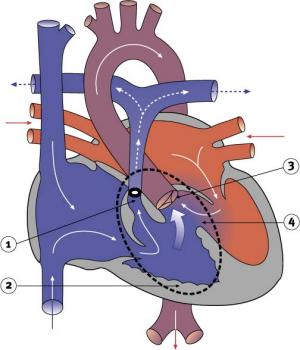
Major Abnormalities: Aortic Stenosis, Atrial Septal Defects, Coarctation of Aorta, Dextrocardia, Hypoplastic Left Heart, Long QT Syndrome, Patent Ductus Arteriosus, Pulmonary Atresia, Pulmonary Stenosis, Tetralogy of Fallot, Transposition of Great Vessels, Tricuspid Atresia, Total Anomalous Pulmonary Venous Connection, Ventricular Septal Defect, Abnormalities of Conducting System.
Links: Cardiovascular Development Abnormalities
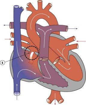
Atrial Septal Defects
|
|
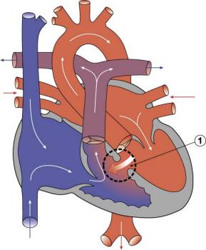
Ventricular Septal Defects
|
|
(V.S.D.)
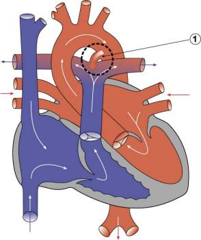
Patent Ductus Arteriosus
|
|
Tetralogy of Fallot
|
|
Movies
| Heart Looping | Heart Realign | Heart Atrial Septation | Heart Outflow Septation |
Link: Flash Movies
Glossary Links
- Glossary: A | B | C | D | E | F | G | H | I | J | K | L | M | N | O | P | Q | R | S | T | U | V | W | X | Y | Z | Numbers | Symbols | Term Link
Course Content 2009
Embryology Introduction | Cell Division/Fertilization | Cell Division/Fertilization | Week 1&2 Development | Week 3 Development | Lab 2 | Mesoderm Development | Ectoderm, Early Neural, Neural Crest | Lab 3 | Early Vascular Development | Placenta | Lab 4 | Endoderm, Early Gastrointestinal | Respiratory Development | Lab 5 | Head Development | Neural Crest Development | Lab 6 | Musculoskeletal Development | Limb Development | Lab 7 | Kidney | Genital | Lab 8 | Sensory - Ear | Integumentary | Lab 9 | Sensory - Eye | Endocrine | Lab 10 | Late Vascular Development | Fetal | Lab 11 | Birth, Postnatal | Revision | Lab 12 | Lecture Audio | Course Timetable
Cite this page: Hill, M.A. (2024, April 30) Embryology 2009 Lecture 21. Retrieved from https://embryology.med.unsw.edu.au/embryology/index.php/2009_Lecture_21
- © Dr Mark Hill 2024, UNSW Embryology ISBN: 978 0 7334 2609 4 - UNSW CRICOS Provider Code No. 00098G
