Category:Histology
From Embryology
Introduction
This Embryology category lists files and media related to histology. Most links are to histological images that relate to tissue structure and development. Many images are sourced from the original UNSW Anatomy Histology slide set and UWA Blue Histology online images.
Subcategories
This category has the following 15 subcategories, out of 15 total.
Pages in category 'Histology'
The following 200 pages are in this category, out of 334 total.
(previous page) (next page)A
- Template:Adrenal Histology
- AE Practical - Neural Histology
- Template:Al. coch.
- Alagille Syndrome
- ANAT2241 Blood
- ANAT2241 Bone, Bone Formation and Joints
- ANAT2241 Cardiovascular System
- ANAT2241 Connective Tissue Components
- ANAT2241 Connective Tissue Types
- ANAT2241 Covering and Lining Epithelia
- ANAT2241 Endocrine System
- ANAT2241 Female Reproductive System
- Template:ANAT2241 footer
- ANAT2241 Gastro-Intestinal System
- ANAT2241 Glandular Epithelia
- ANAT2241 Histology - Basic and Systematic
- Talk:ANAT2241 Histology - Basic and Systematic
- ANAT2241 Integumentary System
- ANAT2241 Liver, Gallbladder, and Pancreas
- ANAT2241 Lymphatic Tissue and Immune System
- ANAT2241 Male Reproductive System
- ANAT2241 Muscle Tissue
- ANAT2241 Nervous Tissue
- ANAT2241 Respiratory System
- ANAT2241 Special Senses The Eye
- ANAT2241 The Virtual Microscope
- ANAT2241 Urinary System
- ANAT2241 Virtual Slides - Example Spot Images
- ANAT2511 - Fundamentals of Anatomy
- ANAT2511 Basic Tissues
- ANAT2511 Bones and Joints
- ANAT2511 Circulatory System
- Template:ANAT2511 footer
- ANAT2511 Gastrointestinal Tract
- ANAT2511 Integumentary System
- ANAT2511 Introduction to Histology
- ANAT2511 Muscle Tissue
- ANAT2511 Nervous Tissue
- ANAT2511 Respiratory System
- ANAT2511 Urinary System
- Template:Artifacts
B
- BGD Lecture - Endocrine Histology
- BGD Lecture - Gastrointestinal System Development
- Talk:BGD Lecture - Gastrointestinal System Development
- BGDA Practical - Female Reproductive Tract Histology
- BGDB Gastrointestinal - Abnormalities
- BGDB Practical - Gastrointestinal System Development
- Talk:BGDB Practical - Gastrointestinal System Development
- BGDB Practical - Upper Gastrointestinal Tract Histology
- Blood Cell Histology Movie
- Template:Blood Histology
- Template:Blood Vessel Histology
- Template:Blood vessel histology
- Template:Blue Histology
- Bone Development
- Bone Histology
- Template:Bone Histology
- Template:Bone histology
- Template:Bone Marrow Histology
- Book - A laboratory guide in histology (1917)
- Book - A Laboratory Text-Book of Embryology 8 (1903)
- Book - A text-book of histology arranged upon an embryological basis (1913)
- Book - A text-book of histology arranged upon an embryological basis (1913) 1
- Book - A text-book of histology arranged upon an embryological basis (1913) 1-1
- Book - A text-book of histology arranged upon an embryological basis (1913) 1-2
- Book - A text-book of histology arranged upon an embryological basis (1913) 1-3
- Book - A text-book of histology arranged upon an embryological basis (1913) 2
- Book - A text-book of histology arranged upon an embryological basis (1913) 2-1
- Book - A text-book of histology arranged upon an embryological basis (1913) 2-2
- Book - A textbook of histology, including microscopic technic (1910) General Histology 1
- Book - A textbook of histology, including microscopic technic (1910) General Histology 2
- Book - A textbook of histology, including microscopic technic (1910) microscopic technic
- Book - A textbook of histology, including microscopic technic (1910) Special Histology 1
- Book - A textbook of histology, including microscopic technic (1910) Special Histology 10
- Book - A textbook of histology, including microscopic technic (1910) Special Histology 2
- Book - A textbook of histology, including microscopic technic (1910) Special Histology 3
- Book - A textbook of histology, including microscopic technic (1910) Special Histology 4
- Book - A textbook of histology, including microscopic technic (1910) Special Histology 5
- Book - A textbook of histology, including microscopic technic (1910) Special Histology 6
- Book - A textbook of histology, including microscopic technic (1910) Special Histology 7
- Book - A textbook of histology, including microscopic technic (1910) Special Histology 8
- Book - A textbook of histology, including microscopic technic (1910) Special Histology 9
- Book - Contributions to Embryology Carnegie Institution No.50
- Book - Histology and Embryology 1941
- Book - Liver development
- Book - Stoehr's Histology (1906)
- Book - Stoehr's Histology 1
- Talk:Book - Stoehr's Histology 1
- Book - Stoehr's Histology 1-1
- Book - Stoehr's Histology 1-2
- Book - Stoehr's Histology 1-3
- Book - Stoehr's Histology 2
- Book - Stoehr's Histology Figures
- Book - The microscopic anatomy of the human body, in health and disease
- Template:Bouin
- Buccopharyngeal membrane
- Template:BöhmDavidoffHuber1910 footer
- Template:BöhmDavidoffHuber1910 TOC
C
- Template:CapillaryEM links
- Template:Cardiac muscle EM
- Cardiac Muscle Histology
- Cardiovascular - Arterial Development
- Cardiovascular System - Blood Development
- Cardiovascular System - Heart Histology
- Cardiovascular System - Spleen Development
- Cartilage Histology
- Template:Cartilage Histology
- Template:Cartilage histology
- Cloaca Development
- Colon Histology 2009
- Template:Common Stains collapsetable
- Template:Common Stains table
E
F
G
- Template:Gall Bladder Histology Images
- Gastrointestinal Tract - Abnormalities
- Gastrointestinal Tract - Carnegie Stage 13
- Gastrointestinal Tract - Carnegie Stage 22
- Gastrointestinal Tract - Colon Histology
- Gastrointestinal Tract - Gall Bladder Development
- Gastrointestinal Tract - Gall Bladder Histology
- Gastrointestinal Tract - Gallbladder Development
- Gastrointestinal Tract - Gallbladder Histology
- Gastrointestinal Tract - Histology
- Gastrointestinal Tract - Intestine Development
- Gastrointestinal Tract - Liver Development
- Gastrointestinal Tract - Liver Histology
- Gastrointestinal Tract - Mesentery Development
- Gastrointestinal Tract - Mouth Development
- Gastrointestinal Tract - Oesophagus Development
- Gastrointestinal Tract - Pancreas Development
- Gastrointestinal Tract - Pancreas Histology
- Gastrointestinal Tract - Postnatal
- Gastrointestinal Tract - Stomach Development
- Gastrointestinal Tract Development
- Template:Gastrointestinal Tract Links
- Template:GIT histology links
H
- Template:Hair histology links
- Template:HE
- Template:Heart histology
- Template:HillH52
- Histology
- Template:Histology
- Histology and Embryology 1941 - Bibliography
- Histology and Embryology 1941 - Embryology
- Histology and Embryology 1941 - Embryology 1
- Histology and Embryology 1941 - Embryology 2
- Histology and Embryology 1941 - Histology
- Histology and Embryology 1941 - Histology 1
- Histology and Embryology 1941 - Histology 2
- Histology and Embryology 1941 - Histology 3
- Histology Artifacts
- Histology Fixatives
- Template:Histology Links
- Histology Stains
- Template:Histology Stains
- HM Practical - Blood Vessel Histology
- HM Practical - Cardiac Histology
- Template:Human follicles lm and em links
- Template:Human ovary - corpus luteum links
- Human System Development
I
L
M
P
- Template:Pancreas Histology Images
- Paper - A case of atresia ani in a human embryo of 26 mm
- Paper - A case of atresia of the esophagus combined with traoheoesophageal fistula in a 9 mm human embryo, and its embryological explanation
- Paper - A case of congenital malformations of the intestinal canal (1923)
- Paper - A Contribution to the Embryology of the Liver and Vascular System in Man
- Paper - A contribution to the morphology and development of the mammalian liver
- Paper - A contribution to the morphology and development of the mammalian liver (1908)
- Paper - A histological investigation of the development and structure of the human lung
- Paper - A model demonstrating the changes in position and peritoneal relations of abdominal viscera during development (1912)
- Paper - A morphological study of the development of the human liver 1
Media in category 'Histology'
The following 116 files are in this category, out of 718 total.
(previous page) (next page) Spinal cord histology 11.jpg 481 × 600; 117 KB
 Spinal cord histology 12.jpg 480 × 600; 130 KB
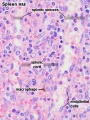
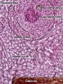
 Spleen histology 01.jpg 450 × 600; 133 KB
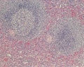
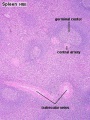 Spleen histology 02.jpg 455 × 606; 142 KB
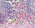
 Spleen histology 03.jpg 450 × 600; 83 KB
 Spleen histology 04.jpg 450 × 600; 108 KB
 Spleen histology 05.jpg 450 × 600; 170 KB
 Spleen histology 06.jpg 1,000 × 800; 408 KB
 Spleen histology 07.jpg 1,000 × 800; 251 KB
 Spleen histology 08.jpg 1,000 × 800; 304 KB
 Spleen histology 09.jpg 1,280 × 1,024; 692 KB
 Spleen histology 10.jpg 1,280 × 1,024; 444 KB
 Spleen histology 11.jpg 1,280 × 1,024; 410 KB
 Spleen histology 12.jpg 600 × 450; 93 KB
 Spleen histology 13.jpg 600 × 450; 80 KB
 Spleen structure 01.jpg 1,200 × 463; 211 KB
 Spleen structure 02.jpg 401 × 463; 46 KB
 Stage11 histology-neural tube roof plate 1.jpg 800 × 594; 134 KB
 Stage11 histology-neural tube roof plate 2.jpg 800 × 552; 145 KB
 Stage11 histology-optic pit.jpg 800 × 510; 157 KB
 Stage11 histology-optic vesicle-hindbrain.jpg 800 × 536; 158 KB
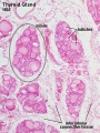
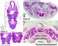 Stage13 and 22 thyroid development a.jpg 800 × 640; 94 KB
 Stage13 and 22 thyroid development.jpg 1,000 × 800; 283 KB
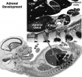
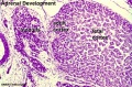 Stage22 adrenal.jpg 596 × 392; 65 KB
 Stage22 HPA1L.jpg 640 × 433; 65 KB
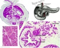
 Stage22 pancreas a.jpg 800 × 640; 99 KB
 Stage22 pancreas b.jpg 600 × 480; 63 KB
 Stage22 pancreas c.jpg 400 × 320; 38 KB
 Stage22 pancreas.jpg 1,000 × 800; 264 KB
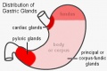 Stomach gastric gland distribution.jpg 500 × 334; 28 KB
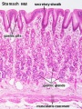 Stomach histology 001.jpg 400 × 533; 87 KB
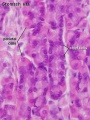 Stomach histology 002.jpg 400 × 532; 53 KB
 Stomach histology 003.jpg 599 × 400; 53 KB
 Stomach histology 004.jpg 1,280 × 1,024; 435 KB
 Stomach histology 005.jpg 1,280 × 1,024; 432 KB
 Stomach histology 006.jpg 1,280 × 1,024; 227 KB
 Stomach histology 007.jpg 1,280 × 1,024; 308 KB
 Stomach histology 008.jpg 1,280 × 1,024; 452 KB
 Stratified epithelia cartoon.jpg 696 × 1,000; 166 KB
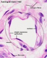
 Sublingual gland histology 02.jpg 500 × 500; 23 KB
 Testis histology 001.jpg 1,280 × 1,024; 574 KB
 Testis histology 002.jpg 1,280 × 1,024; 599 KB
 Testis histology 003.jpg 1,280 × 1,024; 183 KB
 Testis histology 004.jpg 1,280 × 1,024; 396 KB
 Testis histology 005.jpg 1,280 × 1,024; 266 KB
 Testis histology 006.jpg 1,280 × 1,024; 251 KB
 Testis histology 007.jpg 1,280 × 1,024; 256 KB
 Testis histology 008.jpg 1,280 × 1,024; 454 KB
 Testis histology 009.jpg 1,280 × 1,024; 339 KB
 Testis histology 010.jpg 1,280 × 1,024; 422 KB
 Testis histology 011.jpg 1,280 × 1,024; 245 KB
 Testis histology 012.jpg 1,280 × 1,024; 266 KB
 Testis histology 013.jpg 1,280 × 1,024; 418 KB
 Testis histology 014.jpg 1,280 × 1,024; 352 KB
 Testis histology 015.jpg 1,280 × 1,024; 281 KB
 Testis histology 016.jpg 1,280 × 1,024; 322 KB
 Testis histology 017.jpg 1,280 × 1,024; 283 KB
 Testis histology 018.jpg 1,280 × 1,024; 350 KB
 Testis histology 019.jpg 1,280 × 1,024; 239 KB
 Testis histology 02.jpg 246 × 481; 49 KB
 Testis histology 020.jpg 1,300 × 685; 334 KB
 Testis histology 021.jpg 1,200 × 962; 312 KB
 Testis histology 022.jpg 1,229 × 966; 311 KB
 Testis histology 023.jpg 600 × 375; 35 KB
 Testis histology 1.jpg 400 × 500; 113 KB
 Testis histology 2.jpg 400 × 500; 32 KB
 Testis histology.jpg 400 × 500; 54 KB
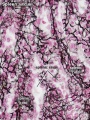
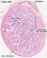
 Testis, young H&E reproductive system, male, convoluted seminiferous tubules x10.jpg 1,280 × 1,024; 396 KB
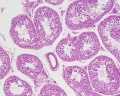
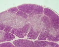
 Thymus - young 01.jpg 450 × 600; 93 KB
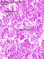 Thymus - young 02.jpg 450 × 600; 91 KB
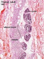 Thymus adult.jpg 450 × 600; 138 KB
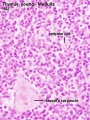
 Thymus histology 01.jpg 1,280 × 1,024; 723 KB
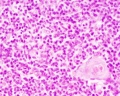 Thymus histology 02.jpg 1,280 × 1,024; 287 KB
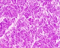 Thymus histology 03.jpg 1,280 × 1,024; 325 KB
 Thymus histology 04.jpg 1,280 × 1,024; 511 KB
 Thymus histology 05.jpg 513 × 385; 41 KB
 Thyroid histology 001.jpg 450 × 600; 96 KB
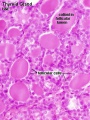 Thyroid histology 002.jpg 450 × 600; 85 KB
 Thyroid histology 003.jpg 1,280 × 1,024; 209 KB
 Thyroid histology 004.jpg 1,280 × 1,024; 351 KB
 Tongue histology 05.jpg 1,280 × 1,024; 418 KB
 Tongue-muscle.jpg 300 × 228; 43 KB
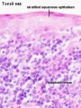
 Tonsil histology 01.jpg 450 × 600; 106 KB
 Tonsil histology 02.jpg 450 × 600; 62 KB
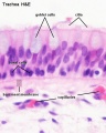 Trachea histology 01.jpg 480 × 600; 47 KB
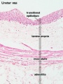 Ureter histology 001.jpg 375 × 500; 50 KB
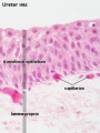 Ureter histology 002.jpg 375 × 500; 34 KB
 Urinary Bladder Histology.jpg 581 × 399; 42 KB
 Uterine gland proliferative phase.jpg 400 × 533; 51 KB
 Uterine gland secretory phase.jpg 400 × 533; 49 KB
 Uterine tube histology 02.jpg 400 × 533; 55 KB
 Uterine tube histology 03.jpg 400 × 533; 34 KB
 Uterine tube histology 04.jpg 1,280 × 1,024; 253 KB
 Uterine tube histology 05.jpg 1,063 × 1,063; 508 KB
 Uterine tube histology.jpg 1,280 × 1,024; 568 KB
 Uterus proliferative phase.jpg 400 × 533; 52 KB
 Uterus secretory phase 01.jpg 1,280 × 1,024; 318 KB
 Uterus secretory phase 02.jpg 1,280 × 1,024; 359 KB
 Uterus secretory phase.jpg 400 × 533; 68 KB
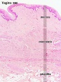 Vagina histology 01.jpg 450 × 600; 92 KB
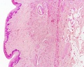 Vagina histology 02.jpg 1,280 × 1,024; 555 KB
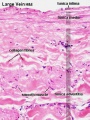
 Vein histology 01.jpg 480 × 600; 57 KB
 Vein histology 02.jpg 400 × 533; 76 KB
 Vein histology 03.jpg 400 × 533; 76 KB
 Vein valve animation.gif 300 × 200; 54 KB
 Week10 adrenal.jpg 600 × 564; 48 KB
 Wheater’s Functional Histology.jpg 425 × 600; 66 KB
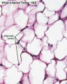
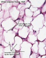 White adipose 01.jpg 500 × 625; 65 KB
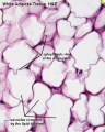
 White adipose 02.jpg 500 × 625; 61 KB
 White adipose histology.jpg 400 × 500; 57 KB
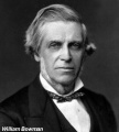 William Bowman.jpg 600 × 665; 49 KB
 Wilms tumor.jpg 776 × 512; 310 KB
 XMRV-infected prostate cells.png 485 × 599; 528 KB
 Zorn2008 fig01.jpg 1,200 × 1,158; 110 KB


