Cardiovascular System - Spleen Development
Introduction

The spleen is located on the left side of the abdomen and has a role initially in blood and then immune system development. The spleen's haematopoietic function (blood cell formation) is lost with embryo development and lymphoid precursor cells migrate into the developing organ. Vascularization of the spleen arises initially by branches from the dorsal aorta. Mesoderm within the dorsal mesogastrium form a long strip of cells adjacent to the forming stomach above the developing pancreas.
| Immune Links: immune | blood | spleen | thymus | lymphatic | lymph node | Antibody | Med Lecture - Lymphatic Structure | Med Practical | Immune Movies | vaccination | bacterial infection | Abnormalities | Category:Immune | ||
|
Some Recent Findings
|
Development Overview

|
The human spleen arises in week 5 within the dorsal mesogastrium as proliferating mesenchyme overlying the dorsal pancreatic endoderm. Cells required for its hemopoietic function arise from the yolk sac wall and near dorsal aorta.
The spleen generates both red and white cells in the 2nd trimester. Note that many embryonic RBCs remain nucleated. |
| D4 Dorsal Mesogastrium (stage 13) |
Spleen Development Movies
Spleen Development in Dorsal Mesogastrium |
This cross-sectional view of the abdomen viewed from above, with dorsal (back) top and ventral (front) bottom of animation.
Later the retroperitoneal position of the developing kidneys is also shown either side of the dorsal (thoracic) aorta.
|
Embryonic Spleen
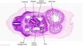
D4 Dorsal Mesogastrium (stage 13)
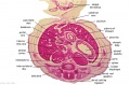
F1 Developing Human Spleen (stage 22)
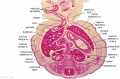
F2 Developing Human Spleen (stage 22)
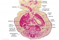
F3 Developing Human Spleen (stage 22)


Fetal Spleen
Reticular framework of white pulp and marginal zone in human fetal spleens from the 17th to 40th week of gestation.[1]
- "The antigenic heterogeneity of the reticular framework of the white pulp (WP) and marginal zone (MZ) is well documented in the human adult spleen. The ontogeny of the WP and MZ of human fetal spleens was examined with special reference to the heterogeneity of the reticular framework. In the spleen of the 17th gestational week (gw), alpha-smooth muscle actin (alpha-SMA)-positive reticulum cells were scattered around the arterioles. From the 20th to 23rd gw, alpha-SMA-positive reticulum cells increased in number and began to form a reticular framework. An accumulation of T and B lymphocytes occurred within the framework, and a primitive WP was observed around the arterioles. At the 24th gw, antigenic diversity of the reticular framework was observed, and T and B lymphocytes were segregated in the framework. T lymphocytes were sorted into the alpha-SMA-positive reticular framework, and the periarteriolar lymphoid sheath (PALS) was formed around the arteriole. B lymphocytes aggregated in eccentric portions to the PALS and formed the lymph follicle (LF). The reticular framework of the LF was alpha-SMA-negative. MZ appeared in the alpha-SMA-positive reticular framework around the WP at the 26th gw. The PALS, LF, and MZ developed with gestational time. The reticular framework of the PALS, LF, and MZ is thus heterogeneous in the fetal spleen, and the development of the heterogeneity is related to the ontogeny of the PALS, LF, and MZ."
Timeline
- week 15 (17 GA) - alpha-smooth muscle actin (alpha-SMA)-positive reticulum cells scattered around the arterioles.
- week 18 to 21 (20 - 23 GA) - alpha-SMA-positive reticulum cells increase in number and began to form a reticular framework. An accumulation of T and B lymphocytes occurred within the framework, and a primitive white pulp was observed around the arterioles.
- week 22 (24 GA) - antigenic diversity of the reticular framework was observed, and T and B lymphocytes were segregated in the framework. T lymphocytes were sorted into the alpha-SMA-positive reticular framework, and the periarteriolar lymphoid sheath (PALS) was formed around the arteriole. B lymphocytes aggregated in eccentric portions to the PALS and formed the lymph follicle (LF). The reticular framework of the LF was alpha-SMA-negative.
- week 24 (26 GA) - marginal zone appeared in the alpha-SMA-positive reticular framework around the white pulp.
Adult Spleen

Adult Spleen and ligamentous attachments.
Abnormalities
Congenital absence of the spleen is usually accompanied by complex cardiac malformations, malposition and maldevelopment of the abdominal organs, and abnormal lobation of the lungs. There are a range of other spleen anatomical development abnormalities, some of which have no effect and others are very rare.
Splenic Lobulation
Accessory Spleen
Clinically no significant efects in most patients, occur as single or multiple and generally found in autopsy or as an an incidental finding. Thought to occur due to a failure of primordia fusion within the dorsal mesogastrium.
Polysplenia
Splenogonadal Fusion
Rare resulting from abnormal fusion of the splenic and gonadal primordia during prenatal development. On the left side and more common in male and adhesion to the gonad, epididymis or ductus deferens and then follows the caudal descent with the gonad. Failure of complete descent can also result in associated intraabdominal cryptorchism.
Two classifications:
- continuous - orthotopic spleen connects to the gonad with a cord of fibrous or splenic tissue.
- discontinuous - no connection between the orthotopic spleen and gonad.
(More? Testis Development)
Ectopic Spleen
A very rare abnormality where the spleen can be found anatomically located in a range of places in the abdominal or thoracic cavity.
Wandering Spleen
Connexin-43 involved with abnormal spleen development (cardiac and lung also).
References
Reviews
<pubmed>17067939</pubmed> <pubmed>16550197</pubmed> <pubmed>15738953</pubmed> <pubmed>15530642</pubmed> <pubmed>14966753</pubmed> <pubmed>10676919</pubmed> <pubmed>7728201</pubmed>
Search PubMed
Search NCBI Bookshelf: Spleen Development
Search PubMed: Search August 2006 "Spleen Development" 13,401 reference articles of which 450 were reviews.
Search term = Spleen Development | Spleen Abnormalities
Glossary Links
- Glossary: A | B | C | D | E | F | G | H | I | J | K | L | M | N | O | P | Q | R | S | T | U | V | W | X | Y | Z | Numbers | Symbols | Term Link
Cite this page: Hill, M.A. (2024, June 26) Embryology Cardiovascular System - Spleen Development. Retrieved from https://embryology.med.unsw.edu.au/embryology/index.php/Cardiovascular_System_-_Spleen_Development
- © Dr Mark Hill 2024, UNSW Embryology ISBN: 978 0 7334 2609 4 - UNSW CRICOS Provider Code No. 00098G