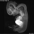
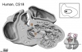
Carnegie stage 18
| Embryology - 10 Jun 2024 |
|---|
| Google Translate - select your language from the list shown below (this will open a new external page) |
|
العربية | català | 中文 | 中國傳統的 | français | Deutsche | עִברִית | हिंदी | bahasa Indonesia | italiano | 日本語 | 한국어 | မြန်မာ | Pilipino | Polskie | português | ਪੰਜਾਬੀ ਦੇ | Română | русский | Español | Swahili | Svensk | ไทย | Türkçe | اردو | ייִדיש | Tiếng Việt These external translations are automated and may not be accurate. (More? About Translations) |
Introduction

Facts
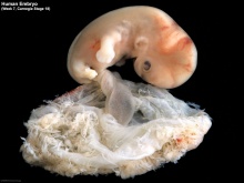
Week 7, 44 - 48 days, 13 - 17 mm
Gestational Age GA - week 9
Summary
- Ectoderm: sensory placodes, lens pit, otocyst,nasal pits moved ventrally, fourth ventricle of brain
- Mesoderm: heart prominence
- Head: 1st, 2nd and 3rd pharyngeal arch, forebrain, eye, auricular hillocks
- Body: heart, liver, umbilical cord
- Limb: upper and lower limb buds, foot plate, wrist, hand plate with digital rays
See also Carnegie stage 18 Events
Features
- Development indices: number of semicircular ducts (1-3) and length of the paramesonephric duct.
- Identify: pigmented eye, eyelid, nasolacrimal groove, external acoustic meatus, heart, digital rays, liver prominance, thigh, ankle, foot plate, umbilical cord
- Links: Week 7 | System Development | Lecture - Limb | Lecture - Head Development | Lecture - Sensory | Science Practical - Head | Science Practical - Sensory | Science Practical - Urogenital | Category:Carnegie Stage 18 | Stage 19
| Week: | 1 | 2 | 3 | 4 | 5 | 6 | 7 | 8 |
| Carnegie stage: | 1 2 3 4 | 5 6 | 7 8 9 | 10 11 12 13 | 14 15 | 16 17 | 18 19 | 20 21 22 23 |
- Carnegie Stages: 1 | 2 | 3 | 4 | 5 | 6 | 7 | 8 | 9 | 10 | 11 | 12 | 13 | 14 | 15 | 16 | 17 | 18 | 19 | 20 | 21 | 22 | 23 | About Stages | Timeline
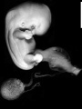
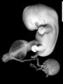
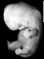
Bright Field

|

|
| Embryo in gestational sac | Embryo open sac |

|

|
| Embryo with placentation (ectopic) | Embryo in amniotic sac |
- Stage 18 Links: Embryo in gestational sac | Embryo open sac | Embryo 2 and gestational sac | Embryo 2 | Carnegie stage 18
Embryo Virtual Slides
|
|

Scanning EM


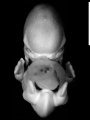
Ventral view of head showing upper lip, maxilla and nasal region.
Image Source: Prof Virginia Diewert
Kyoto Collection

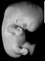
View: This is a dorsolateral view of embryo. Amniotic membrane removed.
Image source: Embryology page Created: 19.03.1999

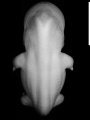
Ventral view of head region (1 mm scale).
- Kyoto Embryo 25783
right+yolk
left+yolk
left
ventral
right
dorsal


Image source: The Kyoto Collection images are reproduced with the permission of Prof. Kohei Shiota and Prof. Shigehito Yamada, Anatomy and Developmental Biology, Kyoto University Graduate School of Medicine, Kyoto, Japan for educational purposes only and cannot be reproduced electronically or in writing without permission.
Carnegie Collection


| Carnegie Collection Embryos - Stage 18 | |||||||||||
|---|---|---|---|---|---|---|---|---|---|---|---|
| Serial No. | Size (mm) | Grade | Fixative | Embedding Medium | Plane | Thinness (µm) | Stain | Semi. ducts | P.-M. duct (mm) | Year | Notes |
| 109 | E., 12.0* Ch.,30 | Poor | Alc. | P | Transverse | 20 | Al. coch. | 1 | 0.4 | 1897 | Tubal Least—advanced third |
| 144 | E., 16.0* Ch, 40x30x30 | Good | Formalin | P | Sagittal | 40 | Al. eoch. | 3 | 0.85 | 1899 | Most—advanced third |
| 175 | E., 13.0 Ch, 30x25x25 | Poor | Alc. | P | Transverse | 20 | Al. coch. | 2 | 0.6 | 1900 | Tubal Partly macerated |
| 296 | E., 17.0 | Poor | Ale. | P | Coronal | 20 | Various | 3 | 0.85 | 1905 | Most—advanced third |
| 317 | E., 16.0 | Good | Formalin | P | Coronal | 20 | (Stain - Haematoxylin Eosin) or. G. | 2 | 0.7 | 1905 | Middle third |
| 351 | E.,14.0* | Good | Formalin | P | Coronal | 250 | Slightly carmine— | 2 | 038 | 1904 | Injected (Berlin blue) |
| 406 | E., 16.0 Ch., 40x40x40 | Good | Formalin | P | Sagittal | 20 | (Stain - Haematoxylin Eosin) | 3 | 0.7 | 1907 | Operative. Most—advanced third |
| 423 | E., 15.2 | Good | Formol—Zenker | P | Transverse | 50 | Carmine | 3 | 0.85 | 1904 | |
| 424 | E., 172 | Good | Formalin | P | Transverse | 50 | Carmine | 3 | 10 | 1904 | Double infection. Advanced |
| 492 | E, 16.8 Ch, 40 x 40 | Exc. | Zenker | P | Coronal | 40 | Al. coch. v | 3 | 0.7 | 1911 | Injected (India ink) |
| 511 | E., 160* Ch., 3?* 32x32 | Good | Ale. | P | Sagittal | 40 | Al. coch. | 3 | 1.1 | 1911 | Head injured. Most advanced in group |
| 670 | E, 12.5 | Poor | Ale. | P | Sagittal | 50 | (Stain - Haematoxylin Eosin) | 3 | 10 | 1913 | Tubal Advanced |
| 719 | E, 15.0 Ch, 50x50x50 | Good | Formalin | P | Trans | 40 | Al. coch. | 2 | 0.6 | 1913 | Median in group |
| 733 | E., Ch., 4Sx40x2S | ISO Poor | Formalin | P | Sagittal | 50 | Al. coch. | 2 | 0.6 | 1913 | Median in group |
| 841 | E. 15.0 Ch., 18 x 16x9 | Good | Formalin | P | Coronal & Trans, 20 | 10 | (Stain - Haematoxylin Eosin), carmine | 2 | 0.32 | 1914 | Operative. Head cut separately |
| 899 | E, 160* Ch. 50 x 18 x IS | Good | Bouin | P | Sagittal | S0 | Al. coch. | 3 | 0,65 | 1914 | Tubal Head injured |
| 991 | E. l?.0 | Good | Formalin | P | Sag | so | R, V, Gieson | 3 | 0.9 | 1914 | Advanced |
| 1909 | E., 14.6 | Good | Formalin | P | Coronal | 20 | Al. coch,or. G. | 1 | 0.3 | 1917 | Less advanced |
| 2673 | E.,15.5 | Good | Formalin | P | Transverse | 40 | Al. coch. | 2 | 0.52 | 1919 | Median in group |
| 4430 | E., 14.0 Ch, 51 x40x21 | Exc. | Corros. acetic | P | Transverse | 15 | Al. coch,or. G. | 3 | 0.9 | 1923 | Most—advanced third |
| 5542B | E., 16.0 Ch, 37x32x25 | Good | Formalin | P | Transverse | 40 | Al. coch. | 2 | 0.7 | 1927 | Other twin abnormal |
| 5747 | E, 15.2 Ch, 32x27x25 | Poor | Alc.—formol | P | Sagittal | 25 | Al. coch. | 2 | 0.25 | 1928 | Least—advanced or middle third |
| 5935A | E, 13.5 Ch, 40x30x30 | Good | Formalin | P | Coronal | 40 | Al. coch. | 1 | 0.38 | 1929 | Other twin stunted |
| 6522 | E, 13.2* | Good | Corros. acetic | C—P | Coronal | 10 | Al. coch. | 3 | 0.8 | 7 | Middle or most—advanced third |
| 6524 | E, 11.7* | Exc. | Corros. acetic | C—P | Transverse | 10 | Al. coch. | 1 | 0.4 | ? | Least—advanced third |
| 6525 | E, 13.8* | Exc. | Corros. acetic | C—P | Sagittal | 8 | Al. coch. | 2 | 0.42 | ? | Weak staining |
| 6527 | E, 14.4* | Exc. | Corros. acetic | C—P | Transverse | 15 | Al. coch. | 2 | 0.67 | ? | Mechanical damage |
| 6528 | E, 13.4* | Exc. | Corros. acetic | C—P | Coronal | 8 | Al. coch. | 1 | 0.33 | ? | Least—advanced third |
| 6529 | E, 15.6* | Good | Corros. acetic | C—P | Coronal | 10 | Al. coch. | 2 | 0.4 .5 | Middle third | |
| 6533 | E, 12.5* | Good | Corros. acetic | C—P | Sagittal | 6, 8, 10 | Al. coch. | 2 | 0.45 | ? | Middle third |
| 6551 | E, 18.0 | Poor | Formalin | p | Coronal | 40 | (Stain - Haematoxylin Eosin) | 3 | 0.8 | 1932 | Tubal |
| 7707 | E, 14.5 Ch ,37x32 | Exc. | Bouin | C—P | Transverse | 10 | (Stain - Haematoxylin Eosin), phlox. | 2 | 0.54 | 1939 | Operative. Middle third |
| 8097 | E, 15.5 Ch, 37x25x21 | Good | Formalin | C—P | Transverse | 10 | (Stain - Haematoxylin Eosin) | 1 | 0.19 | 1942 | Least advanced in group |
| 8172 | E, 16.5 | Exc. | Bouin | C—P | Transverse | 20 | (Stain - Haematoxylin Eosin) | 3 | 0.58 | 1943 | Operative. Very advanced |
| 8235 | E, 14.0* | Good | Bouin | C—P Sagittal | 10 | (Stain - Haematoxylin Eosin) | Mallory | 2 | 0.25 | 1944 | Tubal |
| 8355 | E, 15.0 Ch, 23 | Exc. | Formalin | C—P | Coronal | 10 | Azan | 1946 | Tubal. Duplicated spinal cord caudally | ||
| 8812 | E, 12_9 | Exc | Formalin | C—P | Transverse | 10 | (Stain - Haematoxylin Eosin) | 1950 | Rubella. Medical abortion. Midbrain punctured | ||
| 8945 | E, 13.9 | Good | Zenker | p | Transverse | 8 | Borax, carm. | 1952 | Univ. Chicago No. H 1254 | ||
| 9107 | E, 17.0 Ch, 38x28x22 | Good | Bouin | p | Transverse | 15 | Borax, carm. | 1918 | Univ. Chicago No. H 516 | ||
| 9247 | E, 15.0 | Exc. | Bouin | C—P | Sagittal | 8 | Azan | 1954 | Tubal | ||
Abbreviations
| |||||||||||
| iBook - Carnegie Embryos | |
|---|---|

|
|
Hill Collection

|

|

|

|
| right ventral | right ventrolateral | right ventrolateral | right lateral |

|

|

|
|
| right ventral (smaller) | left lateral (smaller) | right dorsolateral (smaller) |
- Links: Hill Collection
Events
- Vision - Mesenchyme invades the region between the lens epithelium and the surface ectoderm.[1]
- Hearing - otic capsule precartilaginous. Semicircular ducts form in the order anterior, posterior, and lateral from thickened epithelial areas and adjacent epithelial layers fuse. Cochlear duct is now L-shaped. First pharyngeal arch bar begins to chondrify (Meckel’s cartilage), second arch may chondrify (Reichert’s cartilage), stapes and stapedius commence. Auricular hillocks merge to form auricle primordia.
- Bone - lower limb femur and tibia chondrification of cartilage template.[2]
- Cardiovascular
- Coronary circulation connection of the proximal coronary arteries to the aorta.[3]
- Endocrine[4]
- Epiphysis - cellular migration in the pineal body forms a distinct "anterior lobe" in which follicles appear (Stadium 3 of Turkewitsch 1933) (O'Rahilly 1973 a).
- Thymus- thymus makes contact with the thyroid gland and contains a series of canals internally (Weller 1933).
- Thyroid - median thyroid is in contact with "lateral thyroid compo- nents" (Weller 1933) but others have maintained that the telopharyngeal body should not be regarded as a thyroid component (Bejdl and Politzer 1953). The lobes of the thyroid are "composed of series of continuously communicating solid annectent bars" (Weller 1933). This is "the earliest stage of the definitive thyroid" (ibid.). First differentiation occurs in Weller's (1933) "lateral thyroid component," which is beginning to "blend into uniformly constituted thyroid tissue". Weller (1933) illustrated (Fig. 11) a thyroid gland that still showed continuity between its pedicle and the epithelium of the pharynx.
- Adrenal Cortex - gland becomes reorganized. The C1, 2, and 3 cells form cords as sinusoids develop. Cells divide at or near the surface, where new cells are added.[5]
References
- ↑ <pubmed>7364662</pubmed>
- ↑ <pubmed>9185992</pubmed>
- ↑ <pubmed>3286038</pubmed>
- ↑ O'Rahilly R. The timing and sequence of events in the development of the human endocrine system during the embryonic period proper. (1983) Anat. Embryol., 166: 439-451. PMID 6869855
- ↑ Crowder RE. The development of the adrenal gland in man, with special reference to origin and ultimate location of cell types and evidence in favor of the "cell migration" theory. (1957) Contrib. Embryol., Carnegie Inst. Wash. 36, 193-210.
Additional Images
Stage 18 Optical Projection Tomography
Stage 18 Heart MRI

External ear Stages 14-23 and adult


Historic Images
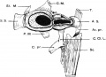
Human embryonic shoulder girdle
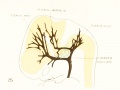
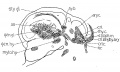
1921. Dural venous system human embryo 15 mm long
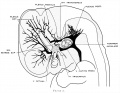
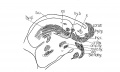
1921 Vascular system of the brain of the human embryo
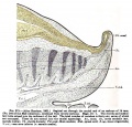
Fig. 273. Sagittal cut caudal end of embryo of 14mm

17 mm Embryo
1911 Cross section to show cricoid cartilage and M. criooarytaenoideus posterior.
1911 Cross section to show thyreoid cartilage, M. criooarytaenoideus lateralis,
1911 Sagittal section to show, especially, M. interarytaenoideus.
1911 Sagittal section to show hyoid bone and thyreoid cartilage, M. cricothyreoideus, and tongue region
1911 Sagittal section


- Carnegie Stages: 1 | 2 | 3 | 4 | 5 | 6 | 7 | 8 | 9 | 10 | 11 | 12 | 13 | 14 | 15 | 16 | 17 | 18 | 19 | 20 | 21 | 22 | 23 | About Stages | Timeline
Cite this page: Hill, M.A. (2024, June 10) Embryology Carnegie stage 18. Retrieved from https://embryology.med.unsw.edu.au/embryology/index.php/Carnegie_stage_18
- © Dr Mark Hill 2024, UNSW Embryology ISBN: 978 0 7334 2609 4 - UNSW CRICOS Provider Code No. 00098G