ANAT2341 Lab 6 - Early Embryo: Difference between revisions
mNo edit summary |
mNo edit summary |
||
| Line 43: | Line 43: | ||
Named by the sensory system and components they will form: otic placode, lens placode and nasal placode. | Named by the sensory system and components they will form: otic placode, lens placode and nasal placode. | ||
{{Chicken Placode Movie 1}} | |||
{{ | |||
|} | |} | ||
| Line 188: | Line 180: | ||
==Additional Information== | ==Additional Information== | ||
===Neural Crest=== | |||
{| | {| | ||
| [[File:Cranial neural crest skeletal fate 01.jpg|600px]] | | [[File:Cranial neural crest skeletal fate 01.jpg|600px]] | ||
| Line 228: | Line 221: | ||
::Our data suggest that in the second trimester of human foetal development, a gene expression signature of neural crest origin still exists in frontal and metopic compartments while gene expression of parietal and sagittal compartments is more similar to mesoderm." | ::Our data suggest that in the second trimester of human foetal development, a gene expression signature of neural crest origin still exists in frontal and metopic compartments while gene expression of parietal and sagittal compartments is more similar to mesoderm." | ||
===Placodes=== | |||
The links below are to '''additional information''' providing background about each of the sensory systems. | |||
{{Hearing Links}} | |||
{{Vision Links}} | |||
{{Smell Links}} | |||
:'''Taste Links:''' [[Sensory - Taste Development|Introduction]] | |||
===Reference=== | ===Reference=== | ||
Revision as of 17:45, 7 September 2016
| Lab 6: Introduction | Trilaminar Embryo | Early Embryo | Late Embryo | Fetal | Postnatal | Abnormalities | Online Assessment |
Week 4
During week 4 a number of features appear visible on the embryo surface:
- At the level of the body heart, liver, somite bulges and limb buds appear.
- At the level of the head sensory placodes and pharyngeal arches appear.
Carnegie Stage 12 to 14
Week 4


This is a scanning EM of the embryo superior dorsal view showing the paired otic placodes sinking into the surface at the level of the hindbrain between day 24 and day 25


Week 5

Human face development (Week 5)


Sensory Placodes
|
|
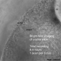
Sensory placodes develop as small patches of ectodermal thickenings. The placodes are laterally paired and contribute key components to sensory structures of the ear, eye and nose. Named by the sensory system and components they will form: otic placode, lens placode and nasal placode.
|
Stage 13
Identify the structure and position of the otic vesicle (otocyst) relative to other head structures.


Pharyngeal Arches


Stage 13 Pharyngeal Arches

A6L A7L B1L B2L B3L B4L B5L B6L
Look through the above cross-sections of the stage 13 embryo observing and identifying structures of the face and ear visible at this stage.

Structures derived from Arches
|
Arch |
Nerve |
Muscles |
Skeletal |
Artery |
|
(maxillary/mandibular) |
|
mastication |
Meckel's cartilage |
maxillary(terminal branches) |
|
(hyoid) |
|
facial expression |
Reichert's cartilage |
stapedial (embryonic) |
|
|
|
stylopharyngeus |
greater cornu of hyoid, lower part of body of hyoid bone |
common carotid, internal carotid (root) |
|
|
|
intrinsic muscles of larynx, pharynx; levator palati |
thyroid, cricoid, arytenoid, corniculate and cuneform cartilages |
4 - aortic arch, right subclavian |
Structures derived from Pouches
|
Pouch |
Overall Structure |
Specific Structures |
|
|
tubotympanic recess |
tympanic membrane, tympanic cavity, mastoid antrum, auditory tube |
|
|
intratonsillar cleft |
crypts of palatine tonsil, lymphatic nodules of palatine tonsil |
|
|
inferior parathyroid gland, thymus gland |
|
|
|
superior parathyroid gland, ultimobranchial body |
|
|
|
becomes part of 4th pouch |
Additional Information
Neural Crest

|
Cranial neural crest contribution to skeletal structures [1]
The embryo figure[1] shows colonization of the head and pharyngeal arches by diencephalic, anterior and posterior mesencephalic, and rhombencephalic neural crest cells (NCCs), as indicated by the colour code. The diagram is representative human embryos, although the NCC migratory pathways might differ slightly in different species. The skull drawings show contributions of NCC populations to cranial skeletal elements of humans, based on NCC fate-mapping studies and on extrapolation of avian and mouse data to known homologues in the human. Some bones, including the squamosal (SQ), alisphenoid (AS), and pterygoid (PT), are shown with mixed contribution from different NCC populations. Note that in mammals the frontal (FR) and parietal (PA) bones have been reported to be of neural crest and mesodermal origin, respectively. (text modified from original figure legend) |
|
|
See also this 2014 Review:
Epigenetic regulation in neural crest development. [2]
- "The neural crest is a migratory and multipotent cell population that plays a crucial role in many aspects of embryonic development. In all vertebrate embryos, these cells emerge from the dorsal neural tube then migrate long distances to different regions of the body, where they contribute to formation of many cell types and structures. These include much of the peripheral nervous system, craniofacial skeleton, smooth muscle, and pigmentation of the skin. The best-studied regulatory events guiding neural crest development are mediated by transcription factors and signaling molecules. In recent years, however, growing evidence supports an important role for epigenetic regulation as an additional mechanism for controlling the timing and level of gene expression at different stages of neural crest development. Here, we summarize the process of neural crest formation, with focus on the role of epigenetic regulation in neural crest specification, migration, and differentiation as well as in neural crest related birth defects and diseases."
Human cranial compartments different embryonic origins[3]
- Our data suggest that in the second trimester of human foetal development, a gene expression signature of neural crest origin still exists in frontal and metopic compartments while gene expression of parietal and sagittal compartments is more similar to mesoderm."
Placodes
The links below are to additional information providing background about each of the sensory systems.
| Vision Links: vision | lens | retina | placode | extraocular muscle | cornea | eyelid | lacrima gland | vision abnormalities | Student project 1 | Student project 2 | Category:Vision | sensory | ||
|
| Smell Links: Introduction | placode | Rhinencephalon | head | respiratory | Student project | taste | sensory | Category:Smell | ||
|
- Taste Links: Introduction
Reference
- ↑ 1.0 1.1 <pubmed>14523380</pubmed>| Nat Rev Neurosci.
- ↑ <pubmed>25446277</pubmed>
- ↑ <pubmed>26188427</pubmed>
| Lab 6: Introduction | Trilaminar Embryo | Early Embryo | Late Embryo | Fetal | Postnatal | Abnormalities | Online Assessment |
Glossary Links
- Glossary: A | B | C | D | E | F | G | H | I | J | K | L | M | N | O | P | Q | R | S | T | U | V | W | X | Y | Z | Numbers | Symbols | Term Link
Cite this page: Hill, M.A. (2026, September 12) Embryology ANAT2341 Lab 6 - Early Embryo. Retrieved from https://embryology.med.unsw.edu.au/embryology/index.php/ANAT2341_Lab_6_-_Early_Embryo
- © Dr Mark Hill 2026, UNSW Embryology ISBN: 978 0 7334 2609 4 - UNSW CRICOS Provider Code No. 00098G


