Cardiovascular System - Heart Valve Development: Difference between revisions
mNo edit summary |
mNo edit summary |
||
| Line 103: | Line 103: | ||
<pubmed>17549728</pubmed> | <pubmed>17549728</pubmed> | ||
<pubmed>16914500</pubmed> | <pubmed>16914500</pubmed> | ||
<pubmed>17104787</pubmed> | |||
===Search PubMed=== | ===Search PubMed=== | ||
Revision as of 15:12, 15 November 2015
| Embryology - 10 Jun 2024 |
|---|
| Google Translate - select your language from the list shown below (this will open a new external page) |
|
العربية | català | 中文 | 中國傳統的 | français | Deutsche | עִברִית | हिंदी | bahasa Indonesia | italiano | 日本語 | 한국어 | မြန်မာ | Pilipino | Polskie | português | ਪੰਜਾਬੀ ਦੇ | Română | русский | Español | Swahili | Svensk | ไทย | Türkçe | اردو | ייִדיש | Tiếng Việt These external translations are automated and may not be accurate. (More? About Translations) |
Introduction


The heart valves form between the atria and ventricles (mitral valve, tricuspid valve) and between the atria and blood vessels (aortic valve, pulmonary valve). The cardiac cushions in the atrioventricular (AV) canal contain cells that are the primordia of the cardiac valves. The atrioventricular valves are attached to papillary muscles by chordae tendineae.
Scleraxis (SCX) is a transcription factor involved in tendon and ligament development and has been identified as also expressed in early heart valve development.[1]
Mitral valve also called the "bicuspid valve".
Some Recent Findings
|
| More recent papers |
|---|
 This table allows an automated computer search of the external PubMed database using the listed "Search term" text link.
More? References | Discussion Page | Journal Searches | 2019 References | 2020 References Search term: Heart Valve Embryology <pubmed limit=5>Heart Valve Embryology</pubmed> |
Textbooks
- Human Embryology (2nd ed.) Larson Ch7 p151-188 Heart
- The Developing Human: Clinically Oriented Embryology (6th ed.) Moore and Persaud Ch14: p304-349
- Before we Are Born (5th ed.) Moore and Persaud Ch12; p241-254
- Essentials of Human Embryology Larson Ch7 p97-122 Heart
- Human Embryology Fitzgerald and Fitzgerald Ch13-17: p77-111
Tutorial Images
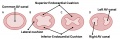
Canal Division (Superior View)
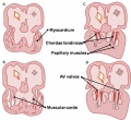
Development of the mitral and tricuspid valves
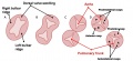
Development of the semilunar valves
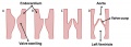
Development of the semilunar cusps


Fetal Heart Valve Sounds
<mp3player>File:Week17 fetal heart rate.mp3</mp3player>
Audio recording of the Second Trimester fetal heart (GA week 17).
The characteristic "lub-dup" sounds are associated with closing of heart valves.
- First sound (lub) occurs as atrioventricular valves close and signifies beginning of systole (contraction)
- Second sound (dup) occurs when semilunar valves close at the beginning of ventricular diastole (relaxation)
- Links: Fetal Heart Sounds Audio
Molecular
Scleraxis (Scx) - basic helix–loop–helix transcription factor expressed in the progenitors and cells of all tendon tissues (mouse).[4]
Periostin - regulates lineage commitment of valve precursor cells (chicken).[5]
Gata4 and Gata6
Tbx5
Images
Historic
| Historic Disclaimer - information about historic embryology pages |
|---|
|
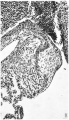
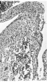
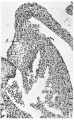
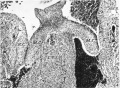
Odgers PNB. The Development of the Atrio-Ventricular Valves in Man (1939) J Anat. 73:643-57. PMID 17104787
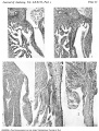
Plate 1
1 right A.-V. orifice in an 11.2 mm. embryo
2 right lateral cusp in a 17.5 mm. embryo
3 left lateral cusp in a 17.5 mm. embryo
4 central cusps in a 23 mm. embryo
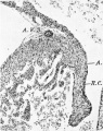
5 right lateral cusp in a 28.5 mm. embryo
Plate 2


Abnormalities
Noonan syndrome
An autosomal dominant single-gene cause of congenital heart disease. Patients also have proportionate short stature, facial abnormalities, and an increased risk of myeloproliferative disease. About half the patients have mutations in PTPN11, encoding the protein tyrosine phosphatase SHP2. A recent study in mice has identified PTPN11 acting in endocardium to enhance endocardial-mesenchymal transformation.[6]
References
Reviews
<pubmed>20809794</pubmed> <pubmed>20201901</pubmed> <pubmed>14567955</pubmed> <pubmed>12768658</pubmed>
Articles
<pubmed>17549728</pubmed> <pubmed>16914500</pubmed> <pubmed>17104787</pubmed>
Search PubMed
Search Pubmed: heart valve development | heart valve morphogenesis | Valvulogenesis
External Links
External Links Notice - The dynamic nature of the internet may mean that some of these listed links may no longer function. If the link no longer works search the web with the link text or name. Links to any external commercial sites are provided for information purposes only and should never be considered an endorsement. UNSW Embryology is provided as an educational resource with no clinical information or commercial affiliation.
Glossary Links
- Glossary: A | B | C | D | E | F | G | H | I | J | K | L | M | N | O | P | Q | R | S | T | U | V | W | X | Y | Z | Numbers | Symbols | Term Link
Cite this page: Hill, M.A. (2024, June 10) Embryology Cardiovascular System - Heart Valve Development. Retrieved from https://embryology.med.unsw.edu.au/embryology/index.php/Cardiovascular_System_-_Heart_Valve_Development
- © Dr Mark Hill 2024, UNSW Embryology ISBN: 978 0 7334 2609 4 - UNSW CRICOS Provider Code No. 00098G