BGD Lecture - Gastrointestinal System Development: Difference between revisions
mNo edit summary |
|||
| (247 intermediate revisions by 3 users not shown) | |||
| Line 1: | Line 1: | ||
{{ | {{Header}} | ||
==Introduction== | ==Introduction== | ||
[[File: | This lecture introduces the early development of the Gastrointestinal Tract (acronym [[:File:Adult_gastrointestinal_tract_cartoon.jpg|'''GIT''']]). Note that the oral cavity and pharynx will be covered in detail in the later [[BGD_Lecture_-_Face_and_Ear_Development|Lecture]] and [[BGDB_Practical_-_Face_and_Ear_Development|Practical]] on head and face development. | ||
{| | |||
| [[File:Endoderm_cartoon.jpg|alt=Endoderm development cartoon|link=Endoderm_Development_Movie]] | |||
| width=5px| | |||
| | |||
{{OneMinuteClock}} | |||
[[One_Minute_Embryology#Endoderm_Development|'''1 Minute Embryology''']]<br>[https://thebox.unsw.edu.au/video/1-minute-embryology-endoderm-development UNSW theBox] | |||
|} | |||
<br> | |||
<br> | |||
{| class="wikitable mw-collapsible mw-collapsed" | |||
! Lecture Archive | |||
|- | |||
| | |||
:'''Links:''' [[Media:BGD Lecture 2019 - Gastrointestinal System Development.pdf|2019 PDF]] | [https://embryology.med.unsw.edu.au/embryology/index.php?title=BGD_Lecture_-_Gastrointestinal_System_Development&oldid=376233 2018] | [[Media:BGD Lecture 2018 - Gastrointestinal System Development.pdf|2018 PDF]] [[Media:BGD Lecture 2017 - Gastrointestinal System Development.pdf|2017 PDF]] | [https://embryology.med.unsw.edu.au/embryology/index.php?title=BGD_Lecture_-_Gastrointestinal_System_Development&oldid=230497 2016] | [[Media:BGD Lecture 2016 - Gastrointestinal System Development.pdf|2016 PDF]] | [https://embryology.med.unsw.edu.au/embryology/index.php?title=BGD_Lecture_-_Gastrointestinal_System_Development&oldid=178801 2015] | [[:Media:BGD Lecture 2015 - Gastrointestinal System Development.pdf|2015 PDF]] | [https://embryology.med.unsw.edu.au/embryology/index.php?title=BGD_Lecture_-_Gastrointestinal_System_Development&oldid=138491 2014] | [[Media:BGD_Lecture_2014_-_Gastrointestinal_System_Development.pdf|2014 PDF]] | [[BGDB_Practical_-_Gastrointestinal_System_Development|BGDB Practical - GIT]] | [http://php.med.unsw.edu.au/embryology/index.php?title=BGD_Lecture_-_Gastrointestinal_System_Development&oldid=122054 2013 Lecture] | [http://php.med.unsw.edu.au/embryology/index.php?title=BGD_Lecture_-_Gastrointestinal_System_Development&oldid=115411 2012 Lecture] | [http://emed.med.unsw.edu.au/Map.nsf/0/D5264C4F39F6E9FCCA257339000628E2?OpenDocument&login Link to Learning Activity] | [[BGDB_Practical_-_Upper_Gastrointestinal_Tract_Histology|Upper GIT Histology - support page]] | |||
|} | |||
==Lecture Objectives== | ==Lecture Objectives== | ||
* Understanding of germ layer contributions | [[File:Bailey304.jpg|alt=Historic drawing of the developing gastrointestinal tract|thumb|300px|Historic drawing of the developing gastrointestinal tract (Kollman)]] | ||
* Understanding of the folding | * Understanding of germ layer contributions | ||
* Understanding of three main | * Understanding of the folding | ||
* Understanding of associated organ | * Understanding of three main embryonic divisions | ||
* Brief understanding of mechanical changes (rotations) | * Understanding of associated organ ({{liver}}, {{pancreas}}, {{spleen}}) development | ||
* Brief understanding of gastrointestinal abnormalities | * Brief understanding of mechanical changes (rotations) | ||
* Brief understanding of gastrointestinal tract abnormalities | |||
==Textbooks | {| class="wikitable mw-collapsible mw-collapsed" | ||
! colspan=2|Textbooks | |||
|- | |||
| [[File: | | [[File:Logo.png|alt=Embryo logo|80px]] | ||
| | | {{Embryo citation}} | ||
{{Gastrointestinal Tract Links}} | |||
|- | |- | ||
| [[File: | | [[File:The Developing Human, 10th edn.jpg|90px]] | ||
| | | {{MPT2015APAcitation}} (links only function with UNSW connection) | ||
Chapter 11 [https://ebookcentral-proquest-com.wwwproxy1.library.unsw.edu.au/lib/unsw/reader.action?docID=2074364&ppg=286 Alimentary System] | |||
{{UNSW textbook - The Developing Human}} | |||
|- | |- | ||
| [[File: | | [[File:Larsen's human embryology 5th ed.jpg|90px]] | ||
| | | {{Larsen2015APAcitation}}(links only function with UNSW connection) | ||
Chapter 14 [https://ebookcentral-proquest-com.wwwproxy1.library.unsw.edu.au/lib/unsw/reader.action?docID=2074524&ppg=359 Development of the Gastrointestinal Tract] | |||
{{UNSW textbook - Larsen's Human Embryology}} | |||
|} | |} | ||
{| class="wikitable mw-collapsible mw-collapsed" | |||
= | ! colspan=4|Gastrointestinal Tract Movies | ||
|- | |||
| valign="bottom"|{{Week 3 mesoderm movie}} | |||
| valign="bottom"|{{Week 3 movie}} | |||
| valign="bottom"|{{Amnion movie}} | |||
| valign="bottom"|{{Endoderm movie}} | |||
|- | |- | ||
| | | valign="bottom"|{{Stomach rotation movie}} | ||
| valign="bottom"|{{GIT growth movie}} | |||
| valign="bottom"|{{Greater omentum movie}} | |||
| valign="bottom"|{{Lesser sac movie}} | |||
| | |||
| | |||
| | |||
| | |||
| | |||
| | |||
|- | |- | ||
| valign="bottom"|{{Urogenital septum movie}} | |||
| valign="bottom"|{{Gastrointestinal stage 13 movie}} | |||
| valign="bottom"|{{Gastrointestinal stage 22 movie}} | |||
| valign="bottom"|{{Ultrasound Gastroschisis}} | |||
|} | |} | ||
== Week 3 == | == Week 3 == | ||
(Gestational age {{GA}} 5 weeks) | |||
===Gastrulation=== | ===Gastrulation=== | ||
[[File:Trilaminar embryo.jpg|thumb|Trilaminar embryo 3 germ layers.]] | |||
Week 3 the term "{{gastrulation}} " means "gut formation" and is the generation of the '''3 germ layers'''. | |||
{| | {| | ||
| | | {{Week 3 mesoderm movie}} | ||
* ''' | | | ||
* ''' | * '''{{endoderm}}''' - epithelium and associated glands, organs | ||
* ''' | * '''{{mesoderm}}''' (splanchnic) - mesentry, connective tissues, smooth muscle, blood vessels, organs | ||
* '''{{ectoderm}}''' ({{neural crest}}) - {{enteric nervous system}} | |||
Both endoderm and mesoderm will contribute to associated organs. | Both endoderm and mesoderm will contribute to associated organs. | ||
|} | |} | ||
===Folding=== | ===Folding=== | ||
{| border='0px' | {| border='0px' | ||
|- | |||
| Week 3to 4 folding of the embryonic disc forms the primitive gut tube. | |||
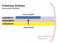
Folding ventrally around the {{notochord}}, running rostro-caudally in the midline. In relation to the notochord: | |||
* '''Laterally''' (either side of the notochord) lies mesoderm. | * '''Laterally''' (either side of the notochord) lies {{mesoderm}}. | ||
* '''Rostrally''' (above the notochord end) lies the buccopharyngeal membrane, above this again is the mesoderm region forming the heart. | * '''Rostrally''' (above the notochord end) lies the buccopharyngeal membrane, above this again is the mesoderm region forming the heart. | ||
* '''Caudally''' (below the notochord end) lies the primitive streak (where gastrulation occurred), below this again is the cloacal membrane. | * '''Caudally''' (below the notochord end) lies the primitive streak (where gastrulation occurred), below this again is the cloacal membrane. | ||
* '''Dorsally''' (above the notochord) lies the neural tube then ectoderm. | * '''Dorsally''' (above the notochord) lies the neural tube then ectoderm. | ||
* '''Ventrally''' (beneath the notochord) lies the mesoderm then endoderm. | * '''Ventrally''' (beneath the notochord) lies the mesoderm then endoderm. | ||
| valign="bottom"|{{Endoderm movie}} | |||
| | | valign="bottom"|{{Amnion movie}} | ||
| | |||
|} | |} | ||
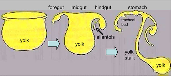
The ventral endoderm (shown yellow) has grown to line a space called the yolk sac. Folding of the embryonic disc "pinches off" part of this yolk sac forming the first primitive gastrointestinal tract. | The ventral endoderm (shown yellow) has grown to line a space called the yolk sac. Folding of the embryonic disc "pinches off" part of this yolk sac forming the first primitive gastrointestinal tract. | ||
==Week 4== | ==Week 4== | ||
[[File:Stage11 bf3.jpg| | (Gestational age {{GA}} 6 weeks) Carnegie stage {{CS11}} | ||
{| | |||
| [[File:Stage11 bf3.jpg|200px]] | |||
| [[File:Stage_11_historic-Atwell1930-1.jpg|240px]] | |||
|- | |||
| Embryo (stage 11 ventral view) | |||
| Embryo (midline section) | |||
|- | |||
| [[File:Stage11_bf9.jpg|300px]] | |||
| [[File:Stage11_sem4.jpg|300px]] | |||
|- | |||
| Stomodeum | |||
| Buccopharyngeal membrane | |||
|} | |||
===Coelomic Cavity=== | ===Coelomic Cavity=== | ||
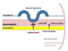
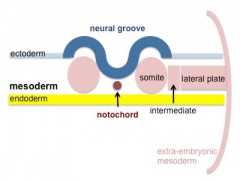
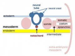
* The mesoderm initially undergoes segmentation to form paraxial, intermediate mesoderm and lateral plate mesoderm. | Mesoderm differentiates and lateral plate cavity forms the 3 main body cavities. | ||
* Paraxial mesoderm segments into somites and lateral plate mesoderm divides into somatic and splanchnic mesoderm. | |||
* The space forming between them is the coelomic cavity, that will form the 3 major body cavities (pericardial, pleural, peritoneal) | * The mesoderm initially undergoes segmentation to form paraxial, intermediate mesoderm and '''lateral plate mesoderm'''. | ||
* Paraxial mesoderm segments into somites and lateral plate mesoderm divides into somatic and '''splanchnic mesoderm'''. | |||
* The space forming between them is the '''coelomic cavity''', that will form the 3 major body cavities (pericardial, pleural, '''peritoneal''') | |||
* Most of the gastrointestinal tract will eventually lie within the peritoneal cavity. | * Most of the gastrointestinal tract will eventually lie within the peritoneal cavity. | ||
<gallery mode="packed-hover" caption="Mesoderm and Ectoderm Cartoons"> | |||
File:Mesoderm-cartoon1.jpg|Trilaminar Embryo | |||
File:Mesoderm-cartoon2.jpg|Paraxial and Lateral Plate | |||
File:Mesoderm-cartoon3.jpg|Somites | |||
File:Mesoderm-cartoon4.jpg|Somatic and Splanchnic | |||
</gallery> | |||
(only the righhand side is shown, lefthand side would be identical) | |||
[[File:Stage11_sem100.jpg| | [[File:Stage11_sem100.jpg|400px]] | ||
Intraembryonic coelom | |||
===Liver Development=== | ===Liver Development=== | ||
[[File:Stage_13_image_073.jpg| | {| | ||
| [[File:Gray0982a.jpg|200px]] | |||
* Stage | | [[File:Stage_13_image_073.jpg|200px|Liver and Stomach]] | ||
* Stage | | [[File:Stage13 bf10.jpg|200px|Stage 13 Embryo]] | ||
* Stage | |} | ||
Both endoderm and splanchnic mesoderm at the level of the transverse septum. Vascular development in mesoderm. (week 4, {{GA}} week 6) | |||
* Stage {{CS11}} - hepatic diverticulum development | |||
* Stage {{CS12}} - cell differentiation, septum transversum (mesoderm) forming liver stroma, hepatic diverticulum (endoderm) forming hepatic trabeculae | |||
* Stage {{CS13}} - epithelial cord proliferation enmeshing stromal capillaries | |||
* '''Size''' - the liver initially occupies the entire anterior body area. | |||
* '''Hepatoblast''' - endoderm the bipotential progenitor for both hepatocytes and cholangiocytes. | |||
* '''Vascular''' - mesoderm blood vessels enter the liver (3 systems: systemic, placental, vitelline) | |||
* '''Sinusoids''' - first blood vessels from vessels in septum transversum mesenchyme. 3 Venous tributaries (right and left placental vein and the single vitelline vein{{#pmid:28786203|PMID28786203}}). Initially continuous endothelium, become fenestrated in fetal period and reticular development ongoing. | |||
{| class="wikitable mw-collapsible mw-collapsed" | |||
! colspan=2|Adult {{liver}} | |||
|- | |||
| [[File:Liver structure cartoon.jpg|400px]] | |||
| Adult Liver Cells | |||
# hepatocytes - form 80% of liver, functional cells | |||
# cholangiocytes - epithelial cells that line the bile ducts | |||
# stellate cells - mesenchymal cells in the space of Disse | |||
# Kupffer cells - liver macrophage in the sinusoids{{#pmid:23720329|PMID23720329}} | |||
[[Gastrointestinal Tract - Liver Histology|Adult Liver Histology]] (covered in Medicine HM) | |||
<html5media>File:HMB2011_Liver_Histology_02.mp3</html5media> | |||
|} | |||
===Stomach=== | ===Stomach=== | ||
{| | {| | ||
| [[File: | | [[File:Gray0982a.jpg|200px]] | ||
| | | [[File:Stage14_stomach.jpg|200px]] | ||
* During week 4 at the level where the stomach will form the tube begins to dilate, forming an enlarged lumen. | | {{Stomach_rotation_movie}} | ||
|} | |||
Contributions - {{endoderm}} (epithelium and glands); {{mesoderm}} (connective tissue, smooth muscle and blood vessels); {{ectoderm}} ({{enteric nervous system}}) | |||
* During week 4 at the level where the {{stomach}} will form the tube begins to dilate, forming an enlarged lumen. | |||
* The dorsal border grows more rapidly than ventral, which establishes the greater curvature of the stomach. | * The dorsal border grows more rapidly than ventral, which establishes the greater curvature of the stomach. | ||
* A second rotation (of 90 degrees) occurs on the longitudinal axis establishing the adult orientation of the stomach. | * A second growth rotation (of 90 degrees) occurs on the longitudinal axis establishing the adult orientation of the stomach. | ||
{| class="wikitable mw-collapsible mw-collapsed" | |||
! Foregut - Week 4 (stage {{CS13}}) | |||
|- | |- | ||
| | | width=600px| Sagittal MRI scan through the human embryo showing the anatomical arrangement of the pharynx, foregut and stomach.<br> | ||
<html5media height="640" width="580">File:Stage 13 MRI_S02.mp4</html5media> | |||
[[Media:Stage 13 EFIC_S02.mp4|'''Click Here''' to play on mobile device]] | |||
|} | |} | ||
==Week 5== | ==Week 5== | ||
({{GA}} 7 weeks) | |||
'''Liver''' - vascular channels enlarge, haematopoietic function | |||
=== Canalization === | === Canalization === | ||
{| border='0px' | {| border='0px' | ||
|- | |- | ||
| | | {{GIT_growth_movie}} | ||
| | | | ||
* Beginning at week 5 endoderm in the GIT wall proliferates | * Beginning at week 5 endoderm in the GIT wall proliferates | ||
| Line 158: | Line 203: | ||
* The process is called recanalization (hollow, then solid, then hollow again) | * The process is called recanalization (hollow, then solid, then hollow again) | ||
* Abnormalities in this process can lead to abnormalities such as atresia, stenosis or duplications. | * Abnormalities in this process can lead to abnormalities such as atresia, stenosis or duplications. | ||
|} | |||
===Mesentery Development=== | |||
[[File:Greater-omentum.jpg|thumb|Greater Omentum]] | |||
{| | |||
| {{Greater_omentum_movie}} {{Lesser sac movie}} | |||
| | |||
* Ventral mesentery lost except at level of stomach and liver. | |||
** contributing the lesser omentum and falciform ligament. | |||
* Dorsal mesentery forms the adult structure along the length of the tract and allows blood vessel, lymph and neural connection. | |||
* At the level of the stomach the dorsal mesogastrium extends as a fold forming the greater omentum | |||
** continues to grow and extend down into the peritoneal cavity and eventually lies anterior to the small intestines. | |||
** This fold of mesentery will also fuse to form a single sheet. | |||
'''Spleen''' | |||
* Mesoderm within the dorsal mesogastrium (week 5) form a long strip of cells adjacent to the forming stomach above the developing pancreas. | |||
* Vascular and immune organ, no direct {{GIT}} function. | |||
|} | |||
[[:File:Stage13 and 22 thyroid development a.jpg|Foregut - thyroid]] | |||
==Week 8 - 10== | |||
({{GA}} 10-12 weeks) | |||
===Intestine Herniation=== | |||
[[File:Stage_22_image_088.jpg|thumb|Week 8 herniated midgut]] | |||
[[File:Human- fetal week 10 sagittal plane D.jpg|thumb|Week 10]] | |||
{| border='0px' | |||
|- | |- | ||
| [[ | | {{Gastrointestinal stage 22 movie}} | ||
| | |||
* '''{{neural crest}}''' migration into the wall forms enteric nervous system (peristalsis, secretion) | |||
* midgut grows in length as a loop extending ventrally, returning as hindgut | |||
* connected by dorsal mesentery | |||
* rotates to form adult anatomical position (abnormalities of rotation) | |||
* continued body growth "engulfs" the intestine by about week 11. | |||
|} | |||
===Intestine Rotation=== | |||
[[File:Normal intestinal rotation cartoon.jpg|500px]] | |||
Normal intestinal rotation (note these are gestational age {{GA}} weeks){{#pmid:20549505|PMID20549505}} | |||
===Hindgut=== | |||
[[File:Stage12_sem9_cloacal_membrane.jpg|thumb|200px|Cloacal membrane (Week 4, Stage 12)]] | |||
{| | |||
| {{Urogenital septum movie}} | |||
| | | | ||
* Initially the '''cloaca''' ({{endoderm}}) forms a common urinary, genital, GIT space | |||
* This is divided by formation of a '''septum''' into anterior urinary and dorsal rectal (superior Tourneux fold; lateral Rathke folds) | |||
* hindgut - distal third transverse colon, descending and sigmoid colon, rectum. | |||
* anal pit - distal third of anorectal canal ({{ectoderm}}) | |||
|} | |||
==Gastrointestinal Tract Divisions== | |||
{| | |||
| During the 4th week the 3 distinct portions (fore-, mid- and hind-gut) extend the length of the embryo and will contribute different components of the GIT. These 3 divisions are also later defined by the vascular (artery) supply to each of theses divisions. | |||
# '''Foregut''' - celiac artery (Adult: pharynx, esophagus, stomach, upper duodenum, respiratory tract, liver, gallbladder pancreas) | |||
# '''Midgut''' - superior mesenteric artery (Adult: lower duodenum, jejunum, ileum, cecum, appendix, ascending colon, half transverse colon) | |||
# '''Hindgut''' - inferior mesenteric artery (Adult: half transverse colon, descending colon, rectum, superior part anal canal) | |||
| [[File:GIT_blood_supply.jpg|400px]] | |||
Gastrointestinal Tract Blood Supply | |||
|} | |||
==Fetal== | |||
{| | |||
| [[File:Fetal small Intestine length growth graph.jpg|300px]] | |||
| [[File:Fetal_liver_weight_growth_graph.jpg|300px]] | |||
|- | |||
| Small Intestine length (mm) | |||
| Liver Growth (weight grams) | |||
|- | |||
| | |||
| 1 to 124 grams (birth) | |||
|- | |||
|} | |||
==Liver== | |||
* Differentiates to form the hepatic diverticulum and hepatic primordium, generates the {{gall bladder}} then divides into right and left hepatic (liver) buds. | |||
* Hepatic Buds - form hepatocytes, produce bile from week 13 (forms meconium of newborn) | |||
** Left Hepatic Bud - left lobe, quadrate, caudate (both q and c anatomically Left) caudate lobe of human liver consists of 3 anatomical parts: Spiegel's lobe, caudate process, and paracaval portion. | |||
** Right Hepatic Bud - right lobe | |||
* Bile duct - 3 connecting stalks (cystic duct, hepatic ducts) which fuse. | |||
* Early liver also involved in '''blood formation''', after the yolk sac and blood islands acting as a primary site. | |||
[[Gastrointestinal_Tract_-_Liver_Development|Liver Development]] | |||
==Pancreas== | |||
[[File:Stage22_pancreas_a.jpg|thumb|Pancreas (week 8)]] | |||
{| class="wikitable mw-collapsible mw-collapsed" | |||
! Pancreas - ventral and dorsal buds | |||
|- | |||
| [[File:Pancreatic duct developing.jpg]] | |||
|} | |||
* Pancreatic buds - endoderm, covered in splanchnic mesoderm | |||
* Pancreatic bud formation – duodenal level endoderm, splanchnic mesoderm forms dorsal and ventral mesentery, '''dorsal bud''' (larger, first), '''ventral bud''' (smaller, later) | |||
* Duodenum growth/rotation – brings ventral and dorsal buds together, fusion of buds, exocrine function (postnatal function) | |||
* Pancreatic duct – ventral bud duct and distal part of dorsal bud | |||
* Pancreatic islets - endocrine function ('''week 10''' onwards) | |||
(Note - covered again in Endocrine Development) | |||
[[File:Pancreas_rotation.jpg|Pancreas rotation cartoon]] | |||
[[Gastrointestinal_Tract_-_Pancreas_Development|Pancreas Development]] | |||
==Spleen== | |||
{| | |||
| | |||
* Mesoderm within the dorsal mesogastrium form a long strip of cells adjacent to the forming stomach above the developing pancreas. | |||
* The spleen is located on the left side of the abdomen and has a role initially in blood and then immune system development. | |||
* The spleen's haematopoietic function (blood cell formation) is lost with embryo development and lymphoid precursor cells migrate into the developing organ. | |||
* Vascularization of the spleen arises initially by branches from the dorsal aorta. | |||
| [[File:Stage 22 image 087.jpg|thumb|300px|Spleen week 8 stage 22 embryo]] | |||
|} | |||
== Gastrointestinal Tract Abnormalities == | |||
[[File:Australian_abnormalities_81-92_git.jpg|thumb|Australian Statistics [[Gastrointestinal Tract - Abnormalities]]]] | |||
{| class="wikitable mw-collapsible mw-collapsed" | |||
! USA Statistics | |||
|- | |||
| | |||
{{USA_Selected_defect_table_2006}} | |||
|} | |||
{{gastrointestinal abnormalities}} | |||
===Lumen Abnormalities=== | |||
{| | |||
| valign=top|There are several types of abnormalities that impact upon the continuity of the gastrointestinal tract lumen, named by by anatomical location and type. | |||
====Atresia==== | |||
* Interruption of the lumen(esophageal atresia, duodenal atresia, extrahepatic biliary atresia, anorectal atresia) | |||
====Stenosis==== | |||
* Narrowing of the lumen (duodenal stenosis, pyloric stenosis) | |||
====Duplication==== | |||
* Incomplete recanalization resulting in parallel lumens, this is really a specialized form of stenosis. | |||
| [[File:Gastrointestinal tract duplication sites.jpg|300px|Gastrointestinal tract duplication sites based upon 78 clinical studies.{{#pmid:718292|PMID718292}}]] | |||
|} | |||
===Meckel's Diverticulum=== | |||
{| | |||
| valign=top| | |||
* This abnormality is a very common (incidence of 1 – 2% in the general population) and results from improper closure and absorption of the vitelline duct during early development. | |||
** vitelline duct (omphalomesenteric duct, yolk stalk) is a transient developmental duct that connects the yolk to the primitive GIT. | |||
| [[File:Meckel%27s_diverticulum_01.jpg|150px]] | |||
Meckel's Diverticulum | |||
|} | |} | ||
== | ===Intestinal Malrotation=== | ||
{| | |||
| Presents clinically in symptomatic malrotation as: | |||
* Neonates - bilious vomiting and bloody stools. | |||
* Newborn - bilious vomiting and failure to thrive. | |||
* Infants - recurrent abdominal pain, intestinal obstruction, malabsorption/diarrhea, peritonitis/septic shock, solid food intolerance, common bile duct obstruction, abdominal distention, and failure to thrive. | |||
'''Ladd's Bands''' - are a series of bands crossing the duodenum which can cause duodenal obstruction. | |||
:Links: [[Gastrointestinal_Tract_-_Abnormalities#Intestinal_Malrotation|Intestinal Malrotation]] | |||
| [[File:Intestinal_malrotation.jpg|200px]] | |||
Intestinal malrotation | |||
|} | |||
' | ===Intestinal Aganglionosis=== | ||
{| | |||
| (intestinal aganglionosis, Hirschsprung's disease, aganglionic colon, megacolon, congenital aganglionic megacolon, congenital megacolon) | |||
* A condition caused by the lack of enteric nervous system (neural ganglia) in the intestinal tract responsible for gastric motility (peristalsis). | |||
* Neural crest cells | |||
** migrate initially into the cranial end of the GIT. | |||
** migrate during embryonic development caudally down the GIT. | |||
* Aganglionosis typically at the anal end of GIT. | |||
** increased severity as it extends cranially. | |||
| [[File:Megacolon_surgery_01.jpg|200px]] | |||
|} | |||
=== Gastroschisis === | |||
{| | |||
| Gastroschisis (omphalocele, paraomphalocele, laparoschisis, abdominoschisis, abdominal hernia) is a congenital abdominal wall defect which results in herniation of fetal abdominal viscera (intestines and/or organs) into the amniotic cavity. | |||
Incidence of gastroschisis has been reported at 1.66/10,000, occuring more frequently in young mothers (less than 20 years old). | |||
By definition, it is a body wall defect, not a gastrointestinal tract defect, which in turn impacts upon GIT development. | |||
This indirect developmental effect (one system impacting upon another) occurs in several other systems. | |||
''' | * [[:File:Omphalocele ruptured.jpg|'''Omphalocele''']] - appears similar to gastroschisis, herniation of the bowel, liver and other organs into the intact umbilical cord, the tissues being '''covered by membranes''' unless the latter are ruptured. | ||
| [[File:Gastroschisis_01.jpg|200px|link=Ultrasound_-_Gastroschisis_Movie_1]] | |||
|- | |||
| | |||
| [[Ultrasound_-_Gastroschisis_Movie_1|Gastroschisis movie page]] | |||
|} | |||
===Polyhydramnios === | |||
{| | |||
| Amniotic fluid volume is regulated in part in the fetus by swallowing and absorption. Gastrointestinal disorders (such as duodenal atresia, esophageal atresia, gastroschisis, and diaphragmatic hernia) can alter this regulation leading to excess or insufficient amniotic fluid levels. | |||
''' | Polyhydramnios (amniotic fluid disorder, hydramnios) refers to an excess volume of amniotic fluid (ICD - O40 Polyhydramnios Incl.: Hydramnios). | ||
| [[File:Fetal polyhydramnios MRI-01.jpg|100px]] | |||
|} | |||
===Final Thoughts- After Birth=== | |||
Remember that the GIT does not function until after birth consider: | |||
* [[:File:Guthrie_card.jpg|'''metabolic disorders''']] discovered by [[Neonatal_Diagnosis|neonatal diagnosis]] | |||
* '''Commensal bacteria''' populating the sterile GIT. | |||
* '''Neonatal feeding''' difficulties due to cleft lip and cleft palate. | |||
* '''Nutrition''' for ongoing postnatal development. | |||
''' | '''Links:''' {{gastrointestinal abnormalities}} | ||
{{GIT terms}} | |||
==Additional Information== | |||
{{Med Prac additional Information}} | |||
<br> | |||
The following concepts were not covered in this lecture. Some will be introduced in the associated practical and some will be covered in the BGD Head Development component: {{mouth}} | {{tooth}} | {{salivary gland}} | |||
<br> | |||
References | |||
<br> | |||
<references/> | |||
=== Terms === | |||
{{Gastrointestinal terms}} | |||
{{ | {{BGDBFooter}} | ||
Latest revision as of 07:59, 29 April 2019
| Embryology - 15 Jun 2024 |
|---|
| Google Translate - select your language from the list shown below (this will open a new external page) |
|
العربية | català | 中文 | 中國傳統的 | français | Deutsche | עִברִית | हिंदी | bahasa Indonesia | italiano | 日本語 | 한국어 | မြန်မာ | Pilipino | Polskie | português | ਪੰਜਾਬੀ ਦੇ | Română | русский | Español | Swahili | Svensk | ไทย | Türkçe | اردو | ייִדיש | Tiếng Việt These external translations are automated and may not be accurate. (More? About Translations) |
Introduction
This lecture introduces the early development of the Gastrointestinal Tract (acronym GIT). Note that the oral cavity and pharynx will be covered in detail in the later Lecture and Practical on head and face development.
|
 |
| Lecture Archive |
|---|
|
Lecture Objectives

- Understanding of germ layer contributions
- Understanding of the folding
- Understanding of three main embryonic divisions
- Understanding of associated organ (liver, pancreas, spleen) development
- Brief understanding of mechanical changes (rotations)
- Brief understanding of gastrointestinal tract abnormalities


| Gastrointestinal Tract Movies | |||||||||||||||
|---|---|---|---|---|---|---|---|---|---|---|---|---|---|---|---|
|
|
|
| ||||||||||||
|
|
|
| ||||||||||||
|
|
|
| ||||||||||||

Week 3
(Gestational age GA 5 weeks)
Gastrulation

Week 3 the term "gastrulation " means "gut formation" and is the generation of the 3 germ layers.
|
Both endoderm and mesoderm will contribute to associated organs. |
Folding
| Week 3to 4 folding of the embryonic disc forms the primitive gut tube.
Folding ventrally around the notochord, running rostro-caudally in the midline. In relation to the notochord:
|
|
|
The ventral endoderm (shown yellow) has grown to line a space called the yolk sac. Folding of the embryonic disc "pinches off" part of this yolk sac forming the first primitive gastrointestinal tract.
Week 4
(Gestational age GA 6 weeks) Carnegie stage 11

|

|
| Embryo (stage 11 ventral view) | Embryo (midline section) |

|

|
| Stomodeum | Buccopharyngeal membrane |
Coelomic Cavity
Mesoderm differentiates and lateral plate cavity forms the 3 main body cavities.
- The mesoderm initially undergoes segmentation to form paraxial, intermediate mesoderm and lateral plate mesoderm.
- Paraxial mesoderm segments into somites and lateral plate mesoderm divides into somatic and splanchnic mesoderm.
- The space forming between them is the coelomic cavity, that will form the 3 major body cavities (pericardial, pleural, peritoneal)
- Most of the gastrointestinal tract will eventually lie within the peritoneal cavity.
- Mesoderm and Ectoderm Cartoons
Trilaminar Embryo
Paraxial and Lateral Plate
Somites
Somatic and Splanchnic


(only the righhand side is shown, lefthand side would be identical)

Intraembryonic coelom
Liver Development

|

|

|
Both endoderm and splanchnic mesoderm at the level of the transverse septum. Vascular development in mesoderm. (week 4, GA week 6)
- Stage 11 - hepatic diverticulum development
- Stage 12 - cell differentiation, septum transversum (mesoderm) forming liver stroma, hepatic diverticulum (endoderm) forming hepatic trabeculae
- Stage 13 - epithelial cord proliferation enmeshing stromal capillaries
- Size - the liver initially occupies the entire anterior body area.
- Hepatoblast - endoderm the bipotential progenitor for both hepatocytes and cholangiocytes.
- Vascular - mesoderm blood vessels enter the liver (3 systems: systemic, placental, vitelline)
- Sinusoids - first blood vessels from vessels in septum transversum mesenchyme. 3 Venous tributaries (right and left placental vein and the single vitelline vein[1]). Initially continuous endothelium, become fenestrated in fetal period and reticular development ongoing.
| Adult liver | |
|---|---|

|
Adult Liver Cells
Adult Liver Histology (covered in Medicine HM) <html5media>File:HMB2011_Liver_Histology_02.mp3</html5media> |
Stomach
|
|

|
|
Contributions - endoderm (epithelium and glands); mesoderm (connective tissue, smooth muscle and blood vessels); ectoderm (enteric nervous system)
- During week 4 at the level where the stomach will form the tube begins to dilate, forming an enlarged lumen.
- The dorsal border grows more rapidly than ventral, which establishes the greater curvature of the stomach.
- A second growth rotation (of 90 degrees) occurs on the longitudinal axis establishing the adult orientation of the stomach.
| Foregut - Week 4 (stage 13) |
|---|
| Sagittal MRI scan through the human embryo showing the anatomical arrangement of the pharynx, foregut and stomach. <html5media height="640" width="580">File:Stage 13 MRI_S02.mp4</html5media> |
Week 5
(GA 7 weeks)
Liver - vascular channels enlarge, haematopoietic function
Canalization
|
|
Mesentery Development

|
Spleen
|
Week 8 - 10
(GA 10-12 weeks)
Intestine Herniation


|
|
Intestine Rotation

Normal intestinal rotation (note these are gestational age GA weeks)[3]
Hindgut

|
|
Gastrointestinal Tract Divisions
| During the 4th week the 3 distinct portions (fore-, mid- and hind-gut) extend the length of the embryo and will contribute different components of the GIT. These 3 divisions are also later defined by the vascular (artery) supply to each of theses divisions.
|

Gastrointestinal Tract Blood Supply |
Fetal

|

|
| Small Intestine length (mm) | Liver Growth (weight grams) |
| 1 to 124 grams (birth) |
Liver
- Differentiates to form the hepatic diverticulum and hepatic primordium, generates the gall bladder then divides into right and left hepatic (liver) buds.
- Hepatic Buds - form hepatocytes, produce bile from week 13 (forms meconium of newborn)
- Left Hepatic Bud - left lobe, quadrate, caudate (both q and c anatomically Left) caudate lobe of human liver consists of 3 anatomical parts: Spiegel's lobe, caudate process, and paracaval portion.
- Right Hepatic Bud - right lobe
- Bile duct - 3 connecting stalks (cystic duct, hepatic ducts) which fuse.
- Early liver also involved in blood formation, after the yolk sac and blood islands acting as a primary site.
Pancreas

| Pancreas - ventral and dorsal buds |
|---|

|
- Pancreatic buds - endoderm, covered in splanchnic mesoderm
- Pancreatic bud formation – duodenal level endoderm, splanchnic mesoderm forms dorsal and ventral mesentery, dorsal bud (larger, first), ventral bud (smaller, later)
- Duodenum growth/rotation – brings ventral and dorsal buds together, fusion of buds, exocrine function (postnatal function)
- Pancreatic duct – ventral bud duct and distal part of dorsal bud
- Pancreatic islets - endocrine function (week 10 onwards)
(Note - covered again in Endocrine Development)

Spleen
|

Gastrointestinal Tract Abnormalities

| USA Statistics | ||||||||||||||||||||||||||||||||||||||||||||||||||||||||||||||||||||||||
|---|---|---|---|---|---|---|---|---|---|---|---|---|---|---|---|---|---|---|---|---|---|---|---|---|---|---|---|---|---|---|---|---|---|---|---|---|---|---|---|---|---|---|---|---|---|---|---|---|---|---|---|---|---|---|---|---|---|---|---|---|---|---|---|---|---|---|---|---|---|---|---|---|
| ||||||||||||||||||||||||||||||||||||||||||||||||||||||||||||||||||||||||
gastrointestinal abnormalities
Lumen Abnormalities
There are several types of abnormalities that impact upon the continuity of the gastrointestinal tract lumen, named by by anatomical location and type.
Atresia
Stenosis
Duplication
|
![Gastrointestinal tract duplication sites based upon 78 clinical studies.[4]](/embryology/index.php?title=File:Gastrointestinal_tract_duplication_sites.jpg)
|
Meckel's Diverticulum
|

Meckel's Diverticulum |
Intestinal Malrotation
Presents clinically in symptomatic malrotation as:
|

Intestinal malrotation |
Intestinal Aganglionosis
(intestinal aganglionosis, Hirschsprung's disease, aganglionic colon, megacolon, congenital aganglionic megacolon, congenital megacolon)
|

|
Gastroschisis
| Gastroschisis (omphalocele, paraomphalocele, laparoschisis, abdominoschisis, abdominal hernia) is a congenital abdominal wall defect which results in herniation of fetal abdominal viscera (intestines and/or organs) into the amniotic cavity.
Incidence of gastroschisis has been reported at 1.66/10,000, occuring more frequently in young mothers (less than 20 years old). By definition, it is a body wall defect, not a gastrointestinal tract defect, which in turn impacts upon GIT development. This indirect developmental effect (one system impacting upon another) occurs in several other systems.
|
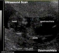
|
| Gastroschisis movie page |
Polyhydramnios
| Amniotic fluid volume is regulated in part in the fetus by swallowing and absorption. Gastrointestinal disorders (such as duodenal atresia, esophageal atresia, gastroschisis, and diaphragmatic hernia) can alter this regulation leading to excess or insufficient amniotic fluid levels.
|

|
Final Thoughts- After Birth
Remember that the GIT does not function until after birth consider:
- metabolic disorders discovered by neonatal diagnosis
- Commensal bacteria populating the sterile GIT.
- Neonatal feeding difficulties due to cleft lip and cleft palate.
- Nutrition for ongoing postnatal development.
Links: gastrointestinal abnormalities
| Gastrointestinal Tract Terms | ||
|---|---|---|
| ||
|
Additional Information
| Additional Information - Content shown under this heading is not part of the material covered in this class. It is provided for those students who would like to know about some concepts or current research in topics related to the current class page. |
The following concepts were not covered in this lecture. Some will be introduced in the associated practical and some will be covered in the BGD Head Development component: mouth | tooth | salivary gland
References
- ↑ Hikspoors JPJM, Peeters MMJP, Mekonen HK, Kruepunga N, Mommen GMC, Cornillie P, Köhler SE & Lamers WH. (2017). The fate of the vitelline and umbilical veins during the development of the human liver. J. Anat. , 231, 718-735. PMID: 28786203 DOI.
- ↑ Dixon LJ, Barnes M, Tang H, Pritchard MT & Nagy LE. (2013). Kupffer cells in the liver. Compr Physiol , 3, 785-97. PMID: 23720329 DOI.
- ↑ Martin V & Shaw-Smith C. (2010). Review of genetic factors in intestinal malrotation. Pediatr. Surg. Int. , 26, 769-81. PMID: 20549505 DOI.
- ↑ Bower RJ, Sieber WK & Kiesewetter WB. (1978). Alimentary tract duplications in children. Ann. Surg. , 188, 669-74. PMID: 718292
Terms
Gastrointestinal Tract Development
- allantois - An extraembryonic membrane, endoderm in origin extension from the early hindgut, then cloaca into the connecting stalk of placental animals, connected to the superior end of developing bladder. In reptiles and birds, acts as a reservoir for wastes and mediates gas exchange. In mammals is associated/incorporated with connecting stalk/placental cord fetal-maternal interface.
- amnion - An extraembryonic membrane]ectoderm and extraembryonic mesoderm in origin and forms the innermost fetal membrane, produces amniotic fluid. This fluid-filled sac initially lies above the trilaminar embryonic disc and with embryoic disc folding this sac is drawn ventrally to enclose (cover) the entire embryo, then fetus. The presence of this membane led to the description of reptiles, bird, and mammals as amniotes.
- amniotic fluid - The fluid that fills amniotic cavity totally encloses and cushions the embryo. Amniotic fluid enters both the gastrointestinal and respiratory tract following rupture of the buccopharyngeal membrane. The late fetus swallows amniotic fluid.
- buccal - (Latin, bucca = cheek) A term used to relate to the mouth (oral cavity).
- buccopharyngeal membrane - (oral membrane) (Latin, bucca = cheek) A membrane which forms the external upper membrane limit (cranial end) of the early gastrointestinal tract (GIT). This membrane develops during gastrulation by ectoderm and endoderm without a middle (intervening) layer of mesoderm. The membrane lies at the floor of the ventral depression (stomodeum) where the oral cavity will open and will breakdown to form the initial "oral opening" of the gastrointestinal tract. The equivilent membrane at the lower end of the gastrointestinal tract is the cloacal membrane.
- cloacal membrane - Forms the external lower membrane limit (caudal end) of the early gastrointestinal tract (GIT). This membrane is formed during gastrulation by ectoderm and endoderm without a middle (intervening) layer of mesoderm. The membrane breaks down to form the initial "anal opening" of the gastrointestinal tract.
- coelom - Term used to describe a space. There are extraembryonic and intraembryonic coeloms that form during vertebrate development. The single intraembryonic coelom will form the 3 major body cavities: pleural, pericardial and peritoneal.
- foregut - The first of the three part/division (foregut - midgut - hindgut) of the early forming gastrointestinal tract. The foregut runs from the buccopharyngeal membrane to the midgut and forms all the tract (esophagus and stomach) from the oral cavity to beneath the stomach. In addition, a ventral bifurcation of the foregut will also form the respiratory tract epithelium.
- gastrula - (Greek, gastrula = little stomach) A stage of an animal embryo in which the three germ layers ([E#endoderm|endoderm]/mesoderm/ectoderm) have just formed.
- gastrulation - The process of differentiation forming a gastrula. Term means literally means "to form a gut" but is more in development, as this process converts the bilaminar embryo (epiblast/hypoblast) into the trilaminar embryo (E#endoderm endoderm/mesoderm/ectoderm) establishing the 3 germ layers that will form all the future tissues of the entire embryo. This process also establishes the the initial body axes.
- hindgut - The last of the three part/division foregut - midgut - hindgut) of the early forming gastrointestinal tract. The hindgut forms all the tract from the distral transverse colon to the cloacal membrane and extends into the connecting stalk (placental cord) as the allantois. In addition, a ventral of the hindgut will also form the urinary tract (bladder, urethra) epithelium.
- intraembryonic coelom - The "horseshoe-shaped" space (cavity) that forms initially in the third week of development in the lateral plate mesoderm that will eventually form the 3 main body cavities: pericardial, pleural, peritoneal. The intraembryonic coelom communicates transiently with the extraembryonic coelom.
- neuralation - The general term used to describe the early formation of the nervous system. It is often used to describe the early events of differentiation of the central ectoderm region to form the neural plate, then neural groove, then neural tube. The nervous system includes the central nervous system (brain and spinal cord) from the neural tube and the peripheral nervous system (peripheral sensory and sympathetic ganglia) from neural crest. In humans, early neuralation begins in week 3 and continues through week 4.
- neural crest - region of cells at the edge of the neural plate that migrates throughout the embryo and contributes to many different tissues. In the gastrointestinal tract it contributes mainly the enteric nervous system within the wall of the gut responsible for peristalsis and secretion.
- pharynx - uppermost end of gastrointestinal and respiratory tract, in the embryo beginning at the buccopharyngeal membrane and forms a major arched cavity within the phrayngeal arches.
- somitogenesis The process of segmentation of the paraxial mesoderm within the trilaminar embryo body to form pairs of somites, or balls of mesoderm. A somite is added either side of the notochord (axial mesoderm) to form a somite pair. The segmentation does not occur in the head region, and begins cranially (head end) and extends caudally (tailward) adding a somite pair at regular time intervals. The process is sequential and therefore used to stage the age of many different species embryos based upon the number visible somite pairs. In humans, the first somite pair appears at day 20 and adds caudally at 1 somite pair/90 minutes until on average 44 pairs eventually form.
- splanchnic mesoderm - Gastrointestinal tract (endoderm) associated mesoderm formed by the separation of the lateral plate mesoderm into two separate components by a cavity, the intraembryonic coelom. Splanchnic mesoderm is the embryonic origin of the gastrointestinal tract connective tissue, smooth muscle, blood vessels and contribute to organ development (pancreas, spleen, liver). The intraembryonic coelom will form the three major body cavities including the space surrounding the gut, the peritoneal cavity. The other half of the lateral plate mesoderm (somatic mesoderm) is associated with the ectoderm of the body wall.
- stomodeum - (stomadeum, stomatodeum) A ventral surface depression on the early embryo head surrounding the buccopharyngeal membrane, which lies at the floor of this depression. This surface depression lies between the maxillary and mandibular components of the first pharyngeal arch.
| Other Terms Lists |
|---|
| Terms Lists: ART | Birth | Bone | Cardiovascular | Cell Division | Endocrine | Gastrointestinal | Genital | Genetic | Head | Hearing | Heart | Immune | Integumentary | Neonatal | Neural | Oocyte | Palate | Placenta | Radiation | Renal | Respiratory | Spermatozoa | Statistics | Tooth | Ultrasound | Vision | Historic | Drugs | Glossary |
BGDB: Lecture - Gastrointestinal System | Practical - Gastrointestinal System | Lecture - Face and Ear | Practical - Face and Ear | Lecture - Endocrine | Lecture - Sexual Differentiation | Practical - Sexual Differentiation | Tutorial
Glossary Links
- Glossary: A | B | C | D | E | F | G | H | I | J | K | L | M | N | O | P | Q | R | S | T | U | V | W | X | Y | Z | Numbers | Symbols | Term Link
Cite this page: Hill, M.A. (2024, June 15) Embryology BGD Lecture - Gastrointestinal System Development. Retrieved from https://embryology.med.unsw.edu.au/embryology/index.php/BGD_Lecture_-_Gastrointestinal_System_Development
- © Dr Mark Hill 2024, UNSW Embryology ISBN: 978 0 7334 2609 4 - UNSW CRICOS Provider Code No. 00098G