Embryonic Development: Difference between revisions
(→Week 8) |
|||
| Line 189: | Line 189: | ||
|} | |} | ||
== Carnegie Stage Table == | |||
{ | {| border="0" | ||
| width="40" | | |||
<center>'''Stage'''</center> | |||
| width="65" | | |||
<center>'''Days''' (approx)</center> | |||
| width="101" | | |||
<center>'''Size'''</center> <center>(mm)</center> | |||
| width="100" | | |||
<center>'''Images<br />'''(not to scale)</center> | |||
| width="400" | | |||
<center>'''Events'''</center> | |||
|- | |||
| width="40" | | |||
<center>'''1'''</center> | |||
| width="65" | | |||
<center> 1 </center>('''week 1''') | |||
| width="101" | | |||
<center>0.1 - 0.15</center> | |||
| width="100" bgcolor="#000000" | | |||
<center>[[File:Human_zygote_two_pronuclei_02.jpg|60px|Link=Carnegie_stage_1]]</center> | |||
| width="400" | | |||
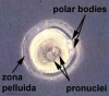
fertilized oocyte, pronuclei | |||
|- | |||
| width="40" | | |||
<center>'''2'''</center> | |||
| width="65" | | |||
<center> 2 - 3</center> | |||
| width="101" | | |||
<center>0.1 - 0.2</center> | |||
| width="100" bgcolor="#000000" | | |||
<center>[[File:Human_embryo_day_3.jpg|60px|Link=Carnegie_stage_2]]</center> | |||
| width="400" | | |||
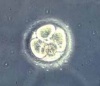
cell division with reduction in cytoplasmic volume, formation of inner and outer cell mass | |||
|- | |||
| width="40" | | |||
<center>'''3'''</center> | |||
| width="65" | | |||
<center> 4 - 5</center> | |||
| width="101" | | |||
<center>0.1 - 0.2</center> | |||
| width="100" bgcolor="#000000" | | |||
<center>[[File:Human_embryo_day_5.jpg|60px|Link=Carnegie_stage_3]]</center> | |||
| width="400" | | |||
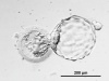
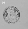
loss of zona pellucida, free blastocyst | |||
|- | |||
| width="40" | | |||
<center>'''4'''</center> | |||
| width="65" | | |||
<center> 5 - 6</center> | |||
| width="101" | | |||
<center>0.1 - 0.2</center> | |||
| width="100" bgcolor="#000000" | | |||
{{Template: | | width="400" | | ||
attaching blastocyst | |||
|- | |||
| width="40" | | |||
<center>'''5'''</center> | |||
| width="65" | | |||
<center> 7 - 12<br /> ('''week 2''')</center> | |||
| width="101" | | |||
<center> 0.1 - 0.2</center> | |||
| width="100" bgcolor="#000000" | | |||
| width="400" | | |||
implantation | |||
|- | |||
| width="40" | | |||
<center>'''6'''</center> | |||
| width="65" | | |||
<center> 13 - 15</center> | |||
| width="101" | | |||
<center> 0.2</center> | |||
| width="100" bgcolor="#000000" | | |||
| width="400" | | |||
extraembryonic mesoderm, primitive streak | |||
|- | |||
| width="40" | | |||
<center>'''7'''</center> | |||
| width="65" | | |||
<center> 15 - 17 </center><center>('''week 3''')</center> | |||
| width="101" | | |||
<center> 0.4</center> | |||
| width="100" bgcolor="#000000" | | |||
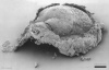
<center>[[File:Stage7_features.jpg|60px|Link=Carnegie_stage_7]]</center> | |||
| width="400" | | |||
gastrulation, notochordal process | |||
|- | |||
| width="40" | | |||
<center>'''8'''</center> | |||
| width="65" | | |||
<center> 17 - 19</center> | |||
| width="101" | | |||
<center> 1.0 - 1.5</center> | |||
| width="100" bgcolor="#000000" | | |||
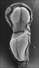
<center>[[File:Stage8_bf4.jpg|60px|Link=Carnegie_stage_8]]</center> | |||
| width="400" | | |||
primitive pit, notochordal canal | |||
|- | |||
| width="40" | | |||
<center>'''9'''</center> | |||
| width="65" | | |||
<center> 19 - 21</center> | |||
| width="101" | | |||
<center> 1.5 - 2.5</center> | |||
| width="100" bgcolor="#000000" | | |||
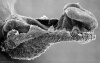
<center>[[File:Stage9_dorsal.jpg|60px|Link=Carnegie_stage_9]]</center> | |||
| width="400" | | |||
'''Somite Number 1 - 3''' neural folds, cardiac primordium, head fold | |||
|- | |||
| width="40" | | |||
<center>'''10'''</center> | |||
| width="65" | | |||
<center> 22 - 23 </center><center>('''week 4''')</center> | |||
| width="101" | | |||
<center> 2 - 3.5</center> | |||
| width="100" bgcolor="#000000" | | |||
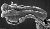
<center>[[File:Stage10_bf4b.jpg|60px|Link=Carnegie_stage_10]]</center> | |||
| width="400" | | |||
'''Somite Number 4 - 12''' neural fold fuses | |||
|- | |||
| width="40" | | |||
<center>'''11'''</center> | |||
| width="65" | | |||
<center> 23 - 26</center> | |||
| width="101" | | |||
<center> 2.5 - 4.5</center> | |||
| width="100" bgcolor="#000000" | | |||
<center>[[File:Stage11 bf7b.jpg|60px|Link=Carnegie_stage_11]]</center> | |||
| width="400" | | |||
'''Somite Number 13 - 20''' rostral neuropore closes | |||
|- | |||
| width="40" | | |||
<center>'''12'''</center> | |||
| width="65" | | |||
<center> 26 - 30</center> | |||
| width="101" | | |||
<center> 3 - 5</center> | |||
| width="100" bgcolor="#000000" | | |||
<center>[[File:Stage12 bf5b.jpg|60px|Link=Carnegie_stage_12]]</center> | |||
| width="400" | | |||
'''Somite Number 21 - 29''' caudal neuropore closes | |||
|- | |||
| width="40" | | |||
<center>'''13'''</center> | |||
| width="65" | | |||
<center> 28 - 32 </center>('''week 5''') | |||
| width="101" | | |||
<center> 4 - 6</center> | |||
| width="100" bgcolor="#000000" | | |||
<center>[[File:Stage13 bf2c.jpg|60px|Link=Carnegie_stage_13]]</center> | |||
| width="400" | | |||
'''Somite Number 30''' leg buds, lens placode, pharyngeal arches | |||
|- bgcolor="#CCFFCC" | |||
| colspan="5" width="376" height="18" | | |||
<center> [[Carnegie_stage_13_-_serial_sections|Stage 13/14 shown in serial embryo sections]] series of Embryology Program</center> | |||
|- | |||
| width="40" | | |||
<center>'''14'''</center> | |||
| width="65" | | |||
<center> 31 - 35</center> | |||
| width="101" | | |||
<center> 5 - 7</center> | |||
| width="100" bgcolor="#000000" | | |||
<center>[[File:Stage14_bf2c.jpg|60px|Link=Carnegie_stage_14]]</center> | |||
| width="400" | | |||
lens pit, optic cup | |||
|- | |||
| width="40" | | |||
<center>'''15'''</center> | |||
| width="65" | | |||
<center> 35 - 38</center> | |||
| width="101" | | |||
<center> 7 - 9</center> | |||
| width="100" bgcolor="#000000" | | |||
<center>[[File:Stage15 bf1c.jpg|60px|Link=Carnegie_stage_15]]</center> | |||
| width="400" | | |||
lens vesicle, nasal pit, hand plate | |||
|- | |||
| width="40" | | |||
<center>'''16'''</center> | |||
| width="65" | | |||
<center> 37 - 42 </center>('''week 6''') | |||
| width="101" | | |||
<center> 8 - 11</center> | |||
| width="100" bgcolor="#000000" | | |||
<center>[[File:Stage16 bf1c.jpg|60px|Link=Carnegie_stage_16]]</center> | |||
| width="400" | | |||
nasal pits moved ventrally, auricular hillocks, foot plate | |||
|- | |||
| width="40" | | |||
<center>'''17'''</center> | |||
| width="65" | | |||
<center> 42 - 101</center> | |||
| width="101" | | |||
<center> 11 - 14</center> | |||
| width="100" bgcolor="#000000" | | |||
<center>[[File:Stage17 bf1c.jpg|60px|Link=Carnegie_stage_17]]</center> | |||
| width="400" | | |||
finger rays | |||
|- | |||
| width="40" | | |||
<center>'''18'''</center> | |||
| width="65" | | |||
<center> 101 - 48 </center>('''week 7''') | |||
| width="101" | | |||
<center> 13 - 17</center> | |||
| width="100" bgcolor="#000000" | | |||
<center>[[File:Stage18 bf1c.jpg|60px|Link=Carnegie_stage_18]]</center> | |||
| width="400" | | |||
ossification commences | |||
|- | |||
| width="40" | | |||
<center>'''19'''</center> | |||
| width="65" | | |||
<center> 48 - 51</center> | |||
| width="101" | | |||
<center> 16 - 18</center> | |||
| width="100" bgcolor="#000000" | | |||
<center>[[File:Stage19 bf1c.jpg|60px|Link=Carnegie_stage_19]]</center> | |||
| width="400" | | |||
straightening of trunk | |||
|- | |||
| width="40" | | |||
<center>'''20'''</center> | |||
| width="65" | | |||
<center> 51 - 53 </center>('''week 8''') | |||
| width="101" | | |||
<center> 18 - 22</center> | |||
| width="100" bgcolor="#000000" | | |||
<center>[[File:Stage20 bf1c.jpg|60px|Link=Carnegie_stage_20]]</center> | |||
| width="400" | | |||
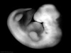
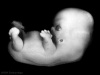
upper limbs longer and bent at elbow | |||
|- | |||
| width="40" | | |||
<center>'''21'''</center> | |||
| width="65" | | |||
<center>53 - 54</center> | |||
| width="101" | | |||
<center>22 - 24</center> | |||
| width="100" bgcolor="#000000" | | |||
<center>[[File:Stage21 bf1c.jpg|60px|Link=Carnegie_stage_21]]</center> | |||
| width="400" | | |||
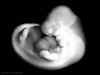
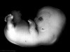
hands and feet turned inward | |||
|- bgcolor="#FFCCCC" | |||
| colspan="5" width="376" | | |||
<center> [[Carnegie_stage_22_-_serial_sections|Stage 22 shown in serial embryo sections series]] of Embryology Program</center> | |||
|- | |||
| width="40" | | |||
<center>'''22'''</center> | |||
| width="65" | | |||
<center> 54 - 56</center> | |||
| width="101" | | |||
<center> 23 - 28</center> | |||
| width="100" bgcolor="#000000" | | |||
<center>[[File:Stage22 bf1c.jpg|60px|Link=Carnegie_stage_22]]</center> | |||
| width="400" | | |||
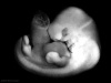
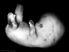
eyelids, external ears | |||
|- | |||
| width="40" | | |||
<center>'''23'''</center> | |||
| width="65" | | |||
<center> 56 - 60</center> | |||
| width="101" | | |||
<center>27 - 31</center> | |||
| width="100" bgcolor="#000000" | | |||
<center>[[File:Stage23 bf1c.jpg|60px|Link=Carnegie_stage_23]]</center> | |||
| width="400" | | |||
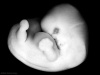
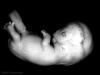
rounded head, body and limbs | |||
|- | |||
| colspan="5" width="376" | | |||
<center>Following this stage [[Fetal Development]] occurs until birth (approx 40 weeks)</center> | |||
|} | |||
{{Template:Carnegie_stages}} | |||
{{Template:Glossary}} | |||
{{Template:Footer}} | |||
[[Category:Human Embryo]] | [[Category:Human Embryo]] | ||
Revision as of 05:27, 13 April 2011
Introduction

| Author Comments |
|---|
It is not so important to memorise the dates, as they are only approximate, but more important to understand growth (size changes) and the development (overall sequence of events) during this period. |
| This page shows some key events of human development during the embryonic period of the first eight weeks (weeks 1 - 8) following fertilization. This period is also considered the organogenic period, when most organs within the embryo have begun to form.
There are links to more detailed descriptions which can be viewed in a week by week format, by the Carnegie stages or integrated into a Timeline of human development. Online resources available include: individual images of all Carnegie stages, scanning electron micrographs of the earlier stages, cross-sections showing internal structures at mid- and late-embryonic, 3D reconstructions of internal structures, animations of processes, ultrasound scans and information about abnormalites of development. Note that there is variability in the actual timing of specific events and at the end of this period fetal development begins. |
<Flowplayer height="300" width="226" autoplay="true">Embryo stages 002.flv</Flowplayer> |
| Quicktime version |
Bookmark with:
![]()
![]()
![]()
![]()
![]()
![]()
- Carnegie Stages: 1 | 2 | 3 | 4 | 5 | 6 | 7 | 8 | 9 | 10 | 11 | 12 | 13 | 14 | 15 | 16 | 17 | 18 | 19 | 20 | 21 | 22 | 23 | About Stages | Timeline

Use the above stage number links to images and information about each specific stage of human development over the first 8 weeks. The links below give a broad overview of developmental events during each week.
Alternatively, look through development week by week.
Embryo Week: Week 1 | Week 2 | Week 3 | Week 4 | Week 5 | Week 6 | Week 7 | Week 8 | Week 9
Week 1

- Week 1 Carnegie stage - 1 | 2 | 3 | 4
- Oocyte | Spermatozoa | Fertilization
- Zygote
- Morula
- Blastocyst
| Carnegie stages | |||
|---|---|---|---|
|
|
|
|
| stage 1 | stage 2 | stage 3 | stage 4 |
Week 2
- Week 2 Carnegie stage - 5 | 6
- Trophoblast - outer cell layer
- Embryoblast - inner cell mass
- Implantation
- Bilaminar embryo
Week 3

| Carnegie stages | ||
|---|---|---|
|
|
|
| stage 7 | stage 8 | stage 9 |
Week 4

| Carnegie stages | |||
|---|---|---|---|
|
|
|
|
| stage 10 | stage 11 | stage 12 | stage 13 |
Week 5

| Carnegie stages | |
|---|---|
|
|
| stage 14 | stage 15 |
Week 6

| Carnegie stages | |
|---|---|
|
|
| stage 16 | stage 17 |
Week 7

| Carnegie stages | |
|---|---|
|
|
| stage 18 | stage 19 |
Week 8

| Carnegie stages | |||
|---|---|---|---|
|
|
|
|
| stage 20 | stage 21 | stage 22 | stage 23 |
Carnegie Stage Table
|
|
|
|
(not to scale) |
|
|
|
|
|
 |
fertilized oocyte, pronuclei |
|
|
|
|
 |
cell division with reduction in cytoplasmic volume, formation of inner and outer cell mass |
|
|
|
|
 |
loss of zona pellucida, free blastocyst |
|
|
|
|
attaching blastocyst | |
|
|
(week 2) |
|
implantation | |
|
|
|
|
extraembryonic mesoderm, primitive streak | |
|
|
|
|
|
gastrulation, notochordal process |
|
|
|
|
|
primitive pit, notochordal canal |
|
|
|
|
 |
Somite Number 1 - 3 neural folds, cardiac primordium, head fold |
|
|
|
|
|
Somite Number 4 - 12 neural fold fuses |
|
|
|
|
|
Somite Number 13 - 20 rostral neuropore closes |
|
|
|
|
|
Somite Number 21 - 29 caudal neuropore closes |
|
|
|
|
|
Somite Number 30 leg buds, lens placode, pharyngeal arches |
|
| ||||
|
|
|
|
|
lens pit, optic cup |
|
|
|
|
|
lens vesicle, nasal pit, hand plate |
|
|
|
|
|
nasal pits moved ventrally, auricular hillocks, foot plate |
|
|
|
|
|
finger rays |
|
|
|
|
|
ossification commences |
|
|
|
|
|
straightening of trunk |
|
|
|
|
|
upper limbs longer and bent at elbow |
|
|
|
|
|
hands and feet turned inward |
|
| ||||
|
|
|
|
|
eyelids, external ears |
|
|
|
|
|
rounded head, body and limbs |
|
| ||||
- Carnegie Stages: 1 | 2 | 3 | 4 | 5 | 6 | 7 | 8 | 9 | 10 | 11 | 12 | 13 | 14 | 15 | 16 | 17 | 18 | 19 | 20 | 21 | 22 | 23 | About Stages | Timeline
Glossary Links
- Glossary: A | B | C | D | E | F | G | H | I | J | K | L | M | N | O | P | Q | R | S | T | U | V | W | X | Y | Z | Numbers | Symbols | Term Link
Cite this page: Hill, M.A. (2024, June 26) Embryology Embryonic Development. Retrieved from https://embryology.med.unsw.edu.au/embryology/index.php/Embryonic_Development
- © Dr Mark Hill 2024, UNSW Embryology ISBN: 978 0 7334 2609 4 - UNSW CRICOS Provider Code No. 00098G