|
|
| Line 45: |
Line 45: |
|
| |
|
| {{Template:Week}} | | {{Template:Week}} |
|
| |
| == Carnegie Stage Table ==
| |
|
| |
| {| border="0"
| |
| | width="40" |
| |
| <center>'''Stage'''</center>
| |
| | width="65" |
| |
| <center>'''Days''' (approx)</center>
| |
| | width="101" |
| |
| <center>'''Size'''</center> <center>(mm)</center>
| |
| | width="100" |
| |
| <center>'''Images<br />'''(not to scale)</center>
| |
| | width="400" |
| |
| <center>'''Events'''</center>
| |
| |-
| |
| | width="40" |
| |
| <center>'''1'''</center>
| |
| | width="65" |
| |
| <center> 1 </center>('''week 1''')
| |
| | width="101" |
| |
| <center>0.1 - 0.15</center>
| |
| | width="100" bgcolor="#000000" |
| |
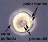
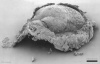
| <center>[[File:Human_zygote_two_pronuclei_02.jpg|60px|Link=Carnegie_stage_1]]</center>
| |
| | width="400" |
| |
| fertilized oocyte, pronuclei
| |
| |-
| |
| | width="40" |
| |
| <center>'''2'''</center>
| |
| | width="65" |
| |
| <center> 2 - 3</center>
| |
| | width="101" |
| |
| <center>0.1 - 0.2</center>
| |
| | width="100" bgcolor="#000000" |
| |
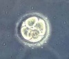
| <center>[[File:Human_embryo_day_3.jpg|60px|Link=Carnegie_stage_2]]</center>
| |
| | width="400" |
| |
| cell division with reduction in cytoplasmic volume, formation of inner and outer cell mass
| |
| |-
| |
| | width="40" |
| |
| <center>'''3'''</center>
| |
| | width="65" |
| |
| <center> 4 - 5</center>
| |
| | width="101" |
| |
| <center>0.1 - 0.2</center>
| |
| | width="100" bgcolor="#000000" |
| |
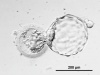
| <center>[[File:Human_embryo_day_5.jpg|60px|Link=Carnegie_stage_3]]</center>
| |
| | width="400" |
| |
| loss of zona pellucida, free blastocyst
| |
| |-
| |
| | width="40" |
| |
| <center>'''4'''</center>
| |
| | width="65" |
| |
| <center> 5 - 6</center>
| |
| | width="101" |
| |
| <center>0.1 - 0.2</center>
| |
| | width="100" bgcolor="#000000" |
| |
|
| |
| | width="400" |
| |
| attaching blastocyst
| |
| |-
| |
| | width="40" |
| |
| <center>'''5'''</center>
| |
| | width="65" |
| |
| <center> 7 - 12<br /> ('''week 2''')</center>
| |
| | width="101" |
| |
| <center> 0.1 - 0.2</center>
| |
| | width="100" bgcolor="#000000" |
| |
|
| |
| | width="400" |
| |
| implantation
| |
| |-
| |
| | width="40" |
| |
| <center>'''6'''</center>
| |
| | width="65" |
| |
| <center> 13 - 15</center>
| |
| | width="101" |
| |
| <center> 0.2</center>
| |
| | width="100" bgcolor="#000000" |
| |
|
| |
| | width="400" |
| |
| extraembryonic mesoderm, primitive streak
| |
| |-
| |
| | width="40" |
| |
| <center>'''7'''</center>
| |
| | width="65" |
| |
| <center> 15 - 17 </center><center>('''week 3''')</center>
| |
| | width="101" |
| |
| <center> 0.4</center>
| |
| | width="100" bgcolor="#000000" |
| |
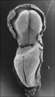
| <center>[[File:Stage7_features.jpg|60px|Link=Carnegie_stage_7]]</center>
| |
| | width="400" |
| |
| gastrulation, notochordal process
| |
| |-
| |
| | width="40" |
| |
| <center>'''8'''</center>
| |
| | width="65" |
| |
| <center> 17 - 19</center>
| |
| | width="101" |
| |
| <center> 1.0 - 1.5</center>
| |
| | width="100" bgcolor="#000000" |
| |
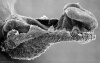
| <center>[[File:Stage8_bf4.jpg|60px|Link=Carnegie_stage_8]]</center>
| |
| | width="400" |
| |
| primitive pit, notochordal canal
| |
| |-
| |
| | width="40" |
| |
| <center>'''9'''</center>
| |
| | width="65" |
| |
| <center> 19 - 21</center>
| |
| | width="101" |
| |
| <center> 1.5 - 2.5</center>
| |
| | width="100" bgcolor="#000000" |
| |
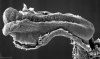
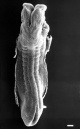
| <center>[[File:Stage9_dorsal.jpg|60px|Link=Carnegie_stage_9]]</center>
| |
| | width="400" |
| |
| '''Somite Number 1 - 3''' neural folds, cardiac primordium, head fold
| |
| |-
| |
| | width="40" |
| |
| <center>'''10'''</center>
| |
| | width="65" |
| |
| <center> 22 - 23 </center><center>('''week 4''')</center>
| |
| | width="101" |
| |
| <center> 2 - 3.5</center>
| |
| | width="100" bgcolor="#000000" |
| |
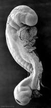
| <center>[[File:Stage10_bf4b.jpg|60px|Link=Carnegie_stage_10]]</center>
| |
| | width="400" |
| |
| '''Somite Number 4 - 12''' neural fold fuses
| |
| |-
| |
| | width="40" |
| |
| <center>'''11'''</center>
| |
| | width="65" |
| |
| <center> 23 - 26</center>
| |
| | width="101" |
| |
| <center> 2.5 - 4.5</center>
| |
| | width="100" bgcolor="#000000" |
| |
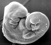
| <center>[[File:Stage11 bf7b.jpg|60px|Link=Carnegie_stage_11]]</center>
| |
| | width="400" |
| |
| '''Somite Number 13 - 20''' rostral neuropore closes
| |
| |-
| |
| | width="40" |
| |
| <center>'''12'''</center>
| |
| | width="65" |
| |
| <center> 26 - 30</center>
| |
| | width="101" |
| |
| <center> 3 - 5</center>
| |
| | width="100" bgcolor="#000000" |
| |
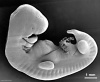
| <center>[[File:Stage12 bf5b.jpg|60px|Link=Carnegie_stage_12]]</center>
| |
| | width="400" |
| |
| '''Somite Number 21 - 29''' caudal neuropore closes
| |
| |-
| |
| | width="40" |
| |
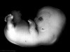
| <center>'''13'''</center>
| |
| | width="65" |
| |
| <center> 28 - 32 </center>('''week 5''')
| |
| | width="101" |
| |
| <center> 4 - 6</center>
| |
| | width="100" bgcolor="#000000" |
| |
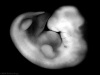
| <center>[[File:Stage13 bf2c.jpg|60px|Link=Carnegie_stage_13]]</center>
| |
| | width="400" |
| |
| '''Somite Number 30''' leg buds, lens placode, pharyngeal arches
| |
| |- bgcolor="#CCFFCC"
| |
| | colspan="5" width="376" height="18" |
| |
| <center> [[Carnegie_stage_13_-_serial_sections|Stage 13/14 shown in serial embryo sections]] series of Embryology Program</center>
| |
| |-
| |
| | width="40" |
| |
| <center>'''14'''</center>
| |
| | width="65" |
| |
| <center> 31 - 35</center>
| |
| | width="101" |
| |
| <center> 5 - 7</center>
| |
| | width="100" bgcolor="#000000" |
| |
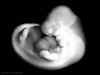
| <center>[[File:Stage14_bf2c.jpg|60px|Link=Carnegie_stage_14]]</center>
| |
| | width="400" |
| |
| lens pit, optic cup
| |
| |-
| |
| | width="40" |
| |
| <center>'''15'''</center>
| |
| | width="65" |
| |
| <center> 35 - 38</center>
| |
| | width="101" |
| |
| <center> 7 - 9</center>
| |
| | width="100" bgcolor="#000000" |
| |
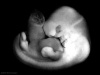
| <center>[[File:Stage15 bf1c.jpg|60px|Link=Carnegie_stage_15]]</center>
| |
| | width="400" |
| |
| lens vesicle, nasal pit, hand plate
| |
| |-
| |
| | width="40" |
| |
| <center>'''16'''</center>
| |
| | width="65" |
| |
| <center> 37 - 42 </center>('''week 6''')
| |
| | width="101" |
| |
| <center> 8 - 11</center>
| |
| | width="100" bgcolor="#000000" |
| |
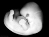
| <center>[[File:Stage16 bf1c.jpg|60px|Link=Carnegie_stage_16]]</center>
| |
| | width="400" |
| |
| nasal pits moved ventrally, auricular hillocks, foot plate
| |
| |-
| |
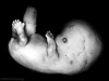
| | width="40" |
| |
| <center>'''17'''</center>
| |
| | width="65" |
| |
| <center> 42 - 101</center>
| |
| | width="101" |
| |
| <center> 11 - 14</center>
| |
| | width="100" bgcolor="#000000" |
| |
| <center>[[File:Stage17 bf1c.jpg|60px|Link=Carnegie_stage_17]]</center>
| |
| | width="400" |
| |
| finger rays
| |
| |-
| |
| | width="40" |
| |
| <center>'''18'''</center>
| |
| | width="65" |
| |
| <center> 101 - 48 </center>('''week 7''')
| |
| | width="101" |
| |
| <center> 13 - 17</center>
| |
| | width="100" bgcolor="#000000" |
| |
| <center>[[File:Stage18 bf1c.jpg|60px|Link=Carnegie_stage_18]]</center>
| |
| | width="400" |
| |
| ossification commences
| |
| |-
| |
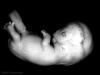
| | width="40" |
| |
| <center>'''19'''</center>
| |
| | width="65" |
| |
| <center> 48 - 51</center>
| |
| | width="101" |
| |
| <center> 16 - 18</center>
| |
| | width="100" bgcolor="#000000" |
| |
| <center>[[File:Stage19 bf1c.jpg|60px|Link=Carnegie_stage_19]]</center>
| |
| | width="400" |
| |
| straightening of trunk
| |
| |-
| |
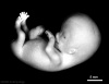
| | width="40" |
| |
| <center>'''20'''</center>
| |
| | width="65" |
| |
| <center> 51 - 53 </center>('''week 8''')
| |
| | width="101" |
| |
| <center> 18 - 22</center>
| |
| | width="100" bgcolor="#000000" |
| |
| <center>[[File:Stage20 bf1c.jpg|60px|Link=Carnegie_stage_20]]</center>
| |
| | width="400" |
| |
| upper limbs longer and bent at elbow
| |
| |-
| |
| | width="40" |
| |
| <center>'''21'''</center>
| |
| | width="65" |
| |
| <center>53 - 54</center>
| |
| | width="101" |
| |
| <center>22 - 24</center>
| |
| | width="100" bgcolor="#000000" |
| |
| <center>[[File:Stage21 bf1c.jpg|60px|Link=Carnegie_stage_21]]</center>
| |
| | width="400" |
| |
| hands and feet turned inward
| |
| |- bgcolor="#FFCCCC"
| |
| | colspan="5" width="376" |
| |
| <center> [[Carnegie_stage_22_-_serial_sections|Stage 22 shown in serial embryo sections series]] of Embryology Program</center>
| |
| |-
| |
| | width="40" |
| |
| <center>'''22'''</center>
| |
| | width="65" |
| |
| <center> 54 - 56</center>
| |
| | width="101" |
| |
| <center> 23 - 28</center>
| |
| | width="100" bgcolor="#000000" |
| |
| <center>[[File:Stage22 bf1c.jpg|60px|Link=Carnegie_stage_22]]</center>
| |
| | width="400" |
| |
| eyelids, external ears
| |
| |-
| |
| | width="40" |
| |
| <center>'''23'''</center>
| |
| | width="65" |
| |
| <center> 56 - 60</center>
| |
| | width="101" |
| |
| <center>27 - 31</center>
| |
| | width="100" bgcolor="#000000" |
| |
| <center>[[File:Stage23 bf1c.jpg|60px|Link=Carnegie_stage_23]]</center>
| |
| | width="400" |
| |
| rounded head, body and limbs
| |
| |-
| |
| | colspan="5" width="376" |
| |
| <center>Following this stage [[Fetal Development]] occurs until birth (approx 40 weeks)</center>
| |
| |}
| |
|
| |
|
| == Week 1 == | | == Week 1 == |


