Lecture - Head Development: Difference between revisions
mNo edit summary |
No edit summary |
||
| (44 intermediate revisions by 2 users not shown) | |||
| Line 1: | Line 1: | ||
{{Header}} | |||
= Head Development= | = Head Development= | ||
== Introduction == | == Introduction == | ||
[[File:Stage11 sem81.jpg|thumb|Human face (Week 4, Stage 11)]] | |||
[[File:Stage16-18 face.jpg|thumb|Human face development (Week 6 to 7)]] | [[File:Stage14_sem2l.jpg|thumb|Human face (Week 5, Stage 14)]] | ||
[[File:Stage16-18 face.jpg|thumb|Human face development (Week 6 to 7, Stage 16-18)]] | |||
The face is the anatomical feature which is truly unique to each human, though the basis of its general development is identical for all humans and similar to that seem for other species. The face has a complex origin arising from a number of head structures and sensitive to a number of teratogens during critical periods of its development. The related structures of upper lip and palate significantly contribute to the majority of face abnormalities. | The face is the anatomical feature which is truly unique to each human, though the basis of its general development is identical for all humans and similar to that seem for other species. The face has a complex origin arising from a number of head structures and sensitive to a number of teratogens during critical periods of its development. The related structures of upper lip and palate significantly contribute to the majority of face abnormalities. | ||
The head and neck structures are more than just the face, and are derived from pharyngeal arches 1 - 6 with the face forming from arch 1 and 2 and the frontonasal prominence. Each arch contains similar Arch components derived from endoderm, mesoderm, neural crest and ectoderm. These components though will form different structures depending on their arch origin. Because the head contains many different structures also review notes on [[Sensory System Development|Special Senses]]), [[Respiratory System Development|Respiratory]], Integumentary (Teeth), [[Endocrine System Development|Endocrine]] (thyroid, parathyroid, pituitary, thymus) and [[Ultrasound]]- Cleft lip/palate. | The head and neck structures are more than just the face, and are derived from pharyngeal arches 1 - 6 with the face forming from arch 1 and 2 and the frontonasal prominence. Each arch contains similar Arch components derived from endoderm, mesoderm, neural crest and ectoderm. These components though will form different structures depending on their arch origin. Because the head contains many different structures also review notes on [[Sensory System Development|Special Senses]]), [[Respiratory System Development|Respiratory]], Integumentary (Teeth), [[Endocrine System Development|Endocrine]] (thyroid, parathyroid, pituitary, thymus) and [[Ultrasound]]- Cleft lip/palate. | ||
{| | |||
| {{OneMinuteClock}} | |||
[[One_Minute_Embryology#Human_Face_and_Palate|'''1 Minute Embryology''']]<br>[https://thebox.unsw.edu.au/video/1-minute-embryology-face-and-palate UNSW theBox] | |||
|} | |||
{| class="wikitable mw-collapsible mw-collapsed" | |||
! 2016 Lecture Video Recording | |||
|- | |||
| Due to a technical issue, the 2017 lecture recording has no sound. | |||
I have therefore included this 2016 lecture video recording that is similar in content to the current 2017 online lecture. | |||
<html5media height="600" width="800">File:2016Lecture-Head.mp4</html5media> | |||
Click to play new window - [[Media:2016Lecture-Head.mp4|'''2016 Lecture Video''']] (48 MB) | |||
|} | |||
==Lecture Objectives== | ==Lecture Objectives== | ||
| Line 18: | Line 37: | ||
==Lecture Resources== | ==Lecture Resources== | ||
[[Media:ANAT2341_Lecture_18_2018_-_Beverdam_-_Head_development.pdf|2018 Lecture PDF]] | |||
{| class="wikitable mw-collapsible mw-collapsed" | {| class="wikitable mw-collapsible mw-collapsed" | ||
! Movies | ! Movies | ||
|- | |- | ||
| valign="bottom"|{{Face movie}} | | valign="bottom"|{{Face movie}} | ||
| Line 34: | Line 55: | ||
|} | |} | ||
{| class="wikitable mw-collapsible mw-collapsed" | {| class="wikitable mw-collapsible mw-collapsed" | ||
! References | ! colspan=2|References | ||
|- | |- | ||
| {{Embryo logocitation}} | | {{Embryo logocitation}} | ||
| | | | ||
{{Head Links}} | {{Head Links}} | ||
* Lecture Archive: | * Lecture Archive: [[Media:ANAT2341_Lecture_13_2017_-_Beverdam_-_Head_development.pdf|2017 PDF]] | [https://embryology.med.unsw.edu.au/embryology/index.php?title=Lecture_-_Head_Development&oldid=197375 2015] | [[Media:2015ANAT2341_Lecture_11_-_Head Development.pdf|2015 PDF]] | [https://embryology.med.unsw.edu.au/embryology/index.php?title=Lecture_-_Head_Development&oldid=143731 2014] | [[Media:ANAT2341_Lecture_11_-_2014_Head_Development.pdf|2014 PDF]] | ||
|- | |- | ||
| {{ | | [[File:The Developing Human, 10th edn.jpg|90px]] | ||
| {{MPT2015APAcitation}} (links only function with UNSW connection) | |||
Chapter 9 [http://ebookcentral.proquest.com.wwwproxy1.library.unsw.edu.au/lib/unsw/reader.action?docID=2074364&ppg=216 Pharyngeal Apparatus, Face, and Neck] | |||
Chapter 18 [http://ebookcentral.proquest.com.wwwproxy1.library.unsw.edu.au/lib/unsw/reader.action?docID=2074364&ppg=553 Development of Eyes and Ears] | |||
{{UNSW textbook - The Developing Human}} | |||
|- | |- | ||
| {{ | | [[File:Larsen's human embryology 5th ed.jpg|90px]] | ||
| {{Larsen2015APAcitation}}(links only function with UNSW connection) | |||
Chapter 17 [http://ebookcentral.proquest.com.wwwproxy1.library.unsw.edu.au/lib/unsw/reader.action?docID=2074524&ppg=447 Development of the Pharyngeal Apparatus and Face] | |||
Chapter 18 [http://ebookcentral.proquest.com.wwwproxy1.library.unsw.edu.au/lib/unsw/reader.action?docID=2074524&ppg=491 Development of the Ears] | |||
Chapter 19 [http://ebookcentral.proquest.com.wwwproxy1.library.unsw.edu.au/lib/unsw/reader.action?docID=2074524&ppg=506 Development of the Eyes] | |||
{{UNSW textbook - Larsen's Human Embryology}} | |||
|} | |} | ||
==Animation of Face Development== | ==Animation of Face Development== | ||
{| | {| | ||
| Line 88: | Line 106: | ||
Note the complex origin of the maxillary region (upper jaw) requiring the fusion of several embryonic elements, abnormalities of this process lead to [[C#cleft lip|cleft lip]] and [[C#cleft palate|cleft palate]]. | Note the complex origin of the maxillary region (upper jaw) requiring the fusion of several embryonic elements, abnormalities of this process lead to [[C#cleft lip|cleft lip]] and [[C#cleft palate|cleft palate]]. | ||
See also the movie | See also the movie showing a similar view of human embryo faces between [[Carnegie stage 16|Carnegie stage 16]] to [[Carnegie stage 18|18]]. | ||
|} | |} | ||
==Buccopharyngeal Membrane== | ==Week 3== | ||
===Buccopharyngeal Membrane=== | |||
These images of the Stage 11 embryo show the breakdown of the buccopharyngeal membrane. | These images of the Stage 11 embryo show the breakdown of the buccopharyngeal membrane. | ||
<gallery> | <gallery> | ||
File:Stage11 | File:Stage11 sem4.jpg|Low power ventral view of the Buccopharyngeal Membrane | ||
File:Stage11 | File:Stage11 sem3.jpg|Higher power ventrolateral view of the Buccopharyngeal Membrane | ||
File:Stage11 | File:Stage11 sem2.jpg|Close up view of the degenerating Buccopharyngeal Membrane | ||
</gallery> | </gallery> | ||
===The Pharynx=== | |||
==The Pharynx== | |||
[[File:Head arches cartoon.jpg|300px]] | [[File:Head arches cartoon.jpg|300px]] | ||
[[File:Stage13 B2 excerpt.gif]] | [[File:Stage13 B2 excerpt.gif]] | ||
| Line 119: | Line 135: | ||
* also contributes to tongue development | * also contributes to tongue development | ||
== Pharyngeal Arch Components == | ==Week 4== | ||
[[File:Stage12 sem1.jpg|thumb|Week 4 ([[Carnegie stage 12|stage 12]])]] | |||
=== Pharyngeal Arch Components === | |||
[[File:Pharyngeal arch structure cartoon.gif]] | [[File:Pharyngeal arch structure cartoon.gif]] | ||
Major features to identify for each: '''arch''', '''pouch''', '''groove''' and '''membrane'''. Contribute to the formation of head and neck and in the human appear at the 4th week. The first arch contributes the majority of upper and lower jaw structures. | Major features to identify for each: '''arch''', '''pouch''', '''groove''' and '''membrane'''. Contribute to the formation of head and neck and in the human appear at the 4th week. The first arch contributes the majority of upper and lower jaw structures. | ||
==Pharyngeal Arch Development== | ===Pharyngeal Arch Development=== | ||
* branchial arch (Greek. ''branchia'' = gill) | |||
* arch consists of all 3 trilaminar embryo layers | |||
* ectoderm- outside | |||
* mesoderm- core of mesenchyme | |||
* endoderm- inside | |||
====Neural Crest ==== | |||
* Mesenchyme invaded by neural crest generating connective tissue components | * Mesenchyme invaded by neural crest generating connective tissue components | ||
* cartilage, bone, ligaments | * cartilage, bone, ligaments | ||
* arises from midbrain and hindbrain region | * arises from midbrain and hindbrain region | ||
=== | {| class="wikitable mw-collapsible mw-collapsed" | ||
!Neural Crest Origins | |||
|- | |||
| [[File:Hindbrain neural crest migration.jpg|400px]] | |||
|} | |||
{| border='0px' | |||
|- | |||
| <html5media height="340" width="410">File:Chicken-neural crest migration 01.mp4</html5media> | |||
Each arch contains: | |||
| valign="top" |[[File:Chicken-neural-crest-migration-01.jpg|100px|right]] | |||
Chicken embryo sequence shows the migration of DiI-labeled neural crest cells towards the branchial arches as the embryo. | |||
White rings indicate migration of individual cells. Each image represents 10 confocal sections separated by 10 microns. | |||
| {{Mouse cranial neural crest movie}} | |||
|- | |||
|} | |||
====Arch Features==== | |||
[[File:Stage13_B2_excerpt.gif|thumb]] | |||
[[File:Stage14 sem2b-limb.jpg|thumb|Pharyngeal arches Week 5 (Stage 14 sensory)]] | |||
Each arch contains: artery, cartilage, nerve, muscular component | |||
Arches and Phanynx Form the face, tongue, lips, jaws, palate, pharynx and neck cranial nerves, sense organ components, glands | Arches and Phanynx Form the face, tongue, lips, jaws, palate, pharynx and neck cranial nerves, sense organ components, glands | ||
* Humans have 5 arches - 1, 2, 3, 4, 6 (Arch 5 does not form or regresses rapidly) | * Humans have 5 arches - 1, 2, 3, 4, 6 (Arch 5 does not form or regresses rapidly) | ||
* | * form in rostro-caudal sequence, Arch 1 to 6 (from week 4 onwards) | ||
* arch 1 and 2 appear at time of closure of cranial neuropore | * arch 1 and 2 appear at time of closure of cranial neuropore | ||
| Line 151: | Line 187: | ||
* Neck components - arch 3 and 4 (arch 4 and 6 fuse) | * Neck components - arch 3 and 4 (arch 4 and 6 fuse) | ||
Pharyngeal Arch 1 (Mandibular Arch) has 2 prominences | * '''arch''' - the entire structure | ||
* '''groove''' - (cleft) externally separates each arch (only first pair persist as external auditory meatus) | |||
* '''pouch''' - internally separates each arch (pockets out from the pharynx) | |||
* '''membrane''' - ectoderm and endoderm contact regions (only first pair persist as tympanic membrane ) | |||
''''Pharyngeal Arch 1''' (Mandibular Arch) has 2 prominences | |||
* smaller upper- maxillary forms maxilla, zygomatic bone and squamous part of temporal | * smaller upper- maxillary forms maxilla, zygomatic bone and squamous part of temporal | ||
* larger lower- mandibular, forms mandible | * larger lower- mandibular, forms mandible | ||
Pharyngeal Arch 2 (Hyoid Arch) | Pharyngeal Arch 2 (Hyoid Arch) | ||
* forms most of hyoid bone | * forms most of hyoid bone | ||
Arch 3 and 4 | |||
* neck structures | |||
===Arch Arteries=== | |||
* | {| | ||
| | |||
* Arch 1 - mainly lost, form part of maxillary artery | |||
* Arch 2 - stapedial arteries | |||
* Arch 3 - common carotid arteries, internal carotid arteries | |||
* Arch 4 - left forms part of aortic arch, right forms part right subclavian artery | |||
* Arch 6 - left forms part of left pulmonary artery , right forms part of right pulmonary artery | |||
placental vein -> liver -> heart -> truncus arteriosus -> aortic sac -> '''arch arteries''' -> dorsal aorta -> placental artery | |||
| [[File:Gray0474.jpg|200px]] | |||
Arch artery fates | |||
|} | |||
===Arch Cartilage=== | |||
[[File:Pharyngeal_arch_cartilages.jpg|thumb|300px|Pharyngeal arch cartilages]] | |||
{| | |||
| [[File:Meckel.jpg|200px]] | |||
Meckel's cartilage, first pharyngeal arch | |||
| | |||
* Arch 1 - Meckel's cartilage, horseshoe shaped | * Arch 1 - Meckel's cartilage, horseshoe shaped | ||
** dorsal ends form malleus and incus | ** dorsal ends form malleus and incus | ||
** midpart forms ligaments (ant. malleus, sphenomandibular) | ** midpart forms ligaments (ant. malleus, sphenomandibular) | ||
** ventral part forms mandible template | ** ventral part forms mandible template | ||
|} | |||
* Arch 2 - Reichert's cartilage | * Arch 2 - Reichert's cartilage | ||
** dorsal ends form | ** dorsal ends form stapes and Temporal bone styloid process | ||
** ventral part ossifies to form | ** ventral part ossifies to form hyoid bone components | ||
** lesser cornu and superior body | |||
* Arch 3 - forms | * Arch 3- forms greater cornu and inferior part of hyoid | ||
* Arch 4&6 - form | * Arch 4&6- form laryngeal cartilages, except epiglottis (from hypobranchial eminence) | ||
==Arch Muscle== | ===Arch Muscle=== | ||
* Arch 1 - | * Arch 1 - muscles of mastication, mylohyoid, tensor tympanic, ant. belly digastric | ||
* Arch 2 - | * Arch 2 - muscles of facial expression, stapedius, stylohyoid, post. belly digastric | ||
* Arch 3 - stylopharyngeus | * Arch 3 - stylopharyngeus | ||
* Arch 4&6 - crycothyroid, pharynx constrictors, larynx muscles, oesophagus (st. muscle) | * Arch 4&6 - crycothyroid, pharynx constrictors, larynx muscles, oesophagus (st. muscle) | ||
==Arch Nerve== | ===Arch Nerve=== | ||
* Arch 1 - | * Arch 1 - CN V trigeminal, caudal 2/3 maxillary and mandibular, cranial 1/3 sensory nerve of heaad and neck, mastication motor | ||
* Arch 2 - | * Arch 2 - CN VII facial | ||
* Arch 3 - | * Arch 3 - CN IX glossopharyngeal | ||
* Arch 4&6 - | * Arch 4&6 - CN X vagus, arch 4- superior laryngeal, arch 6- recurrent laryngeal | ||
==Arch Pouches== | ===Arch Pouches=== | ||
* Arch 1 - elongates to form '''tubotympanic recess''', tympanic cavity, mastoid antrum, eustachian tube | * Arch 1 - elongates to form '''tubotympanic recess''', tympanic cavity, mastoid antrum, eustachian tube | ||
* Arch 2 - forms tonsillar sinus, mostly oblierated by palatine tonsil | * Arch 2 - forms '''tonsillar sinus''', mostly oblierated by palatine tonsil | ||
* Arch 3 - forms '''inferior parathyroid''' and '''thymus''' | * Arch 3 - forms '''inferior parathyroid''' and '''thymus''' | ||
* Arch 4 - forms '''superior parathyroid''', | * Arch 4 - forms '''superior parathyroid''', parafollicular cells of thyroid | ||
==Thyroid Gland == | ==Endocrine== | ||
Note endocrine development will be covered in detail in a later lecture. | |||
===Thyroid Gland === | |||
{| | |||
| | |||
* not a pouch structure | * not a pouch structure | ||
* first endocrine organ to develop day 24 | * first endocrine organ to develop day 24 | ||
* from | * from floor of pharynx | ||
* descends thyroglossal duct (which closes) | * descends thyroglossal duct (which closes) | ||
* upper end at foramen cecum | * upper end at foramen cecum | ||
| [[File:Thyroid-development-cartoon.jpg|300px]] | |||
==Anterior Pituitary== | |} | ||
===Anterior Pituitary=== | |||
{| | |||
| | |||
* not a pouch structure | * not a pouch structure | ||
* boundary epitheilal | * boundary epitheilal ectoderm in the roof of the pharynx | ||
* forms a pocket (Rathke's pouch) that comes into contact with the ectoderm of developing brain. | * forms a pocket (Rathke's pouch) that comes into contact with the ectoderm of developing brain. | ||
** Rathke's pouch is named after German embryologist and anatomist Martin Heinrich Rathke (1793 | ** Rathke's pouch is named after German embryologist and anatomist Martin Heinrich Rathke (1793 — 1860). | ||
| [[File:Historic-pituitary.jpg|300px]] | |||
|} | |||
==Face Development== | ==Face Development== | ||
Begins week 4 centered around stomodeum, external depression at oral membrane | Begins week 4 centered around stomodeum, external depression at oral membrane | ||
5 initial primordia from neural crest mesenchyme | 5 initial primordia from neural crest mesenchyme (week 4) | ||
* single frontonasal prominence (FNP) - forms forehead, nose dorsum and apex | * '''single frontonasal prominence''' (FNP) - forms forehead, nose dorsum and apex | ||
* nasal placodes develop later bilateral, pushed medially | ** nasal placodes develop later bilateral, pushed medially | ||
* paired maxillary prominences - form upper cheek and upper lip | * '''paired maxillary prominences''' - form upper cheek and upper lip | ||
* paired mandibular prominences - lower cheek, chin and lower lip | * '''paired mandibular prominences''' - lower cheek, chin and lower lip | ||
<gallery> | |||
File:Stage11 sem4.jpg|Stage 11 (25 days) | |||
File:Stage12_sem2a.jpg|Stage 12 (26 days) | |||
File:Stage13_sem2.jpg|Stage 13 (28 days) | |||
File:Stage14_sem2.jpg|Stage 14 (32 days) | |||
</gallery> | |||
[[:File:Face animation.gif]] | [[:File:Face animation.gif|Week 4 onward]] | [[:File:Stage16-18 face animation.gif|Week 6-7]] | ||
==Head/Skull== | ==Head/Skull== | ||
[[File:Fetal_head_medial.jpg|thumb|300px|Fetal skull (week 12)]] | |||
[[File:Human_skull_lateral_simplified.png|thumb|300px|Adult skull]] | |||
* Cranium (Neurocranium) surrounds brain. | |||
** dermatocranium (membranous) - skull calvarial vault develops from '''intramembranous ossification''' | |||
** chondrocranium (cartilaginous) - skull base develops from '''endochondral ossification''' | |||
** 8 bones - occipital, 2 parietals, frontal, 2 temporals, sphenoidal, ethmoidal. | |||
* Face (Viscerocranium) development of the facial bones | |||
** 14 bones - 2 nasals, 2 maxillæ, 2 lacrimals, 2 zygomatics, 2 palatines, 2 inferior nasal conchæ, vomer, mandible. | |||
[[File:Fetal_head_growth_circumference_graph01.jpg|thumb|300px|Fetal head growth circumference]] | |||
Calveria - bone has no cartilage (direct ossification of mesenchyme) | |||
* bones do not fuse, fibrous sutures | |||
# allow distortion to pass through birth canal | |||
# allow growth of the brain | |||
* 6 fontanelles - posterior closes at 3 months, anterior closes at 18 months | |||
<gallery> | |||
File:Skull anterior.gif|:Skull anterior (anterior fontenelle, sutures, mandible) | |||
File:Skull_superior.gif|Skull_superior (anterior fontenelle, sutures) | |||
File:Skull lateral view.gif|Skull lateral view ( suture, mandible) | |||
File:Skull_CT_normal_sutures.jpg|Developing overview {{CT}} | |||
File:Skull_CT_normal_sutures_01.jpg|Developing vertex and lateral {{CT}} | |||
File:Skull_CT_normal_sutures_02.jpg|Developing endocranial and vertex {{CT}} | |||
</gallery> | |||
:Links: [[Musculoskeletal_System_-_Skull_Development|Skull Development]] | |||
==Sensory Placodes == | ==Sensory Placodes == | ||
[[File:Stage14 sem2b-limb.jpg|thumb|Stage 14 sensory placodes]] | |||
* Sensory development will be covered in detail in a later lecture. | |||
* During week 4 a series of thickened surface ectodermal patches form in pairs rostro-caudally in the head region. | * During week 4 a series of thickened surface ectodermal patches form in pairs rostro-caudally in the head region. | ||
* Recent research suggests that all sensory placodes may arise from common panplacodal primordium origin around the neural plate, and then differentiate to eventually have different developmental fates. | |||
* These sensory placodes will later contribute key components of each of our special senses (vision, hearing and smell). | * These sensory placodes will later contribute key components of each of our special senses (vision, hearing and smell). Other species have a number of additional placodes which form other sensory structures (fish, lateral line receptor). Note that their initial postion on the developing head is significantly different to their final position in the future sensory system | ||
===Otic | ===Otic Placode=== | ||
* | * [[Carnegie stage 12]] still visible on embryo surface. | ||
* Carnegie stage 13/14 embryo (shown below) the otic placode has sunk from the surface ectoderm to form a hollow epithelial ball, the otocyst, which now lies beneath the surface surrounded by mesenchyme (mesoderm). The epithelia of this ball varies in thickness and has begun to distort, it will eventually form the inner ear membranous labyrinth. | |||
[[File:Adult hearing embryonic origins.jpg|600px]] | |||
===Lens Placode=== | |||
* (optic placode) lies on the surface, adjacent to the outpocketing of the nervous system (which will for the retina) and will form the lens. | |||
===Nasal Placode=== | |||
* Has 2 components (medial and lateral) and will form the nose olefactory epithelium. | |||
===Nasal | |||
==Head Growth== | ==Head Growth== | ||
| Line 298: | Line 342: | ||
* bone plates remain unfused to allow growth, puberty growth of face | * bone plates remain unfused to allow growth, puberty growth of face | ||
=== | ==Palate== | ||
The palate has both an embryonic and fetal developmental component. | |||
=== Embryonic=== | |||
{| | |||
| Primary palate, fusion in the human embryo between week 6-7 (stage 17 and 18, GA Week 8-9), from an epithelial seam to the mesenchymal bridge. | |||
[[File:Stage17-18 Primary palate.gif]] | |||
| [[File:Stage18_em11.jpg|300px]] | |||
|} | |} | ||
==Palate | <gallery mode="packed-hover" caption="Embryonic Palate"> | ||
File:Stage16 em01.jpg|Week 5 [[Carnegie stage 16|stage 16]] | |||
File:Stage17 em01.jpg|Week 6 [[Carnegie stage 17|stage 17]] | |||
File:Stage18 em01.jpg|Week 6.5 [[Carnegie stage 18|stage 18]] | |||
File:Stage19 em01.jpg|Week 7 [[Carnegie stage 19|stage 19]] | |||
File:Stage_22_image_061.jpg|Week 8 [[Carnegie stage 22|stage 22]] | |||
File:Stage23 embryo oral cavity 03.jpg|Week 8 [[Carnegie stage 23|stage 23]] | |||
File:Stage23 embryo oral cavity 04.jpg|Week 8 [[Carnegie stage 23|stage 23]] | |||
</gallery> | |||
===Fetal=== | ===Fetal=== | ||
{| | |||
Secondary palate, fusion in the human embryo in week 9. This requires the early palatal shelves growth, elevation and fusion during the early embryonic period. The fusion event is to both each other and the primary palate. [[:File:Palatal shelves animation.gif|palatal shelf elevation]] | [[:File:palate.gif|secondary palate]] | | Secondary palate, fusion in the human embryo in week 9 (GA week 11). This requires the early palatal shelves growth, elevation and fusion during the early embryonic period. The fusion event is to both each other and the primary palate. [[:File:Palatal shelves animation.gif|palatal shelf elevation]] | [[:File:palate.gif|secondary palate]] | ||
| [[File:Stage_22_image_061.jpg|300px]] | |||
Week 8 palatal shelves | |||
|} | |||
{{Palate 1}} | |||
{{Palate 2}} | |||
==Tongue Development== | ==Tongue Development== | ||
[[File:Tongue1.png|200px]] [[File:Tongue2.png|200px]] [[File:Tongue3.png|200px]] | [[File:Tongue1.png|200px]] [[File:Tongue2.png|200px]] [[File:Tongue3.png|200px]] | ||
* | * Ectoderm of the first arch surrounding the stomodeum forms the epithelium lining the buccal cavity. | ||
* Also the salivary glands, enamel of the teeth, epithelium of the body of the tongue. | |||
* | ** As the tongue develops "inside" the floor of the oral cavity, it is not readily visible in the external views of the embryonic (Carnegie) stages of development. | ||
{| | |||
| Contributions from all arches, which changes with time, begins as swelling rostral to foramen cecum, '''median tongue bud''' | |||
* | * Arch 1 - oral part of tongue (ant 3/2) | ||
* | * Arch 2 - initial contribution to surface is lost | ||
* Arch 3 - pharyngeal part of tongue (post 1/3) | |||
* Arch 4 - epiglottis and adjacent regions | |||
| {{Tongue movie}} | |||
[[:File:tongue.gif|tongue development animation]] | |||
|} | |||
===Tongue Muscle=== | |||
[[:File:tongue.gif|tongue development animation]] | |||
===Tongue | |||
[[File:Tongue-muscle.jpg|thumb|tongue muscle]] | [[File:Tongue-muscle.jpg|thumb|tongue muscle]] | ||
* Tongue muscles originate from the somites. Tongue muscles develop before masticatory muscles and is completed by birth. | * Tongue muscles originate from the somites. Tongue muscles develop before masticatory muscles and is completed by birth. | ||
| Line 377: | Line 403: | ||
== Abnormalities == | == Abnormalities == | ||
===Cleft Lip and Palate=== | ===Cleft Lip and Palate=== | ||
{| | |||
| [[File:Cleft_palate.jpg|Cleft_palate.jpg]] | |||
| [[File:Unilateral_cleft_palate.jpg]] | |||
| [[File:Bilateral cleft palate.jpg]] | |||
|- | |||
| cleft palate | |||
| unilateral cleft lip and palate | |||
| bilateral cleft lip and palate | |||
|} | |||
* 300+ different abnormalities, different cleft forms and extent, upper lip and ant. maxilla, hard and soft palate | * 300+ different abnormalities, different cleft forms and extent, upper lip and ant. maxilla, hard and soft palate | ||
{| class="wikitable mw-collapsible mw-collapsed" | |||
! Statistics | |||
|- | |||
====Victoria==== | |||
The ten most frequently reported birth defects in Victoria between 2003-2004. | |||
# [[Genital_Abnormality_-_Hypospadia|Hypospadias]] | |||
# [[Renal_System_-_Abnormalities#Obstructive_Renal_Pelvis_Defect|Obstructive Defects of the Renal Pelvis]] or [[Renal_System_-_Abnormalities|Obstructive Genitourinary Defects]] | |||
# [[Cardiovascular_System_-_Ventricular_Septal_Defects|Ventricular Septal Defect]] | |||
# [[Musculoskeletal_System_-_Abnormalities#Developmental_Dysplasia_of_the_Hip |Congenital Dislocated Hip]] | |||
# [[Trisomy 21]] or Down syndrome | |||
# [[Abnormal_Development_-_Congenital_Hydrocephalus |Hydrocephalus]] | |||
# [[Palate Development|Cleft Palate]] | |||
# [[Trisomy 18]] or Edward Syndrome - multiple abnormalities of the heart, diaphragm, lungs, kidneys, ureters and palate 86% discontinued. | |||
# [[Renal_System_-_Abnormalities#Renal_Agenesis_or_Dysgenesis|Renal Agenesis/Dysgenesis]] - reduction in neonatal death and stillbirth since 1993 may be due to the more severe cases being identified in utero and being represented amongst the increased proportion of terminations (approximately 31%). | |||
# [[Palate Development|Cleft Lip]] and Palate - occur with another defect in 33.7% of cases. | |||
|- | |||
|} | |||
====Cleft Palate==== | ====Cleft Palate==== | ||
| Line 386: | Line 440: | ||
* The International Classification of Diseases code 749.1 for isolated cleft lip and 749.2 for cleft lip with cleft palate. | * The International Classification of Diseases code 749.1 for isolated cleft lip and 749.2 for cleft lip with cleft palate. | ||
* In Australia the national rate (1982-1992) for this abnormalitity was 8.1 - 9.9 /10,000 births. Of 2,465 infants 6.2% were stillborn and 7.8% liveborn died during neonatal period and the rate was similar in singleton and twin births. | * In Australia the national rate (1982-1992) for this abnormalitity was 8.1 - 9.9 /10,000 births. Of 2,465 infants 6.2% were stillborn and 7.8% liveborn died during neonatal period and the rate was similar in singleton and twin births. | ||
:'''Links:''' [[Palate Development]] | |||
===First Arch Syndrome === | ===First Arch Syndrome === | ||
There are 2 major types of associated first arch syndromes, Treacher Collins (Mandibulofacial dysostosis) and Pierre Robin (Pierre Robin complex or sequence), both result in extensive facial, sensory and palate abnormalites. | |||
===DiGeorge Syndrome=== | ===DiGeorge Syndrome=== | ||
| Line 404: | Line 454: | ||
=== Facial Clefts=== | === Facial Clefts=== | ||
extremely rare | |||
* Holoprosencephaly | * Holoprosencephaly - shh abnormality | ||
===Maternal Effects === | ===Maternal Effects === | ||
* Retinoic Acid - present in skin ointments | * Retinoic Acid - present in skin ointments | ||
*1988 associated with facial developmental abnormalities | * 1988 associated with facial developmental abnormalities | ||
===Fetal Alcohol Syndrome=== | ===Fetal Alcohol Syndrome=== | ||
| Line 427: | Line 476: | ||
Exposure of embryos in vitro to ethanol simulates premature differentiation of prechondrogenic mesenchyme of the facial primordia (1999) | Exposure of embryos in vitro to ethanol simulates premature differentiation of prechondrogenic mesenchyme of the facial primordia (1999) | ||
[ | :'''Links:''' [[Abnormal Development - Fetal Alcohol Syndrome|Fetal Alcohol Syndrome]] | ||
== Table - Structures derived from Arches == | == Table - Structures derived from Arches == | ||
{ | {{Pharyngeal Arch table}} | ||
{ | |||
=== Structures derived from Pouches === | === Structures derived from Pouches === | ||
| Line 564: | Line 523: | ||
=== Structures derived from Membranes === | === Structures derived from Membranes === | ||
At the bottom of each groove lies the membrane which is formed from the contact region of ectodermal groove and endodermal pouch. Only the '''first''' '''membrane''' differentiates into an adult structure and forms the tympanic membrane. | At the bottom of each groove lies the membrane which is formed from the contact region of ectodermal groove and endodermal pouch. Only the '''first''' '''membrane''' differentiates into an adult structure and forms the tympanic membrane. | ||
==Terms== | ==Terms== | ||
* palate - The roof of the mouth (oral cavity) a structure which separates the oral from the nasal cavity. Develops as two lateral palatal shelves which grow and fuse in the midline. Initally a primary palate forms with fusion of the maxillary processes with the nasal processes in early face formation. Later the secondary palate forms the anterior [[H#hard_palate|hard palate]] which will ossify and separate the oral and nasal cavities. The posterior part of the palate is called the soft palate (velum, muscular palate) and contains no bone. Abnormalities of palatal shelf fusion can lead to [[C#cleft_palate|cleft palate]]. (More? [[Palate Development]] | [[Head Development|Head]] | [[Head_Development_-_Abnormalities|Head Abnormalities]]) | |||
* palate - The roof of the mouth (oral cavity) a structure which separates the oral from the nasal cavity. Develops as two lateral palatal shelves which grow and fuse in the midline. Initally a primary palate forms with fusion of the maxillary processes with the nasal processes in early face formation. Later the secondary palate forms the anterior [[H#hard_palate|hard palate]] which will ossify and separate the oral and nasal cavities. The posterior part of the palate is called the soft palate (velum, muscular palate) and contains no bone. Abnormalities of palatal shelf fusion can lead to [[C#cleft_palate|cleft palate]]. (More? [[Head Development|Head]] | [[Head_Development_-_Abnormalities|Head Abnormalities] | * palatogenesis - The process of palate formation, divided into primary and secondary palate development. (More? [[Palate Development]] | [[Head Development|Head]] | [[Head_Development_-_Abnormalities|Head Abnormalities]]) | ||
* palatogenesis - The process of palate formation, divided into primary and secondary palate development. (More? [[Head Development|Head]] | [[Head_Development_-_Abnormalities|Head Abnormalities] | |||
* pharyngeal arch - ([[B#branchial arch|branchial arch]], Greek, ''branchial'' = gill) These are a series of externally visible anterior tissue bands lying under the early brain that give rise to the structures of the head and neck. In humans, five arches form (1,2,3,4 and 6) but only four are externally visible on the [[E#embryo|embryo]]. Each arch has initially identical structures: an internal endodermal pouch, a mesenchymal ([[M#mesoderm|mesoderm]] and [[N#neural crest|neural crest]]) core, a membrane ([[E#endoderm|endoderm]] and [[E#ectoderm|ectoderm]]) and external cleft ([[E#ectoderm|ectoderm]]). Each arch mesenchymal core also contains similar components: blood vessel, nerve, muscular, cartilage. Each arch though initially formed from similar components will differentiate to form different head and neck structures. (More? | [[Head Development]] | [[Endocrine System Development|Endocrine]] | [[Neural Crest Development|Neural Crest]]) | * pharyngeal arch - ([[B#branchial arch|branchial arch]], Greek, ''branchial'' = gill) These are a series of externally visible anterior tissue bands lying under the early brain that give rise to the structures of the head and neck. In humans, five arches form (1,2,3,4 and 6) but only four are externally visible on the [[E#embryo|embryo]]. Each arch has initially identical structures: an internal endodermal pouch, a mesenchymal ([[M#mesoderm|mesoderm]] and [[N#neural crest|neural crest]]) core, a membrane ([[E#endoderm|endoderm]] and [[E#ectoderm|ectoderm]]) and external cleft ([[E#ectoderm|ectoderm]]). Each arch mesenchymal core also contains similar components: blood vessel, nerve, muscular, cartilage. Each arch though initially formed from similar components will differentiate to form different head and neck structures. (More? | [[Head Development]] | [[Endocrine System Development|Endocrine]] | [[Neural Crest Development|Neural Crest]]) | ||
* pharyngeal arch artery - Each early developing pharyngeal arch contains a lateral pair of arteries arising from the aortic sac, above the heart, and running into the dorsal aorta. later in development these arch arteries are extensively remodelled to form specific components of the vascular system. Pharyngeal Arch 1 arteries are mainly lost and forms part of maxillary artery. Pharyngeal Arch 2 arteries remains to form the stapedial arteries. Pharyngeal Arch 3 arteries forms the common carotid arteries, internal carotid arteries in the neck. Pharyngeal Arch 4 arteries will form part of aortic arch (left arch artery) and part right subclavian artery (right arch artery) Pharyngeal Arch 6 arteries form part of left pulmonary artery (left arch artery) and part of right pulmonary artery (right arch artery). (More? | [[Head Development]] | [[Cardiovascular System Development|Cardiovascular]]) | * pharyngeal arch artery - Each early developing pharyngeal arch contains a lateral pair of arteries arising from the aortic sac, above the heart, and running into the dorsal aorta. later in development these arch arteries are extensively remodelled to form specific components of the vascular system. Pharyngeal Arch 1 arteries are mainly lost and forms part of maxillary artery. Pharyngeal Arch 2 arteries remains to form the stapedial arteries. Pharyngeal Arch 3 arteries forms the common carotid arteries, internal carotid arteries in the neck. Pharyngeal Arch 4 arteries will form part of aortic arch (left arch artery) and part right subclavian artery (right arch artery) Pharyngeal Arch 6 arteries form part of left pulmonary artery (left arch artery) and part of right pulmonary artery (right arch artery). (More? | [[Head Development]] | [[Cardiovascular System Development|Cardiovascular]]) | ||
* pharyngeal arch cartilage - Each early developing pharyngeal arch contains a horseshoe shaped band of cartilage that acts as a template and contributes to the development of head and neck bony and cartilagenous features, including the middle ear bones. Pharyngeal Arch 1 cartilage (Meckel’s cartilage) dorsal ends form malleus and incus midpart forms ligaments (ant. malleus, sphenomandibular) ventral part forms mandible template. Pharyngeal Arch 2 cartilage (Reichert’s cartilage) dorsal ends form stapes and Temporal bone styloid process, ventral part ossifies to form hyoid bone components, lesser cornu and superior body. Pharyngeal Arch 3 cartilage forms hyoid components, greater cornu and inferior part of hyoid. Pharyngeal Arch 4 and 6 cartilage forms laryngeal cartilages except epiglottis (from hypobranchial eminence). (More? [[Head Development]] | [[Hearing_-_Middle_Ear_Development|Middle Ear]]) | * pharyngeal arch cartilage - Each early developing pharyngeal arch contains a horseshoe shaped band of cartilage that acts as a template and contributes to the development of head and neck bony and cartilagenous features, including the middle ear bones. Pharyngeal Arch 1 cartilage (Meckel’s cartilage) dorsal ends form malleus and incus midpart forms ligaments (ant. malleus, sphenomandibular) ventral part forms mandible template. Pharyngeal Arch 2 cartilage (Reichert’s cartilage) dorsal ends form stapes and Temporal bone styloid process, ventral part ossifies to form hyoid bone components, lesser cornu and superior body. Pharyngeal Arch 3 cartilage forms hyoid components, greater cornu and inferior part of hyoid. Pharyngeal Arch 4 and 6 cartilage forms laryngeal cartilages except epiglottis (from hypobranchial eminence). (More? [[Head Development]] | [[Hearing_-_Middle_Ear_Development|Middle Ear]]) | ||
* pharyngeal arch nerve - Each early developing pharyngeal arch contains the developing cranial nerves, as a pair, within the arch mesenchyme. Each cranial nerve is numbered (roman numeral) in rostrocaudal sequence and also has a specific name. The cranial nerve within each arch often relates to the other structures formed from taht arch. Pharyngeal Arch 1 contains the trigeminal nerve (CN V, cranial nerve 5). Pharyngeal Arch 2 contains the facial nerve (CN VII, cranial nerve 7). Pharyngeal Arch 3 contains the glossopharyngeal nerve (CN IX, cranial nerve 9) Pharyngeal Arch 4 and 6 contains the Vagus (CN X cranial nerve 10), forming the adult superior laryngeal and recurrent laryngeal branches. (More? | [[Head Development]] | [[Neural System Development|Neural]] | [[Neural Crest Development|Neural Crest]]) | * pharyngeal arch nerve - Each early developing pharyngeal arch contains the developing cranial nerves, as a pair, within the arch mesenchyme. Each cranial nerve is numbered (roman numeral) in rostrocaudal sequence and also has a specific name. The cranial nerve within each arch often relates to the other structures formed from taht arch. Pharyngeal Arch 1 contains the trigeminal nerve (CN V, cranial nerve 5). Pharyngeal Arch 2 contains the facial nerve (CN VII, cranial nerve 7). Pharyngeal Arch 3 contains the glossopharyngeal nerve (CN IX, cranial nerve 9) Pharyngeal Arch 4 and 6 contains the Vagus (CN X cranial nerve 10), forming the adult superior laryngeal and recurrent laryngeal branches. (More? | [[Head Development]] | [[Neural System Development|Neural]] | [[Neural Crest Development|Neural Crest]]) | ||
* pharyngeal arch pouch - An out-pocketing of the [[E#endoderm|endoderm]] lined pharynx occurring between each developing pharyngeal arch. Each of the pharyngeal arch pouches contributes different components of the head and neck, either cavities or endocrine tissues. Pharyngeal Arch 1 pouch elongates to form tubotympanic recess tympanic cavity, mastoid antrum and auditory tube (Eustachian tube). Pharyngeal Arch 2 pouch forms the tonsillar sinus and is later mostly oblierated by palatine tonsil. Pharyngeal Arch 3 pouch forms the inferior parathyroid and thymus. Pharyngeal Arch 4 pouch forms the superior parathyroid, parafollicular cells of Thyroid. (More? [[Hearing_-_Middle_Ear_Development|Middle Ear]] | [[Endocrine - Thyroid Development|Thyroid]] | [[Endocrine - Parathyroid Development|Parathyroid]] | [[Endocrine - Thymus Development|Thymus]] | [[Endocrine System Development|Endocrine]] | [[Head Development]] | * pharyngeal arch pouch - An out-pocketing of the [[E#endoderm|endoderm]] lined pharynx occurring between each developing pharyngeal arch. Each of the pharyngeal arch pouches contributes different components of the head and neck, either cavities or endocrine tissues. Pharyngeal Arch 1 pouch elongates to form tubotympanic recess tympanic cavity, mastoid antrum and auditory tube (Eustachian tube). Pharyngeal Arch 2 pouch forms the tonsillar sinus and is later mostly oblierated by palatine tonsil. Pharyngeal Arch 3 pouch forms the inferior parathyroid and thymus. Pharyngeal Arch 4 pouch forms the superior parathyroid, parafollicular cells of Thyroid. (More? [[Hearing_-_Middle_Ear_Development|Middle Ear]] | [[Endocrine - Thyroid Development|Thyroid]] | [[Endocrine - Parathyroid Development|Parathyroid]] | [[Endocrine - Thymus Development|Thymus]] | [[Endocrine System Development|Endocrine]] | [[Head Development]] | ||
* pharyngotympanic tube - ([[A#auditory tube|auditory tube]], [[E#eustachian tube|eustachian tube]], [[O#otopharyngeal tube|otopharyngeal tube]]) A narrow canal connecting the [[M#middle ear|middle ear]] space to the back of the oral cavity. The tube allows ventilation, protection and clearance for the middle ear cavity. Ventilation is the pressure equalization in the middle ear. Clearance is to allow fluid drainage from the middle ear. Embryonic origin is from the first pharyngeal pouch. In development, the canal is initially both horizontal, short and very narrow leading to poor drainage and easy blockage. (More? [[Hearing_-_Middle_Ear_Development|Middle Ear]] | [[Sensory_-_Hearing_and_Balance_Development|Hearing]] | [[Sensory_-_Hearing_Abnormalities|Hearing Abnormalities]]) | * pharyngotympanic tube - ([[A#auditory tube|auditory tube]], [[E#eustachian tube|eustachian tube]], [[O#otopharyngeal tube|otopharyngeal tube]]) A narrow canal connecting the [[M#middle ear|middle ear]] space to the back of the oral cavity. The tube allows ventilation, protection and clearance for the middle ear cavity. Ventilation is the pressure equalization in the middle ear. Clearance is to allow fluid drainage from the middle ear. Embryonic origin is from the first pharyngeal pouch. In development, the canal is initially both horizontal, short and very narrow leading to poor drainage and easy blockage. (More? [[Hearing_-_Middle_Ear_Development|Middle Ear]] | [[Sensory_-_Hearing_and_Balance_Development|Hearing]] | [[Sensory_-_Hearing_Abnormalities|Hearing Abnormalities]]) | ||
* pharynx - (throat) Forms the initial segment of the upper respiratory tract divided anatomically into three regions: nasopharynx, oropharynx, and laryngopharynx (hypopharynx). Anatomically extends from the base of the skull to the level of the sixth cervical vertebra. (More? [[Respiratory System Development]]) | |||
{{ | {{2016ANAT2341}} | ||
Latest revision as of 09:48, 18 September 2018
| Embryology - 20 Jun 2024 |
|---|
| Google Translate - select your language from the list shown below (this will open a new external page) |
|
العربية | català | 中文 | 中國傳統的 | français | Deutsche | עִברִית | हिंदी | bahasa Indonesia | italiano | 日本語 | 한국어 | မြန်မာ | Pilipino | Polskie | português | ਪੰਜਾਬੀ ਦੇ | Română | русский | Español | Swahili | Svensk | ไทย | Türkçe | اردو | ייִדיש | Tiếng Việt These external translations are automated and may not be accurate. (More? About Translations) |
Head Development
Introduction


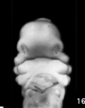
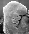
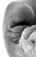
The face is the anatomical feature which is truly unique to each human, though the basis of its general development is identical for all humans and similar to that seem for other species. The face has a complex origin arising from a number of head structures and sensitive to a number of teratogens during critical periods of its development. The related structures of upper lip and palate significantly contribute to the majority of face abnormalities.
The head and neck structures are more than just the face, and are derived from pharyngeal arches 1 - 6 with the face forming from arch 1 and 2 and the frontonasal prominence. Each arch contains similar Arch components derived from endoderm, mesoderm, neural crest and ectoderm. These components though will form different structures depending on their arch origin. Because the head contains many different structures also review notes on Special Senses), Respiratory, Integumentary (Teeth), Endocrine (thyroid, parathyroid, pituitary, thymus) and Ultrasound- Cleft lip/palate.
 |
| 2016 Lecture Video Recording |
|---|
| Due to a technical issue, the 2017 lecture recording has no sound.
I have therefore included this 2016 lecture video recording that is similar in content to the current 2017 online lecture. <html5media height="600" width="800">File:2016Lecture-Head.mp4</html5media> Click to play new window - 2016 Lecture Video (48 MB) |
Lecture Objectives
- Understand the main structures derived from the pharyngeal arches, pouches and clefts.
- Understand the stages and structures involved in the development of the face.
- Understand the development of palate and tongue.
- Briefly understand special sensory early development.
- Briefly understand the abnormal development of the face and palate.


Lecture Resources
| Movies | |||||||||||||||||||||||
|---|---|---|---|---|---|---|---|---|---|---|---|---|---|---|---|---|---|---|---|---|---|---|---|
|
|
|
|
|
| ||||||||||||||||||
|
|
|
|


Animation of Face Development
| <html5media height="500" width="420">File:Face_001.mp4</html5media> |
Development of the Face
The separate embryonic components that contribute to the face have been colour coded.
The stomodeum is the primordial mouth region and a surface central depression lying between the forebrain bulge and the heart bulge. At the floor of the stomodeum indentation is the buccopharyngeal membrane (oral membrane). Note the complex origin of the maxillary region (upper jaw) requiring the fusion of several embryonic elements, abnormalities of this process lead to cleft lip and cleft palate. See also the movie showing a similar view of human embryo faces between Carnegie stage 16 to 18.
|
Week 3
Buccopharyngeal Membrane
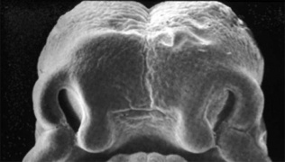
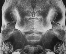
These images of the Stage 11 embryo show the breakdown of the buccopharyngeal membrane.

Low power ventral view of the Buccopharyngeal Membrane

Higher power ventrolateral view of the Buccopharyngeal Membrane

Close up view of the degenerating Buccopharyngeal Membrane


The Pharynx


The cavity within the pharyngeal arches forms the pharynx.
- begins at the buccopharyngeal membrane (oral membrane), apposition of ectoderm with endoderm (no mesoderm between)
- expands behind pharyngeal arches
- narrows at glottis and bifurcation of gastrointestinal (oesophagus) and respiratory (trachea) systems
- regions on roof, walls and floor have important contributions to endocrine in oral and neck regions
- also contributes to tongue development
Week 4

Pharyngeal Arch Components
Major features to identify for each: arch, pouch, groove and membrane. Contribute to the formation of head and neck and in the human appear at the 4th week. The first arch contributes the majority of upper and lower jaw structures.
Pharyngeal Arch Development
- branchial arch (Greek. branchia = gill)
- arch consists of all 3 trilaminar embryo layers
- ectoderm- outside
- mesoderm- core of mesenchyme
- endoderm- inside
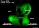
Neural Crest
- Mesenchyme invaded by neural crest generating connective tissue components
- cartilage, bone, ligaments
- arises from midbrain and hindbrain region
| Neural Crest Origins |
|---|

|
| <html5media height="340" width="410">File:Chicken-neural crest migration 01.mp4</html5media>
|
 Chicken embryo sequence shows the migration of DiI-labeled neural crest cells towards the branchial arches as the embryo. White rings indicate migration of individual cells. Each image represents 10 confocal sections separated by 10 microns. |
|
Arch Features

Each arch contains: artery, cartilage, nerve, muscular component
Arches and Phanynx Form the face, tongue, lips, jaws, palate, pharynx and neck cranial nerves, sense organ components, glands
- Humans have 5 arches - 1, 2, 3, 4, 6 (Arch 5 does not form or regresses rapidly)
- form in rostro-caudal sequence, Arch 1 to 6 (from week 4 onwards)
- arch 1 and 2 appear at time of closure of cranial neuropore
- Face - mainly arch 1 and 2
- Neck components - arch 3 and 4 (arch 4 and 6 fuse)
- arch - the entire structure
- groove - (cleft) externally separates each arch (only first pair persist as external auditory meatus)
- pouch - internally separates each arch (pockets out from the pharynx)
- membrane - ectoderm and endoderm contact regions (only first pair persist as tympanic membrane )
'Pharyngeal Arch 1 (Mandibular Arch) has 2 prominences
- smaller upper- maxillary forms maxilla, zygomatic bone and squamous part of temporal
- larger lower- mandibular, forms mandible
Pharyngeal Arch 2 (Hyoid Arch)
- forms most of hyoid bone
Arch 3 and 4
- neck structures
Arch Arteries
placental vein -> liver -> heart -> truncus arteriosus -> aortic sac -> arch arteries -> dorsal aorta -> placental artery |

Arch artery fates |
Arch Cartilage


Meckel's cartilage, first pharyngeal arch |
|
- Arch 2 - Reichert's cartilage
- dorsal ends form stapes and Temporal bone styloid process
- ventral part ossifies to form hyoid bone components
- lesser cornu and superior body
- Arch 3- forms greater cornu and inferior part of hyoid
- Arch 4&6- form laryngeal cartilages, except epiglottis (from hypobranchial eminence)
Arch Muscle
- Arch 1 - muscles of mastication, mylohyoid, tensor tympanic, ant. belly digastric
- Arch 2 - muscles of facial expression, stapedius, stylohyoid, post. belly digastric
- Arch 3 - stylopharyngeus
- Arch 4&6 - crycothyroid, pharynx constrictors, larynx muscles, oesophagus (st. muscle)
Arch Nerve
- Arch 1 - CN V trigeminal, caudal 2/3 maxillary and mandibular, cranial 1/3 sensory nerve of heaad and neck, mastication motor
- Arch 2 - CN VII facial
- Arch 3 - CN IX glossopharyngeal
- Arch 4&6 - CN X vagus, arch 4- superior laryngeal, arch 6- recurrent laryngeal
Arch Pouches
- Arch 1 - elongates to form tubotympanic recess, tympanic cavity, mastoid antrum, eustachian tube
- Arch 2 - forms tonsillar sinus, mostly oblierated by palatine tonsil
- Arch 3 - forms inferior parathyroid and thymus
- Arch 4 - forms superior parathyroid, parafollicular cells of thyroid
Endocrine
Note endocrine development will be covered in detail in a later lecture.
Thyroid Gland
|

|
Anterior Pituitary
|

|
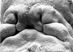
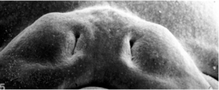
Face Development
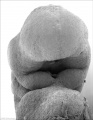
Begins week 4 centered around stomodeum, external depression at oral membrane
5 initial primordia from neural crest mesenchyme (week 4)
- single frontonasal prominence (FNP) - forms forehead, nose dorsum and apex
- nasal placodes develop later bilateral, pushed medially
- paired maxillary prominences - form upper cheek and upper lip
- paired mandibular prominences - lower cheek, chin and lower lip
Stage 11 (25 days)
Stage 12 (26 days)
Stage 13 (28 days)
Stage 14 (32 days)


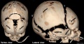
Head/Skull


- Cranium (Neurocranium) surrounds brain.
- dermatocranium (membranous) - skull calvarial vault develops from intramembranous ossification
- chondrocranium (cartilaginous) - skull base develops from endochondral ossification
- 8 bones - occipital, 2 parietals, frontal, 2 temporals, sphenoidal, ethmoidal.
- Face (Viscerocranium) development of the facial bones
- 14 bones - 2 nasals, 2 maxillæ, 2 lacrimals, 2 zygomatics, 2 palatines, 2 inferior nasal conchæ, vomer, mandible.

Calveria - bone has no cartilage (direct ossification of mesenchyme)
- bones do not fuse, fibrous sutures
- allow distortion to pass through birth canal
- allow growth of the brain
- 6 fontanelles - posterior closes at 3 months, anterior closes at 18 months
- Skull anterior (anterior fontenelle, sutures, mandible)
Skull_superior (anterior fontenelle, sutures)
Skull lateral view ( suture, mandible)
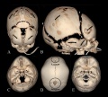
Developing overview CT
Developing vertex and lateral CT
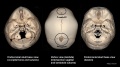
Developing endocranial and vertex CT


- Links: Skull Development
Sensory Placodes
- Sensory development will be covered in detail in a later lecture.
- During week 4 a series of thickened surface ectodermal patches form in pairs rostro-caudally in the head region.
- Recent research suggests that all sensory placodes may arise from common panplacodal primordium origin around the neural plate, and then differentiate to eventually have different developmental fates.
- These sensory placodes will later contribute key components of each of our special senses (vision, hearing and smell). Other species have a number of additional placodes which form other sensory structures (fish, lateral line receptor). Note that their initial postion on the developing head is significantly different to their final position in the future sensory system
Otic Placode
- Carnegie stage 12 still visible on embryo surface.
- Carnegie stage 13/14 embryo (shown below) the otic placode has sunk from the surface ectoderm to form a hollow epithelial ball, the otocyst, which now lies beneath the surface surrounded by mesenchyme (mesoderm). The epithelia of this ball varies in thickness and has begun to distort, it will eventually form the inner ear membranous labyrinth.

Lens Placode
- (optic placode) lies on the surface, adjacent to the outpocketing of the nervous system (which will for the retina) and will form the lens.
Nasal Placode
- Has 2 components (medial and lateral) and will form the nose olefactory epithelium.
Head Growth

- continues postnatally - fontanelle allow head distortion on birth and early growth
- bone plates remain unfused to allow growth, puberty growth of face
Palate
The palate has both an embryonic and fetal developmental component.
Embryonic
| Primary palate, fusion in the human embryo between week 6-7 (stage 17 and 18, GA Week 8-9), from an epithelial seam to the mesenchymal bridge.
|

|

- Embryonic Palate
Week 5 stage 16
Week 6 stage 17
Week 6.5 stage 18
Week 7 stage 19
Week 8 stage 22
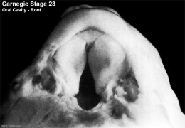
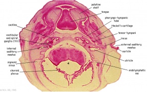
Week 8 stage 23

Week 8 stage 23


Fetal
| Secondary palate, fusion in the human embryo in week 9 (GA week 11). This requires the early palatal shelves growth, elevation and fusion during the early embryonic period. The fusion event is to both each other and the primary palate. palatal shelf elevation | secondary palate | 
Week 8 palatal shelves |
| Palate (oral view) |
| Page | Play |
| Palate (front view) |
| Page | Play |
Tongue Development


- Ectoderm of the first arch surrounding the stomodeum forms the epithelium lining the buccal cavity.
- Also the salivary glands, enamel of the teeth, epithelium of the body of the tongue.
- As the tongue develops "inside" the floor of the oral cavity, it is not readily visible in the external views of the embryonic (Carnegie) stages of development.
Contributions from all arches, which changes with time, begins as swelling rostral to foramen cecum, median tongue bud
|
|
Tongue Muscle

- Tongue muscles originate from the somites. Tongue muscles develop before masticatory muscles and is completed by birth.
- Masticatory muscles originate from the somitomeres. These muscles develop late and are not complete even at birth.
Salivary Glands
- epithelial buds in oral cavity (wk 6-7) extend into mesenchyme
- parotid, submandibular, sublingual
Abnormalities
Cleft Lip and Palate

|

|

|
| cleft palate | unilateral cleft lip and palate | bilateral cleft lip and palate |
- 300+ different abnormalities, different cleft forms and extent, upper lip and ant. maxilla, hard and soft palate
Victoria
The ten most frequently reported birth defects in Victoria between 2003-2004.
- Hypospadias
- Obstructive Defects of the Renal Pelvis or Obstructive Genitourinary Defects
- Ventricular Septal Defect
- Congenital Dislocated Hip
- Trisomy 21 or Down syndrome
- Hydrocephalus
- Cleft Palate
- Trisomy 18 or Edward Syndrome - multiple abnormalities of the heart, diaphragm, lungs, kidneys, ureters and palate 86% discontinued.
- Renal Agenesis/Dysgenesis - reduction in neonatal death and stillbirth since 1993 may be due to the more severe cases being identified in utero and being represented amongst the increased proportion of terminations (approximately 31%).
- Cleft Lip and Palate - occur with another defect in 33.7% of cases.
| Statistics |
|---|
Cleft Palate
- Cleft palate has the International Classification of Diseases code 749.0.
- In Australia the national rate (1982-1992) for this abnormalitity in births was 4.8 - 6/10,000 births, which represented 1,530 infants 5.5% were stillborn and 11.5% liveborn died during neonatal period and slightly more common in twin births than singleton.
Cleft Lip
- The International Classification of Diseases code 749.1 for isolated cleft lip and 749.2 for cleft lip with cleft palate.
- In Australia the national rate (1982-1992) for this abnormalitity was 8.1 - 9.9 /10,000 births. Of 2,465 infants 6.2% were stillborn and 7.8% liveborn died during neonatal period and the rate was similar in singleton and twin births.
- Links: Palate Development
First Arch Syndrome
There are 2 major types of associated first arch syndromes, Treacher Collins (Mandibulofacial dysostosis) and Pierre Robin (Pierre Robin complex or sequence), both result in extensive facial, sensory and palate abnormalites.
DiGeorge Syndrome
- absence of thymus and parathyroid glands, 3rd and 4th pouch do not form
- disturbance of cervical neural crest migration
Cysts
- Many different types
Facial Clefts
extremely rare
- Holoprosencephaly - shh abnormality
Maternal Effects
- Retinoic Acid - present in skin ointments
- 1988 associated with facial developmental abnormalities
Fetal Alcohol Syndrome

Due to alcohol in early development (week 3+) leading to both facial and neurological abnormalities
- lowered ears, small face, mild+ retardation
- Microcephaly - leads to small head circumference
- Short Palpebral fissure - opening of eye
- Epicanthal folds - fold of skin at inside of corner of eye
- Flat midface
- Low nasal bridge
- Indistinct Philtrum - vertical grooves between nose and mouth
- Thin upper lip
- Micrognathia - small jaw
Exposure of embryos in vitro to ethanol simulates premature differentiation of prechondrogenic mesenchyme of the facial primordia (1999)
- Links: Fetal Alcohol Syndrome
Table - Structures derived from Arches
| Pharyngeal Arch | Nerve | Artery | Neural Crest (Skeletal Structures) |
Muscles | Ligaments |
|---|---|---|---|---|---|
| 1 (maxillary/mandibular) |
trigeminal (CN V) | maxillary artery (terminal branches) | mandible, maxilla, malleus, incus | muscles of mastication, mylohyoid, tensor tympanic, ant. belly digastric | ant lig of malleus, sphenomandibular ligament |
| 2 (hyoid) |
facial (CN VII) | stapedial (embryonic) corticotympanic (adult) |
stapes, styloid process, lesser cornu of hyoid, upper part of body of hyoid bone | muscles of facial expression, stapedius, stylohyoid, post. belly digastric | stylohyoid ligament |
| 3 | glossopharyngeal (CN IX) | common carotid, internal carotid arteries | greater cornu of hyoid, lower part of body of hyoid bone | stylopharyngeus | |
| 4 | vagus (CN X) superior laryngeal branch | part of aortic arch (left), part right subclavian artery (right) | thyroid, cricoid, arytenoid, corniculate and cuneform cartilages | crycothyroid, soft palate levator veli palatini (not tensor veli palatini) | |
| 6 | vagus (CN X) recurrent laryngeal branch | part of left pulmonary artery (left), part of right pulmonary artery (right) | thyroid, cricoid, arytenoid, corniculate and cuneform cartilages | larynx intrinsic muscles (not cricothyroid muscle) |
Structures derived from Pouches
Each pouch is lined with endoderm and generates specific structures.
| Overall Structure | Specific Structures | |
| tubotympanic recess | tympanic membrane, tympanic cavity, mastoid antrum, auditory tube | |
| intratonsillar cleft | crypts of palatine tonsil, lymphatic nodules of palatine tonsil | |
| inferior parathyroid gland, thymus gland | ||
| superior parathyroid gland, ultimobranchial body | ||
| becomes part of 4th pouch |
Structures derived from Grooves
Only the first groove differentiates into an adult structure and forms part of the external acoustic meatus.
Structures derived from Membranes
At the bottom of each groove lies the membrane which is formed from the contact region of ectodermal groove and endodermal pouch. Only the first membrane differentiates into an adult structure and forms the tympanic membrane.
Terms
- palate - The roof of the mouth (oral cavity) a structure which separates the oral from the nasal cavity. Develops as two lateral palatal shelves which grow and fuse in the midline. Initally a primary palate forms with fusion of the maxillary processes with the nasal processes in early face formation. Later the secondary palate forms the anterior hard palate which will ossify and separate the oral and nasal cavities. The posterior part of the palate is called the soft palate (velum, muscular palate) and contains no bone. Abnormalities of palatal shelf fusion can lead to cleft palate. (More? Palate Development | Head | Head Abnormalities)
- palatogenesis - The process of palate formation, divided into primary and secondary palate development. (More? Palate Development | Head | Head Abnormalities)
- pharyngeal arch - (branchial arch, Greek, branchial = gill) These are a series of externally visible anterior tissue bands lying under the early brain that give rise to the structures of the head and neck. In humans, five arches form (1,2,3,4 and 6) but only four are externally visible on the embryo. Each arch has initially identical structures: an internal endodermal pouch, a mesenchymal (mesoderm and neural crest) core, a membrane (endoderm and ectoderm) and external cleft (ectoderm). Each arch mesenchymal core also contains similar components: blood vessel, nerve, muscular, cartilage. Each arch though initially formed from similar components will differentiate to form different head and neck structures. (More? | Head Development | Endocrine | Neural Crest)
- pharyngeal arch artery - Each early developing pharyngeal arch contains a lateral pair of arteries arising from the aortic sac, above the heart, and running into the dorsal aorta. later in development these arch arteries are extensively remodelled to form specific components of the vascular system. Pharyngeal Arch 1 arteries are mainly lost and forms part of maxillary artery. Pharyngeal Arch 2 arteries remains to form the stapedial arteries. Pharyngeal Arch 3 arteries forms the common carotid arteries, internal carotid arteries in the neck. Pharyngeal Arch 4 arteries will form part of aortic arch (left arch artery) and part right subclavian artery (right arch artery) Pharyngeal Arch 6 arteries form part of left pulmonary artery (left arch artery) and part of right pulmonary artery (right arch artery). (More? | Head Development | Cardiovascular)
- pharyngeal arch cartilage - Each early developing pharyngeal arch contains a horseshoe shaped band of cartilage that acts as a template and contributes to the development of head and neck bony and cartilagenous features, including the middle ear bones. Pharyngeal Arch 1 cartilage (Meckel’s cartilage) dorsal ends form malleus and incus midpart forms ligaments (ant. malleus, sphenomandibular) ventral part forms mandible template. Pharyngeal Arch 2 cartilage (Reichert’s cartilage) dorsal ends form stapes and Temporal bone styloid process, ventral part ossifies to form hyoid bone components, lesser cornu and superior body. Pharyngeal Arch 3 cartilage forms hyoid components, greater cornu and inferior part of hyoid. Pharyngeal Arch 4 and 6 cartilage forms laryngeal cartilages except epiglottis (from hypobranchial eminence). (More? Head Development | Middle Ear)
- pharyngeal arch nerve - Each early developing pharyngeal arch contains the developing cranial nerves, as a pair, within the arch mesenchyme. Each cranial nerve is numbered (roman numeral) in rostrocaudal sequence and also has a specific name. The cranial nerve within each arch often relates to the other structures formed from taht arch. Pharyngeal Arch 1 contains the trigeminal nerve (CN V, cranial nerve 5). Pharyngeal Arch 2 contains the facial nerve (CN VII, cranial nerve 7). Pharyngeal Arch 3 contains the glossopharyngeal nerve (CN IX, cranial nerve 9) Pharyngeal Arch 4 and 6 contains the Vagus (CN X cranial nerve 10), forming the adult superior laryngeal and recurrent laryngeal branches. (More? | Head Development | Neural | Neural Crest)
- pharyngeal arch pouch - An out-pocketing of the endoderm lined pharynx occurring between each developing pharyngeal arch. Each of the pharyngeal arch pouches contributes different components of the head and neck, either cavities or endocrine tissues. Pharyngeal Arch 1 pouch elongates to form tubotympanic recess tympanic cavity, mastoid antrum and auditory tube (Eustachian tube). Pharyngeal Arch 2 pouch forms the tonsillar sinus and is later mostly oblierated by palatine tonsil. Pharyngeal Arch 3 pouch forms the inferior parathyroid and thymus. Pharyngeal Arch 4 pouch forms the superior parathyroid, parafollicular cells of Thyroid. (More? Middle Ear | Thyroid | Parathyroid | Thymus | Endocrine | Head Development
- pharyngotympanic tube - (auditory tube, eustachian tube, otopharyngeal tube) A narrow canal connecting the middle ear space to the back of the oral cavity. The tube allows ventilation, protection and clearance for the middle ear cavity. Ventilation is the pressure equalization in the middle ear. Clearance is to allow fluid drainage from the middle ear. Embryonic origin is from the first pharyngeal pouch. In development, the canal is initially both horizontal, short and very narrow leading to poor drainage and easy blockage. (More? Middle Ear | Hearing | Hearing Abnormalities)
- pharynx - (throat) Forms the initial segment of the upper respiratory tract divided anatomically into three regions: nasopharynx, oropharynx, and laryngopharynx (hypopharynx). Anatomically extends from the base of the skull to the level of the sixth cervical vertebra. (More? Respiratory System Development)
| ANAT2341 Course Timetable | |||
|---|---|---|---|
| Week (Mon) | Lecture 1 (Mon 1-2pm) | Lecture 2 (Tue 3-4pm) | Practical (Fri 1-3pm) |
| Week 2 (1 Aug) | Introduction | Fertilization | Lab 1 |
| Week 3 (8 Aug) | Week 1 and 2 | Week 3 | Lab 2 |
| Week 4 (15 Aug) | Mesoderm | Ectoderm | Lab 3 |
| Week 5 (22 Aug) | Early Vascular | Placenta | Lab 4 |
| Week 6 (29 Aug) | Gastrointestinal | Respiratory | Lab 5 |
| Week 7 (5 Sep) | Head | Neural Crest | Lab 6 |
| Week 8 (12 Sep) | Musculoskeletal | Limb Development | Lab 7 |
| Week 9 (19 Sep) | Renal | Genital | Lab 8 |
| Mid-semester break | |||
| Week 10 (3 Oct) | Public Holiday | Stem Cells | Lab 9 |
| Week 11 (10 Oct) | Integumentary | Endocrine | Lab 10 |
| Week 12 (17 Oct) | Heart | Sensory | Lab 11 |
| Week 13 (24 Oct) | Fetal | Birth and Revision | Lab 12 |
|
ANAT2341 2016: Moodle page | ECHO360 | Textbooks | Students 2016 | Projects 2016 | |||
Glossary Links
- Glossary: A | B | C | D | E | F | G | H | I | J | K | L | M | N | O | P | Q | R | S | T | U | V | W | X | Y | Z | Numbers | Symbols | Term Link
Cite this page: Hill, M.A. (2024, June 20) Embryology Lecture - Head Development. Retrieved from https://embryology.med.unsw.edu.au/embryology/index.php/Lecture_-_Head_Development
- © Dr Mark Hill 2024, UNSW Embryology ISBN: 978 0 7334 2609 4 - UNSW CRICOS Provider Code No. 00098G