Cardiovascular System - Lymphatic Development: Difference between revisions
mNo edit summary |
|||
| Line 17: | Line 17: | ||
|-bgcolor="F5FAFF" | |-bgcolor="F5FAFF" | ||
| | | | ||
* '''Development of the mammalian lymphatic vasculature'''<ref name=PMID24590273><pubmed>24590273</pubmed></ref> "The two vascular systems of our body are the blood and lymphatic vasculature. Our understanding of the cellular and molecular processes controlling the development of the lymphatic vasculature has progressed significantly in the last decade. In mammals, this is a stepwise process that starts in the embryonic veins, where lymphatic EC (LEC) progenitors are initially specified. The differentiation and maturation of these progenitors continues as they bud from the veins to produce scattered primitive lymph sacs, from which most of the lymphatic vasculature is derived. Here, we summarize our current understanding of the key steps leading to the formation of a functional lymphatic vasculature." | |||
* '''prox1b Activity is essential in zebrafish lymphangiogenesis'''<ref><pubmed>20976189</pubmed></ref> "The lymphatic vascular system, draining interstitial fluids from most tissues and organs, exerts crucial functions in several physiological and pathological processes. Lymphatic system development depends on Prox1, the first marker to be expressed in the endothelial cells of the cardinal vein from where lymph vessels originate." | * '''prox1b Activity is essential in zebrafish lymphangiogenesis'''<ref><pubmed>20976189</pubmed></ref> "The lymphatic vascular system, draining interstitial fluids from most tissues and organs, exerts crucial functions in several physiological and pathological processes. Lymphatic system development depends on Prox1, the first marker to be expressed in the endothelial cells of the cardinal vein from where lymph vessels originate." | ||
* '''Lineage tracing demonstrates the venous origin of the mammalian lymphatic vasculature'''<ref name="PMID17908929"><pubmed>17908929</pubmed></ref> "These studies, together with the analysis of Runx1-mutant embryos lacking definitive hematopoiesis, conclusively determined that from venous-derived lymph sacs, lymphatic endothelial cells sprouted, proliferated, and migrated to give rise to the entire lymphatic vasculature, and that hematopoietic cells did not contribute to the developing lymph sacs. We conclude that the mammalian lymphatic system has a solely venous origin." | * '''Lineage tracing demonstrates the venous origin of the mammalian lymphatic vasculature'''<ref name="PMID17908929"><pubmed>17908929</pubmed></ref> "These studies, together with the analysis of Runx1-mutant embryos lacking definitive hematopoiesis, conclusively determined that from venous-derived lymph sacs, lymphatic endothelial cells sprouted, proliferated, and migrated to give rise to the entire lymphatic vasculature, and that hematopoietic cells did not contribute to the developing lymph sacs. We conclude that the mammalian lymphatic system has a solely venous origin." | ||
Revision as of 13:16, 26 February 2015
Introduction


An important part of the cardiovascular system is the lymphatic vasculature, which functions to both return interstitial fluid (lymph) to the bloodstream and also as part of the immune system. In the embryo, lymphatic development begins at the cardinal vein, where venous endothelial cells differentiate (express Prox1) to form lymphatic endothelial cells that out-pocket and bud to form lymph sacs. During development these lymph sacs remodel to form both the lymphatic space within future nodes, formed by engulfed connective tissue, and the associated afferent and efferent vessel network.
This system was first identified by Aselli G. (1627) in a paper "De Lacteibus sive Lacteis Venis", Quarto Vasorum Mesarai corum Genere novo invento. Milan: Mediolani. Then postulated by Sabin (1902)[1] as venous in origin, it required a recent 2007 lineage tracing study to confirm this theory.[2] Only vertebrates possess a true lymphatic vascular system, with primitive fish possessing a lymphatic-like secondary vascular system that also contains blood. Clinically, important for roles in immune surveillance and oncogenic (cancer) processes.
| Cardiovascular Links: cardiovascular | Heart Tutorial | Lecture - Early Vascular | Lecture - Heart | Movies | 2016 Cardiac Review | heart | coronary circulation | heart valve | heart rate | Circulation | blood | blood vessel | blood vessel histology | heart histology | Lymphatic | ductus venosus | spleen | Stage 22 | cardiovascular abnormalities | OMIM | 2012 ECHO Meeting | Category:Cardiovascular | ||
|
Some Recent Findings
|
Lymphatic Vessels

- Lymph capillaries - begin as blind-ending tubes in connective tissue, larger than blood capillaries, very irregularly shaped.
- Lymph collecting vessels - larger and form valves, morphology similar to lymph capillaries.
- Lymph ducts - 1 or 2 layers of smooth muscle cells in wall.
- (Remember the anatomy acronym NAVL = Nerve, Artery, Vein and Lymph)
Lymphatic Capillaries
- single-cell layer of overlapping endothelial cells
- lack a basement membrane
- lack smooth muscle cells or pericytes (pre-collecting and collecting trunks contain both)
- linked by discontinuous endothelial cell-cell junctions (button-like).
- junctions open in response to increased interstitial fluid pressure.

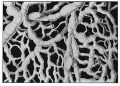
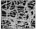
Lymphatic microvasculature model[6]
Lymphatic Vessel Development

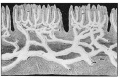
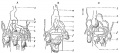
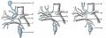
Tunneling model of lymphatic vessel formation. |
The model shown here is from a recent paper[7] and is based on ultrastructural observations performed in in vitro and in vivo models of lymphangiogenesis. Lymphatic endothelial cells (LEC) display tight junctions and interdigitations, and are connected to the surrounding collagen fibers by anchoring filaments.
Note that this postnatal model may differ from developmental lymphatic vessel development.
|
Lymphatic Vessel Contraction
Lymphatic vessels undergo spontaneous rhythmic contractions which aid lymph flow. This is most easily demonstrated in models based upon mesentry lymphatics of the gastrointestinal tract. Contractile activity is regulated by physical factors (transmural pressure) and neurological (alpha-adrenergic, histamine, bradykinin) acting on lymphatic smooth muscle. Contractility and receptor expression may also be different in different parts of the lymphatic system.
Alpha-adrenergic - alpha 1- and not alpha 2-adrenoceptors.
Histamine - lymphatic smooth muscle via stimulation of H(1) (and in some vessels H(2)) receptors.
Bradykinin - chronotropic but not inotropic effects on lymphatic pump activity via stimulation of B1 receptors.

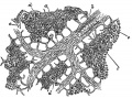
Lymphatic Vasculature Organization[8]
Molecular Development
Angiopoietins (Ang1–Ang4)
Notch probably mediates choice of fate between arterial and venous.
Prox1 Prospero-related Homeobox 1 - expressed in a subpopulation of blood endothelial cells that then generate, by both budding and sprouting, cells of the lymphatic vascular system. Triggers the molecular program leading to the formation of the lymphatic system. (OMIM - PROSPERO-RELATED HOMEOBOX 1; PROX1)
Tie (Tie1 and Tie2) tyrosine kinase receptors.
Vascular endothelial growth factor (VEGF) family of proteins and angiopoietin/Tie, Notch, and ephrin/Eph pathways play major roles in eary vessel development. (VEGFR-3)
LYVE-1
Podoplanin
Abnormalities
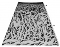
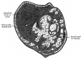
Lymphangioma
Dysplasia of childhood form lymphatic capillaries or collectors, which form fluid-filled cysts.
- lymphatic spaces lined by endothelium
- smooth muscle fascicles in the septa between the lymphatic spaces
- lymphoid aggregates in the delicate collagenous stroma
Additional Images
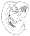
Human embryo measuring 8 mm and 9 mm
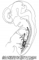
Human embryo measuring 10.5 mm
Human embryo measuring 11 mm
Human embryo measuring 22 mm
Human embryo measuring 30 mm


Gray's Anatomy - Lymphatic
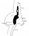
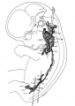
592 Primary lymph sacs.
593 Lymph capillaries of the human conjunctiva.
594 Lymph capillaries from the human scrotum.
595 Lymph capillaries of the sole of the human foot.
596 Section through human tongue.
597 Lymph gland (Node).
598 Lymph gland tissue.
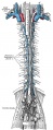
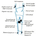
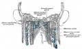
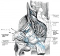
599 Thoracic and right lymphatic ducts.
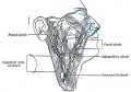
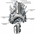
600 Modes of origin of thoracic duct.
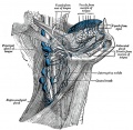
601 Terminal collecting trunks of right side.
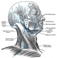
602 Lymph glands of the head.
603 Lymphatics of pharynx.
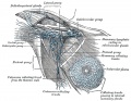
604 Lymphatics of the face.
605 Lymphatics of the Tongue.
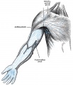
606 Lymph glands of the upper extremity.
607 Lymphatics of the mamma.
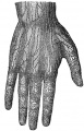
608 Lymphatic vessels of the dorsal hand surface.
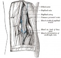
609 Lymph glands of popliteal fossa.
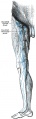
610 Superficial lymph glands and vessels of the lower extremity.
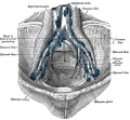
611 Parietal lymph glands of the pelvis.
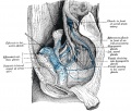
612 Iliopelvic glands.
613 Lymphatics of stomach.
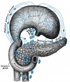
614 Lymphatics of stomach.
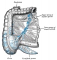
615 Lymphatics of cecum and vermiform process.
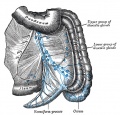
616 Lymphatics of cecum and vermiform process.
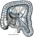
617 Lymphatics of Colon.
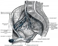
618 Lymphatic of the Bladder.
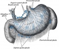
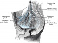
619 Lymphatics of the Prostate.
620 Lymphatics of the Uterus.
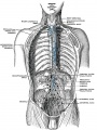
621 Lymphatics of the thorax and abdomen.
622 Tracheobronchial Lymph Glands


References
- ↑ Sabin, F. R. On the origin of the lymphatic system from the veins and the development of the lymph hearts and thoracic duct in the pig. (1902) Am. J. Anat. 1, 367-389.
- ↑ 2.0 2.1 <pubmed>17908929</pubmed>
- ↑ <pubmed>24590273</pubmed>
- ↑ <pubmed>20976189</pubmed>
- ↑ <pubmed>16212503</pubmed>
- ↑ <pubmed>14581448</pubmed>| JCB
- ↑ <pubmed>21702933</pubmed>| PMC3141733 | BMC Cell Biol.
- ↑ <pubmed>21576390</pubmed>| PMC3166860 | J Cell Biol.
Journals
- Lymphology - Journal Homepage
Reviews
<pubmed>18519960</pubmed> <pubmed>17036631</pubmed> <pubmed>16212503</pubmed> <pubmed>15293565</pubmed> <pubmed>14704766</pubmed> <pubmed>12543725</pubmed>
Articles
<pubmed>19056883</pubmed> <pubmed>17908929</pubmed> <pubmed>17202268</pubmed> <pubmed>16291864</pubmed> <pubmed>16877368</pubmed> <pubmed>11937485</pubmed>
Lymphatic endothelial cell identity is reversible and its maintenance requires Prox1 activity Nicole C. Johnson, Miriam E. Dillard, Peter Baluk, Donald M. McDonald, Natasha L. Harvey, Sharon L. Frase, and Guillermo Oliver Genes Dev. 2008;22 3282-3291
Search Pubmed
Search NLM Bookshelf: lymphatic
Search PubMed: lymphatic development | lymphology
External Links
External Links Notice - The dynamic nature of the internet may mean that some of these listed links may no longer function. If the link no longer works search the web with the link text or name. Links to any external commercial sites are provided for information purposes only and should never be considered an endorsement. UNSW Embryology is provided as an educational resource with no clinical information or commercial affiliation.
- International Society of Lymphology ISL | Lymphology Journal
- Australian Lymphology Association ALA
- American Society of Lymphology ASL
- British Lymphology Society BLS
- Embryology original page
Glossary Links
- Glossary: A | B | C | D | E | F | G | H | I | J | K | L | M | N | O | P | Q | R | S | T | U | V | W | X | Y | Z | Numbers | Symbols | Term Link
Cite this page: Hill, M.A. (2026, February 27) Embryology Cardiovascular System - Lymphatic Development. Retrieved from https://embryology.med.unsw.edu.au/embryology/index.php/Cardiovascular_System_-_Lymphatic_Development
- © Dr Mark Hill 2026, UNSW Embryology ISBN: 978 0 7334 2609 4 - UNSW CRICOS Provider Code No. 00098G