Sensory - Vision Development
| Embryology - 5 Jul 2026 |
|---|
| Google Translate - select your language from the list shown below (this will open a new external page) |
|
العربية | català | 中文 | 中國傳統的 | français | Deutsche | עִברִית | हिंदी | bahasa Indonesia | italiano | 日本語 | 한국어 | မြန်မာ | Pilipino | Polskie | português | ਪੰਜਾਬੀ ਦੇ | Română | русский | Español | Swahili | Svensk | ไทย | Türkçe | اردو | ייִדיש | Tiếng Việt These external translations are automated and may not be accurate. (More? About Translations) |
Introduction


These notes introduce vision development of the eye: induction and regional specification of the eye structures, maturation and formation of retina and optic tectum neuronal connections.
The adult eye has contributions from several different embryonic layers eventually forming neuronal, supportive connective tissue, optical structures, and muscular tissues. Historically determined that at birth, the human eyeball volume is about 3.25 cc and the estimated weight is about 3.40 grams.[2]
There are additional pages shown in the vision links, covering specific topics of vision development.
| Senses Links: Introduction | placode | Hearing and Balance hearing | balance | vision | smell | taste | touch | Stage 22 | Category:Sensory |
Some Recent Findings
|
| More recent papers |
|---|
 This table allows an automated computer search of the external PubMed database using the listed "Search term" text link.
More? References | Discussion Page | Journal Searches | 2019 References | 2020 References Search term: Vision Development | Vision Embryology |
| Older papers |
|---|
| These papers originally appeared in the Some Recent Findings table, but as that list grew in length have now been shuffled down to this collapsible table.
See also the Discussion Page for other references listed by year and References on this current page.
|
Timeline
|
Embryonic Development
|

|
| Carnegie Stage | Event | ||||||||||||||||||
|---|---|---|---|---|---|---|---|---|---|---|---|---|---|---|---|---|---|---|---|
| 10 | optic primordia appear. | ||||||||||||||||||
| 11 | Right and left optic primordia meet at the optic chiasma forming a U-shaped rim. | ||||||||||||||||||
| 12 | optic neural crest reaches its maximum extent and the optic vesicle becomes covered by a complete sheath, | ||||||||||||||||||
| 13 | By the end of the fourth week the optic vesicle lies close to the surface ectoderm. Optic evagination differentiation allows identification of optic part of retina, future pigmented layer of retina, and optic stalk. The surface ectoderm overlying the optic vesicle, in response to this contact, has thickened to form the lense placode. | ||||||||||||||||||
| 14 | (about 32 days) Lens placode is indented by the lens pit, cup-shaped and still communicates with the surface by a narrowing pore. | ||||||||||||||||||
| 15 | (about 33 days) Lens pit is closed. The lens vesicle and optic cup lie close to the surface ectoderm and appear to press against the surface. | ||||||||||||||||||
| 16 | (37 days) Growth of the lens body results in a D-shaped lens cavity. Perilental blood vessels (tunica vasculosa lentis) are visible. Prior to the development of the eyelids, one small sulcus or groove forms above the eye (eyelid groove) and another below it. | ||||||||||||||||||
| 17 - 19 | Retinal pigment is visible and the retinal fissure is largely closed. Eyelids grooves deepen, eyelid folds develop, first below, and then above, the eye. | ||||||||||||||||||
| 18 | Mesenchyme invades the region between the lens epithelium and the surface ectoderm. | ||||||||||||||||||
| 19 - 22 | Eyelid folds develop into the eyelids and cover more of the eye as the palpebral fissure takes shape. The upper and the lower eyelids meet at the outer canthus in Stage 19. | ||||||||||||||||||
| 20 | Lens cavity is lost and a lens suture begins to form. The inner canthus is established. | ||||||||||||||||||
| 23 | retina comprises the pigmented layer, external limiting membrane, proliferative zone, external neuroblastic layer, transient fiber layer, internal neuroblastic layer, nerve fiber layer, and internal limiting membrane. Eyelids closure is complete (Note - shown as still open in the Kyoto embryo). | ||||||||||||||||||
Data from a study of human embryonic carnegie stages[9] and other sources.
| |||||||||||||||||||
See below the drawings of sections of the whole eye from week 8 of development.[10]
Optic Nerve
- Carnegie stage 19 - Optic nerve small, slender. Lumen practically whole length of stalk. Few or no fibers.
- Carnegie stage 20 - Ependymal arrangement partially retained along stalk. Remnant of ependyma along whole length of stalk. Hyaloid groove at bulbar end. A few fibers arriving at brain.
- Carnegie stage 21 - Remnant of ependyma present.
- Carnegie stage 22 - Sheath layer beginning to form. Vascular canal present.
- Carnegie stage 23 - Early nerve sheath. Reticular spongioblastic framework, striate arrangement of nuclei, bundles of fibers. Definite nerve sheath.
- Eye and Optic Nerve
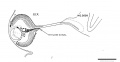
Carnegie stage 19
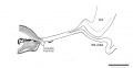
Carnegie stage 20
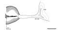
Carnegie stage 21
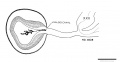
Carnegie stage 22
Carnegie stage 23


Lens

The lens or crystalline lens or aquula (Latin, aquula = a little stream) has a key role in focussing light (with the cornea) upon the neural retina. The lens embryonic origin is from surface ectoderm of the sensory placodes that form in the head region (More? Week 4 - Placodes). The lens focusses by refracting light as it passes through the biconvex lens, which can be altered in shape (accommodation) by surrounding ciliary muscles. These ciliary muscles are activated (contracted) by parasympathetic innervation from the ciliary ganglion itself innervated by the oculomotor nerve (Cranial Nerve III) (More? Cranial Nerves).
surface ectoderm -> lens placode -> lens pit -> lens vesicle -> lens fibres -> lens capsule and embryonic/fetal nucleus.
- Links: Vision - Lens Development
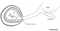
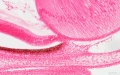
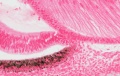
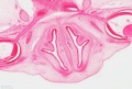
Stage 22 Eye
The images below link to virtual slides of the human developing eye at Carnegie stage 22. Click on the image to open or select specific regions from the regions of interest links.
|
|
Virtual Slide - Regions of Interest
|


Links: Embryo Virtual Slides
Retinotopic Map
This neuroscience term describes how the developing retina is precisely "mapped" onto the visual cortex through a series of signaling and activity dependent mechanisms. This follows from Hubel and Wiesel (1981 Nobel Prize in Physiology or Medicine) key discoveries (1959-70) of how in development system matching occurs in the visual system. The topographic map establishes an ordered neuronal connection between sensory structures and the central nervous system.
The retinotectal map (eye to brain) of birds (lower vertebrates):
- temporal (posterior) retina is connected to the rostral (anterior) part of the contralateral optic tectum
- nasal (anterior) retina to the caudal (posterior) tectum
- ventral retina to the dorsal (medial) tectum
- dorsal ventral (lateral) tectum
Retinal waves a form of coordinated spontaneous activity that occurs in the developing retina. These waves of electrical activity (action potentials) are thought to have a role in establishing the initial retinotopic map by correlating/coordinating the activity of neighbouring retinal ganglion cells.
EphA/ephrin-A molecular signaling also thought to have a role in establishing the initial retinotopic map.
Neural Crest

Mouse eye neural crest[11] |

Mouse eye TGF-beta model[11] |
- Links: Image - Mouse eye neural crest | Image - Mouse eye TGF-beta model | Vision Development | Neural Crest Development | Head Development
Schlemm's canal

Schematic showing the stages of Schlemm's canal development in the postnatal mouse by the novel process of canalogenesis.[12] (Cartoons have been drawn for clarity and are not intended to suggest that most early sprouts arise from the LVP.)
Extraocular Muscles
Extraocular muscles are required to move the eye within the orbit. Their embryonic origin requires an interaction between the cranial mesoderm and the migrating neural crest cells.
The following is from a recent paper comparing human to zebrafish muscle development.[13]
| About the Muscles | Legend | |
|---|---|---|
|
|

|
- Links: Extraocular Muscles
Additional Images
Human stage 22 developing iris region
Human stage 22 developing iris region
Human stage 22 overview of optic nerve
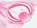
Human stage 22 overview of eye
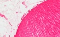
Human stage 22 lens and hyaloid vessels
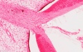
Human stage 22 optic nerve (stalk)
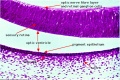
Human stage 22 retina
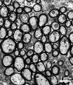
Mouse adult optic nerve axons
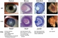
Pax6 eye phenotypes


Historic Images
| Historic Disclaimer - information about historic embryology pages |
|---|
|
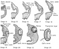
Frog eye development.
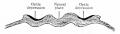
Fig. 456. Location of optic areas before the closure of the neural groove.
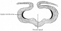
Fig. 457. Location of areas shown in Fig. 456 after the formation of the neural canal.
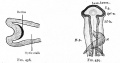
Fig. 458. Location of the optic area after the beginning of the formation of the optic cup and optic stalk. Fig. 459. Dorsal view of head of chick of 58 hours' incubation.
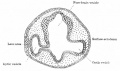
Fig. 460. Section through head of chick of two days' incubation.
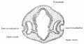
Fig. 461. Section through head of chick of three days' incubation.
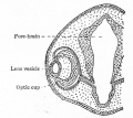
Fig. 462. Later stage in development of optic cup and lens than is shown in Fig. 461.
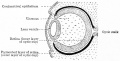
Fig. 463. Developing lens and optic cup.
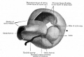
Fig. 464. Model showing lens and formation of optic cup.

Fig. 465. Stages in the development of the lens in the rabbit embryo.
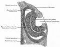
Fig. 466. Section through optic cup and lens invagination of chick of fifty-four hours' incubation.
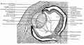
Fig. 467. Section through eye of human embryo of 13-14 weeks.
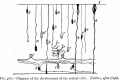
Fig. 468. Development of the retinal cells.
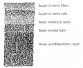
Fig. 469. Vertical section through retina of a four months' human embryo.
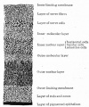
Fig. 470. Vertical section through retina of a five and one-half months' human embryo.
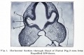
Fig. 1. Section through head of pig, 2 mm long.
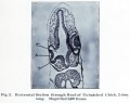
Fig. 2. Section through head of chick, 2 mm long.
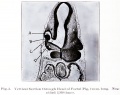
Fig. 3. Section through head of Foetal Pig, 2 mm long.
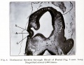
Fig. 4. Section through head of Foetal Pig, 3 mm long.
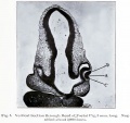
Fig. 5. Section through head of Foetal Pig, 3 mm long.
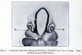
Fig. 6. Section through head of Foetal Pig, 4 mm long.
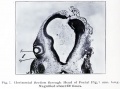
Fig. 7. Section through head of Foetal Pig, 7 mm long.
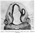
Fig. 8. Section through head of pig, 8 mm long.
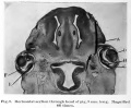
Fig. 9. Section through head of pig, 9 mm long.
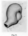
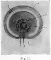
Fig. 11. colobomba of the fundus in the adult and means a lack of development.


References
- ↑ Swaroop A & Zack DJ. (2002). Transcriptome analysis of the retina. Genome Biol. , 3, REVIEWS1022. PMID: 12186651
- ↑ Scammon RE. and Armstrong EL. On the growth of the human eyeball and optic nerve. (1925) J. Comp. Neurol. 33(2):165-219.
- ↑ Cheng S, Butrus S, Tan L, Xu R, Sagireddy S, Trachtenberg JT, Shekhar K & Zipursky SL. (2022). Vision-dependent specification of cell types and function in the developing cortex. Cell , 185, 311-327.e24. PMID: 35063073 DOI.
- ↑ Mackin RD, Frey RA, Gutierrez C, Farre AA, Kawamura S, Mitchell DM & Stenkamp DL. (2019). Endocrine regulation of multichromatic color vision. Proc. Natl. Acad. Sci. U.S.A. , 116, 16882-16891. PMID: 31383755 DOI.
- ↑ Zhang Z, Lin X, Yu Q, Teng G, Zang F, Wang X, Liu S & Hou Z. (2019). Fetal ocular development in the second trimester of pregnancy documented by 7.0 T postmortem Magnetic Resonance Imaging. PLoS ONE , 14, e0214939. PMID: 30947240 DOI.
- ↑ 6.0 6.1 Ribas VT, Gonçalves BS, Linden R & Chiarini LB. (2012). Activation of c-Jun N-terminal kinase (JNK) during mitosis in retinal progenitor cells. PLoS ONE , 7, e34483. PMID: 22496813 DOI.
- ↑ Hayakawa I & Kawasaki H. (2010). Rearrangement of retinogeniculate projection patterns after eye-specific segregation in mice. PLoS ONE , 5, e11001. PMID: 20544023 DOI.
- ↑ Rapicavoli NA, Poth EM & Blackshaw S. (2010). The long noncoding RNA RNCR2 directs mouse retinal cell specification. BMC Dev. Biol. , 10, 49. PMID: 20459797 DOI.
- ↑ Pearson AA. (1980). The development of the eyelids. Part I. External features. J. Anat. , 130, 33-42. PMID: 7364662
- ↑ Streeter GL. Developmental Horizons In Human Embryos Description Or Age Groups XIX, XX, XXI, XXII, And XXIII, Being The Fifth Issue Of A Survey Of The Carnegie Collection. (1957) Carnegie Instn. Wash. Publ. 611, Contrib. Embryol., 36: 167-196.
- ↑ 11.0 11.1 Ittner LM, Wurdak H, Schwerdtfeger K, Kunz T, Ille F, Leveen P, Hjalt TA, Suter U, Karlsson S, Hafezi F, Born W & Sommer L. (2005). Compound developmental eye disorders following inactivation of TGFbeta signaling in neural-crest stem cells. J. Biol. , 4, 11. PMID: 16403239 DOI.
- ↑ Kizhatil K, Ryan M, Marchant JK, Henrich S & John SW. (2014). Schlemm's canal is a unique vessel with a combination of blood vascular and lymphatic phenotypes that forms by a novel developmental process. PLoS Biol. , 12, e1001912. PMID: 25051267 DOI.
- ↑ Kasprick DS, Kish PE, Junttila TL, Ward LA, Bohnsack BL & Kahana A. (2011). Microanatomy of adult zebrafish extraocular muscles. PLoS ONE , 6, e27095. PMID: 22132088 DOI.
Online Textbooks
- Kolb H, Fernandez E, Nelson R, editors. Webvision: The Organization of the Retina and Visual System [Internet]. Salt Lake City (UT): University of Utah Health Sciences Center; 1995-. Available from: http://www.ncbi.nlm.nih.gov/books/NBK11530/
- Gilbert SF. Developmental Biology. 6th edition. Sunderland (MA): Sinauer Associates; 2000. Development of the Vertebrate Eye. Available from: https://www.ncbi.nlm.nih.gov/books/NBK10024/
- Evolution of the mammalian middle ear bones from the reptilian jaw | Chick embryo rhombomere neural crest cells | Some derivatives of the pharyngeal arches | Formation of the Neural Tube | Differentiation of the Neural Tube | Tissue Architecture of the Central Nervous System | Neuronal Types | Snapshot Summary: Central Nervous System and Epidermis
- Neuroscience Purves, Dale; Augustine, George J.; Fitzpatrick, David; Katz, Lawrence C.; LaMantia, Anthony-Samuel; McNamara, James O.; Williams, S. Mark. Sunderland (MA): Sinauer Associates, Inc. ; c2001 The Auditory System | The Inner Ear | The Middle Ear | The External Ear | Early Brain Development | Construction of Neural Circuits | Modification of Brain Circuits as a Result of Experience
- Molecular Biology of the Cell (4th Edn) Alberts, Bruce; Johnson, Alexander; Lewis, Julian; Raff, Martin; Roberts, Keith; Walter, Peter. New York: Garland Publishing; 2002. Neural Development | The three phases of neural development
- Clinical Methods 63. Cranial Nerves IX and X: The Glossopharyngeal and Vagus Nerves | The Tongue | 126. The Ear and Auditory System | An Overview of the Head and Neck - Ears and Hearing | Audiometry
- Health Services/Technology Assessment Text (HSTAT) Bethesda (MD): National Library of Medicine (US), 2003 Oct. Developmental Disorders Associated with Failure to Thrive
- Eurekah Bioscience Collection Cranial Neural Crest and Development of the Head Skeleton
- Webvision: The Organization of the Retina and Visual System. Kolb H, Fernandez E, Nelson R, editors. Salt Lake City (UT): University of Utah Health Sciences Center; 1995-.
Reviews
Beby F & Lamonerie T. (2013). The homeobox gene Otx2 in development and disease. Exp. Eye Res. , 111, 9-16. PMID: 23523800 DOI.
Sung CH & Chuang JZ. (2010). The cell biology of vision. J. Cell Biol. , 190, 953-63. PMID: 20855501 DOI.
The International Journal of Developmental Biology Vol. 48 Nos. 8/9 (2004) Eye Development
Articles
Paquette LB, Jackson HA, Tavaré CJ, Miller DA & Panigrahy A. (2009). In utero eye development documented by fetal MR imaging. AJNR Am J Neuroradiol , 30, 1787-91. PMID: 19541779 DOI.
Bookshelf vision development
Search Pubmed
Search Pubmed: vision development | eye development | eye embryology | retina embryology | lens embryology
Search Entrez: vision development | eye development | eye embryology | retina embryology | lens embryology
Terms
| Vision Terms | ||
|---|---|---|
|
External Links
External Links Notice - The dynamic nature of the internet may mean that some of these listed links may no longer function. If the link no longer works search the web with the link text or name. Links to any external commercial sites are provided for information purposes only and should never be considered an endorsement. UNSW Embryology is provided as an educational resource with no clinical information or commercial affiliation.
Glossary Links
- Glossary: A | B | C | D | E | F | G | H | I | J | K | L | M | N | O | P | Q | R | S | T | U | V | W | X | Y | Z | Numbers | Symbols | Term Link
Cite this page: Hill, M.A. (2026, July 5) Embryology Sensory - Vision Development. Retrieved from https://embryology.med.unsw.edu.au/embryology/index.php/Sensory_-_Vision_Development
- © Dr Mark Hill 2026, UNSW Embryology ISBN: 978 0 7334 2609 4 - UNSW CRICOS Provider Code No. 00098G