Book - Contributions to Embryology Carnegie Institution No.36
| Embryology - 5 May 2024 |
|---|
| Google Translate - select your language from the list shown below (this will open a new external page) |
|
العربية | català | 中文 | 中國傳統的 | français | Deutsche | עִברִית | हिंदी | bahasa Indonesia | italiano | 日本語 | 한국어 | မြန်မာ | Pilipino | Polskie | português | ਪੰਜਾਬੀ ਦੇ | Română | русский | Español | Swahili | Svensk | ไทย | Türkçe | اردو | ייִדיש | Tiếng Việt These external translations are automated and may not be accurate. (More? About Translations) |
Sabin FR. Studies on the origin of blood-vessels and of red blood-corpuscles as seen in the living blastoderm of chicks during the second day of incubation. (1920) Contrib. Embryol., Carnegie Inst. Wash. No. 9 36: 213-262.
| Historic Disclaimer - information about historic embryology pages |
|---|
 |
Studies On The Origin Of Blood-Vessels And Of Red Blood-Corpuscles As Seen In The Living Blastoderm Of Chicks During The Second Day Of Incubation
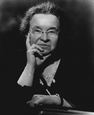
Professor of Histology in the Johns Hopkins University.
With six plates and one text-figure.
Introduction
In the study of the origin of endothelium and blood-cells it is obvious that any extension of the method of observing them in hving form i.s an advance. Until recently the tadpole's tail constituted the main subject for the study of growth of the vascular system in a living form. From the time of the discovery by Kolhker that it contains two kinds of capillaries, those carrjong blood and those carrying lymph, the tadpole's tail has been studied over and over again in connection with the question of the growth of vessels. The final studies on the living tadpole, made by E. R. Clark (1909, 1918), have demonstrated conclusively that at certain stages both blood-capillaries and lymph-capillaries in the area of the tail grow by the method of sprouting; that is, by the division of and increase in the cytoplasm of the endothehum lining the vessels, and not by the addition of new mesenchjTQal cells to their walls.
An extension of the study of the vascular system to a living form was then made by Stockard (1915), who watched blood-vessels arise in developing fish embryos. This author showed that the first sign of blood-vessels is the elongation of certain mesenchymal cells into spindle-shaped cells which could be identified as angioblasts. This I believe to be a fundamental point — that the vascular system begins by the differentiation of certain cells which we may term angioblasts, or according to Ranvier, vasoformative cells. Stockard then subjected the fish to experimental conditions, by which he made an analy.sis of the place of origin of red blood-cells. In fish embryos that had been treated by certain chemicals he produced an abnormality consisting of a lack of union between the venous end of the heart and the vitelUne veins, so that there was no circulation and consequently no moving of the blood from place to place. From these experiments he concluded that in the fish all of the cells that form red blood-cells are located in the
intermediate cell-mass in the posterior part of the embryo, and that none of them arise in the head. In this conclusion his results have been questioned by Reagan (1917) in a study on the same form. Certainly in the chick it can be definitely proved that the endothehum of the vessels gives rise to red blood-cells, and that this process can be seen to take place not only in every part of the area vasculosa, but within the embryo itself, the process having been observed in the aorta in the living chick.
Observations on blood-vessels in living chicks are by no means new; in fact
it is obvious that many of the conclusions in the monograph of His, published in 1S6S, arc based upon studies of liviM<i; clucks immediately after removing them
from the shell; wliile Duval in his atlas, published in 1889, called attention to the
advantage of studying the beating of the heart in living chicks in a watch crystal.
Indeed this advantage has been quite apparent to the whole group of embryologists
using the same material. As a result of the develoi)ment of different artificial solutions ujwn which the method of tissue culture depends, there has naturally followed
a marked increase in the studies of Uving embryos of the higher forms; for example,
the series of observations made by E. R. and E. L. Clark (1912, 1915) on the superficial lymphatics in chicks held in the shell and kept alive by Rusch solution, and
the demonstration by Brachet (1913) that a mammaUan egg could be kept aUve
for at least 48 hours in blood-plasma. These studies, though depending upon the
development of the media wliich are used in tissue-culture, are not specifically
tissue-cultures, for by that term is meant the growth of tissues on a coverslip in artificial media, so arranged that the development of their cells can be watched under the microscope.
The first direct appUcation of the method of tissue-culture to the entire blastoderm of the chick — that is, the study of the blastoderm on a coverslip so that its
cells could be clearlj- followed — was made by McWhorter and Whip])le in 1912.
These investigators followed the technique of Burrows and Carrel, developed from
the work of Harrison, using clotted chicken-plasma in which to grow the specimens.
In this medium they kept chicks under observation for a considerable period of
time — namely, from the stage of about 2 or 3 somites up to the stage of 17 or 18
somites. In my experiments I have followed the method of W. H. and M. R.
Lewis, using Locke-Lewis solution instead of the clotted plasma. This tcchnique
is much more simple and, I beheve, offers many advantages, in spite of the fact that
the embryos do not Uve nearly as long. I have made no attempt to determine the
possible duration of life of the specimens in the media, but have watched them on
an average of 4 to 5 hours, during which time the maximum number of new somites
is 3, with an average of 2. The greater ease of the method renders possible much
more extensive observations. My series numbers 266.
With this method of studying the blastoderm of the chick, it is po.ssible to trace
the formation of blood-vessels back to their very beginning and to ob.serve them
developing by differentiation from mesoderm to a new tyi)e of cell, the angioblast.
These angioblasts form soUd clumps, then bands or cords of cells, wliich soon unite
in a plexus by the well-known method of sprouting. The central part of the substance, either of the isolated clumps or of the plexus of angioblasts, then liquefies,
leaving the rim to form the endothelial wall of the vessel. In this hciuefaction both
cytoplasm and nuclei are involved, and the resulting fluid constitutes the first blood-
plasma. Moreover, angiol)lasts not only give rise to endothelium and blood-plasma,
but also produce the red blood-coriniscles. Thus in the chick angioblasts are cells
which differentiate from mesoderm and form endothehum, blood-plasma, and red blood-cells.
That angioblasts differentiate in the area vasculosa of the chick has long been known; indeed, the idea is involved in our knowledge of the origin of the so-called blood-islands dating l)ack to the early embryologists, notably Wolff and Pander. In this study I propose to sharpen the conception of the differentiation of angioblasts and to Hmit the term blood-island to those masses of cells which actually produce hemoglobin and become red blood-corpuscles. It will be shown that though angioblasts, immediately after their differentiation, begin to sjjread by sprouting, the primitive vessels do not invade the embryo in the manner beheved by His, but that there is a progressive differentiation of angioblasts in silu, starting in the area opaca of the cliick and gradually extending toward the embryo. The heart, most of the dorsal aorta, and even a part of the ventral aorta of the head, can be seen in the living chick to differentiate in situ. Thus the.se studies in the living blastoderm confirm the experiments of Hahn (1909), in which he destroyed the area vasculosa on one side of a very young blastoderm and subsequently found an aorta on that side. These experiments were confirmed later by McWhorter and INIillcr (1914) and the .same point was brought out by Reagan (1915), who i.solated the head-fold of a young embryo and found vessels in the isolated segment. It may thus be considered as settled that blood-vessels arise from cells which differentiate from mesoderm, and that these cells (angioblasts) differentiate not only throughout the area vasculo.sa, but also throughout the body of the embryo itself. The ultimate i)eriod at which angioblasts cease to differentiate from mesenchyme must be regarded as still unknown.
Content to be added----
| Historic Disclaimer - information about historic embryology pages |
|---|
|
Glossary Links
- Glossary: A | B | C | D | E | F | G | H | I | J | K | L | M | N | O | P | Q | R | S | T | U | V | W | X | Y | Z | Numbers | Symbols | Term Link
Cite this page: Hill, M.A. (2024, May 5) Embryology Book - Contributions to Embryology Carnegie Institution No.36. Retrieved from https://embryology.med.unsw.edu.au/embryology/index.php/Book_-_Contributions_to_Embryology_Carnegie_Institution_No.36
- © Dr Mark Hill 2024, UNSW Embryology ISBN: 978 0 7334 2609 4 - UNSW CRICOS Provider Code No. 00098G