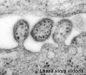
Abnormal Development - Lassa Virus
| Embryology - 27 Feb 2026 |
|---|
| Google Translate - select your language from the list shown below (this will open a new external page) |
|
العربية | català | 中文 | 中國傳統的 | français | Deutsche | עִברִית | हिंदी | bahasa Indonesia | italiano | 日本語 | 한국어 | မြန်မာ | Pilipino | Polskie | português | ਪੰਜਾਬੀ ਦੇ | Română | русский | Español | Swahili | Svensk | ไทย | Türkçe | اردو | ייִדיש | Tiếng Việt These external translations are automated and may not be accurate. (More? About Translations) |
Introduction
Lassa virus is a member of the virus family Arenaviridae, a single-stranded RNA virus. The virus is the causative agent of a hemorrhagic fever and can be transmitted between species (zoonotic). Discovered in 1969 when two missionary nurses died in Nigeria and today still occurs mainly in West Africa.
Death rates are high for women in the third trimester of pregnancy. Fetal death (95%) occurs in uterus of infected pregnant mothers.
Some Recent Findings
|
Pathology
Pathogenesis of Lassa fever in cynomolgus macaques
Virol J. 2011 May 6;8:205.
Hensley LE, Smith MA, Geisbert JB, Fritz EA, Daddario-DiCaprio KM, Larsen T, Geisbert TW. Source Virology, US Army Medical Research Institute of Infectious Diseases, Fort Detrick, MD, USA.
Abstract
BACKGROUND: Lassa virus (LASV) infection causes an acute and sometimes fatal hemorrhagic disease in humans and nonhuman primates; however, little is known about the development of Lassa fever. Here, we performed a pilot study to begin to understand the progression of LASV infection in nonhuman primates.
METHODS: Six cynomolgus monkeys were experimentally infected with LASV. Tissues from three animals were examined at an early- to mid-stage of disease and compared with tissues from three animals collected at terminal stages of disease.
RESULTS: Dendritic cells were identified as a prominent target of LASV infection in a variety of tissues in all animals at day 7 while Kupffer cells, hepatocytes, adrenal cortical cells, and endothelial cells were more frequently infected with LASV in tissues of terminal animals (days 13.5-17). Meningoencephalitis and neuronal necrosis were noteworthy findings in terminal animals. Evidence of coagulopathy was noted; however, the degree of fibrin deposition in tissues was less prominent than has been reported in other viral hemorrhagic fevers.
CONCLUSION: The sequence of pathogenic events identified in this study begins to shed light on the development of disease processes during Lassa fever and also may provide new targets for rational prophylactic and chemotherapeutic interventions.
PMID 21548931
http://www.virologyj.com/content/8/1/205
References
Reviews
<pubmed></pubmed>
Articles
Search Pubmed
June 2010
Search Pubmed: Lassa Virus |
| Environmental Links: Introduction | low folic acid | iodine deficiency | Nutrition | Drugs | Australian Drug Categories | USA Drug Categories | thalidomide | herbal drugs | Illegal Drugs | smoking | Fetal Alcohol Syndrome | TORCH | viral infection | bacterial infection | fungal infection | zoonotic infection | toxoplasmosis | Malaria | maternal diabetes | maternal hypertension | maternal hyperthermia | Maternal Inflammation | Maternal Obesity | hypoxia | biological toxins | chemicals | heavy metals | air pollution | radiation | Prenatal Diagnosis | Neonatal Diagnosis | International Classification of Diseases | Fetal Origins Hypothesis |
External Links
External Links Notice - The dynamic nature of the internet may mean that some of these listed links may no longer function. If the link no longer works search the web with the link text or name. Links to any external commercial sites are provided for information purposes only and should never be considered an endorsement. UNSW Embryology is provided as an educational resource with no clinical information or commercial affiliation.
Glossary Links
- Glossary: A | B | C | D | E | F | G | H | I | J | K | L | M | N | O | P | Q | R | S | T | U | V | W | X | Y | Z | Numbers | Symbols | Term Link
Cite this page: Hill, M.A. (2026, February 27) Embryology Abnormal Development - Lassa Virus. Retrieved from https://embryology.med.unsw.edu.au/embryology/index.php/Abnormal_Development_-_Lassa_Virus
- © Dr Mark Hill 2026, UNSW Embryology ISBN: 978 0 7334 2609 4 - UNSW CRICOS Provider Code No. 00098G
