Abnormal Development - Folic Acid and Neural Tube Defects

From Embryology
Embryology - 19 Mar 2024    Facebook link Pinterest link Twitter link  Expand to Translate  
Google Translate - select your language from the list shown below (this will open a new external page)

العربية | català | 中文 | 中國傳統的 | français | Deutsche | עִברִית | हिंदी | bahasa Indonesia | italiano | 日本語 | 한국어 | မြန်မာ | Pilipino | Polskie | português | ਪੰਜਾਬੀ ਦੇ | Română | русский | Español | Swahili | Svensk | ไทย | Türkçe | اردو | ייִדיש | Tiếng Việt    These external translations are automated and may not be accurate. (More? About Translations)

 ICD-11

5B5E Folate deficiency - Between days 21 and 27 post-conception, the neural plate closes to form what will eventually be the spinal cord and cranium. Spina bifida, anencephaly, and other similar conditions are collectively called NTDs. They result from improper closure of the spinal cord and cranium, respectively, and are the most common congenital abnormalities associated with folate deficiency.

LA00-LA0Z Structural developmental anomalies of the nervous system - LA00.0 Anencephaly LA00.1 Iniencephaly LA00.2 Acephaly LA00.3 Amyelencephaly LA02 Spina bifida - LA02.0 Spina bifida cystica LA02.00 Myelomeningocele with hydrocephalus LA02.01 Myelomeningocele without hydrocephalus LA02.02 Myelocystocele LA02.1 Spina bifida aperta

Introduction

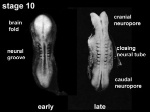
Neural groove closing to neural tube, early week 4, embryo stage 10, Gestational Age GA week 6).

In 2001, the Australian estimated birth prevalence of neural tube defects was 0.5 per 1,000 births (National Perinatal Statistics Unit). Low maternal dietary folic acid (folate) has been shown to be associated with the development of neural tube defects. In September 2009, mandatory folic acid fortification of bread flour was introduced in Australia.

Research over the last 20 years had suggested a relationship between maternal diet and the birth of an affected infant. Recent evidence has confirmed that folic acid, a water soluble vitamin (vitamin B9) found in many fruits (particularly oranges, berries and bananas), leafy green vegetables, cereals and legumes, may prevent the majority of neural tube defects.

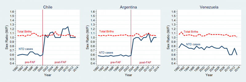
This class of abnormalities was historically more associated with Female infants and a recent study of Latin American countries has identified a substantial decrease in this ratio following folate fortification.[1]


Folatefruit.jpg
Folate.jpg
Fruits
Folic Acid

In the U.S.A., the Food and Drug Administration in 1996 authorized that all enriched cereal grain products be fortified with folic acid, with optional fortification beginning in March 1996 and mandatory fortification in January 1998.


In Australia, from 2009 the mandatory folic acid fortification standard required addition of folic acid to all wheat flour for bread making, with the exception of organic bread, within the prescribed range of 200–300 μg per 100 g of flour.


The March of Dimes Folic Acid Campaign (a major US charity group) has as one of its major objectives to reduce neural tube defects by 30% by 2001 using community programs, professional education, and mass media information.


Nutrition Links: nutrition | Vitamin A | Vitamin B | Vitamin C | Vitamin D | Vitamin E | Vitamin K | folate | iodine deficiency | neural abnormalities | Axial Skeleton Abnormalities


Environmental Links: Introduction | low folic acid | iodine deficiency | Nutrition | Drugs | Australian Drug Categories | USA Drug Categories | thalidomide | herbal drugs | Illegal Drugs | smoking | Fetal Alcohol Syndrome | TORCH | viral infection | bacterial infection | fungal infection | zoonotic infection | toxoplasmosis | Malaria | maternal diabetes | maternal hypertension | maternal hyperthermia | Maternal Inflammation | Maternal Obesity | hypoxia | biological toxins | chemicals | heavy metals | air pollution | radiation | Prenatal Diagnosis | Neonatal Diagnosis | International Classification of Diseases | Fetal Origins Hypothesis

Some Recent Findings

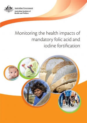
AIHW Report - Folic acid and iodine fortification (2016)
Folic acid and iodine fortification (2016)
  • Maternal folic acid depletion during early pregnancy increases sensitivity to squamous tumor formation in the offspring in mice[2] "Gestational nutrition is widely recognized to affect an offspring's future risk of lifestyle-related diseases, suggesting the involvement of epigenetic mechanisms. As folic acid (FA) is a nutrient essential for modulating DNA methylation, we sought to determine how maternal FA intake during early pregnancy might influence tumor sensitivity in an offspring. Dams were maintained on a FA-depleted (FA(-)) or normal (2 mg FA/kg; FA(+)) diet from 2 to 3 days before mating to 7 days post-conception, and their offspring were challenged with chemical tumorigenesis using 7,12-dimethylbenz[a)anthracene and phorbol 12-myristate 13-acetate for skin and 4-nitroquinoline N-oxide for tongue. In both squamous tissues, tumorigenesis was more progressive in the offspring from FA(-) than FA(+) dams. Notably, in the skin of FA(-) offspring, the expression and activity of cylindromatosis (Cyld) were decreased due to the altered DNA methylation status in its promoter region, which contributed to increased tumorigenesis coupled with inflammation in the FA(-) offspring. Thus, we conclude that maternal FA insufficiency during early pregnancy is able to promote neoplasm progression in the offspring through modulating DNA methylation, such as Cyld. Moreover, we propose, for the first time, "innate" utero nutrition as the third cause of tumorigenesis besides the known causes-hereditary predisposition and acquired environmental factors."
  • Network correlation analysis revealed potential new mechanisms for neural tube defects beyond folic acid[3] " METHODS: Microarray data of GSE51285 was downloaded from the NCBI GEO database, which contains the RNA expression profiles of livers from five NTD mouse mutants (heterozygous females) and their corresponding wildtype (WT) controls. RESULTS: In total, we identified 18 genes related to the pathogenesis of NTDs, as well as 55 genes related to FA responsiveness. Eight more candidate genes (Abcc3, Gsr, Gclc, Mthfd1, Gart, Bche, Slc25a32, and Slc44a2) were identified by examining the DEGs of those genes involved in the extended folate metabolic pathway between FA-responsive and FA-resistant mutants. CONCLUSIONS: Those genes are involved in mitochondrial choline metabolism, de novo purine synthesis, and glutathione generation, suggesting that formate, choline, and manipulating antioxidant levels may be effective interventions in FA-resistant NTDs."
  • Neural tube defects: Sex ratio changes after fortification with folic acid[1] "Historically, neural tube defects (NTDs) have predominated in Female infants but the reasons remain unclear. In South America, the pre- folic acid fortification (FAF) rates of NTDs were around 18/10,000 births for females and 12/10,000 births for males, with an estimated sex ratio (male/female) of 0.67. During the post-FAF period, unpublished routine reports have indicated changes in the sex ratio for these defects while some descriptive reports are controversial. To date and to our knowledge, however, no studies specifically focusing on these changes to test this hypothesis directly have been undertaken. The aim of this study was to analyze changes in the sex ratio of infants with NTDs after FAF in South American countries. MATERIALS AND METHODS: With a descriptive cross-sectional study design, 2,597 infants with isolated NTDs born between 1990 and 2013 in 3 countries participating in the Latin American Collaborative Study of Congenital Malformations (ECLAMC) network were included: (Chile N = 521 and Argentina N = 1,619 [with FAF policies]; Venezuela N = 457 [without FAF policies; used as control]; total births = 2,229,561). The differences-in-differences method and Poisson regressions were used to evaluate the sex ratio shift from female to male before vs. after FAF, and to assess whether these differences were related to the fortification. RESULTS AND CONCLUSIONS: In Chile and Argentina the prevalence of NTDs, particularly anencephaly and cervico-thoracic spina bifida, showed a greater reduction rate in females than in males after FAF, resulting in a change of the sex ratio of infants with NTDs."
  • Novel Mutation of LRP6 Identified in Chinese Han Population Links Canonical WNT Signaling to Neural Tube Defects[4] "Three rare missense mutations (c.1514A>G, p.Y505C); c.2984A>G, p.D995G; and c.4280C>A, p.P1427Q) of the LRP6 gene were identified in Chinese NTD patients. The Y505C mutation is a loss-of-function mutation on both WNT/β-catenin and PCP signaling. The D995G mutation only partially lost inhibition on PCP signaling without affecting WNT/β-catenin signaling. The P1427Q mutation dramatically increased WNT/β-catenin signaling but only mildly loss of inhibition on PCP signaling. All three mutations failed to rescue CE defects caused by lrp6 morpholino oligos knockdown in zebrafish. Of interest, when overexpressed, D995G did not induce any defects, but Y505C and P1427Q caused more severe CE defects in zebrafish. CONCLUSION: Our results suggested that over-active canonical WNT signaling induced by gain-of-function mutation in LRP6 could also contribute to human NTDs, and a balanced WNT/β-catenin and PCP signaling is probably required for proper neural tube development." WNT
More recent papers  
Mark Hill.jpg
PubMed logo.gif

This table allows an automated computer search of the external PubMed database using the listed "Search term" text link.

  • This search now requires a manual link as the original PubMed extension has been disabled.
  • The displayed list of references do not reflect any editorial selection of material based on content or relevance.
  • References also appear on this list based upon the date of the actual page viewing.


References listed on the rest of the content page and the associated discussion page (listed under the publication year sub-headings) do include some editorial selection based upon both relevance and availability.

More? References | Discussion Page | Journal Searches | 2019 References | 2020 References

Search term: Neural Tube Defect | Spina bifida | Spina bifida cystica | | Spina bifida aperta | Myelomeningocele | Myelocystocele

Older papers  
These papers originally appeared in the Some Recent Findings table, but as that list grew in length have now been shuffled down to this collapsible table.

See also the Discussion Page for other references listed by year and References on this current page.

  • Genetic contribution of retinoid related genes to neural tube defects[5] "Rare variants are considered underlying causes of complex diseases. The complex and severe group of disorders called neural tube defects (NTDs) results from failure of the neural tube to close during early embryogenesis. Neural tube closure requires the coordination of numerous signaling pathways, including the precise regulation of retinoic acid (RA) concentration which is controlled by enzymes involved in RA synthesis and degradation. Here we used a case-control mutation screen study to reveal rare variants in retinoid related genes in a Han Chinese NTD population by sequencing six genes in 355 NTD cases and 225 controls. NTD-specific rare variants were found in exonic regions and upstream regions. The RA-responsive genes CYP26A1, CRABP1 and ALDH1A2 harbored NTD-specific rare variants in their upstream regions. Unexpectedly, the majority of missense variants in NTD cases were found in CYP26B1 which encodes a RA degradation enzyme, whereas no missense variants in this gene were found in controls. Functional analysis indicated that the CYP26B1 NTD variants were inefficient in the degradation of RA using assays of RA-induced transcription and RA-initiated neuronal differentiation." retinoic acid
  • Australia - Decrease in neural tube defects since folic acid added to bread[6]"Mandatory fortification of bread with folic acid (in Australia) and iodine (in Australia and New Zealand) was introduced in 2009 to address two important public health issues: to reduce the prevalence of neural tube defects (serious birth defects such as spina bifida) in Australia and to deal with the re-emergence of iodine deficiency in both Australia and New Zealand. There was a significant (14.4%) overall decrease in the rate of neural tube defects (NTDs) in Australia following mandatory folic acid fortification. However among teenagers, the rate of NTDs decreased even more, by almost 55%, and for Aboriginal and Torres Strait Islander women, the rate of NTDs decreased by 74%."
  • Preconception folic acid supplementation and risk for chromosome 21 nondisjunction: a report from the US National Down Syndrome Project[7] "Both a lack of maternal folic acid supplementation and the presence of genetic variants that reduce enzyme activity in folate pathway genes have been linked to meiotic nondisjunction of chromosome 21; however, the findings in this area of research have been inconsistent. To better understand these inconsistencies, we asked whether maternal use of a folic acid-containing supplement before conception reduces risk for chromosome 21 nondisjunction. ...These data suggest that lack of folic acid supplementation may be associated specifically with MII errors in the aging oocyte. If confirmed, these results could account for inconsistencies among previous studies, as each study sample may vary by maternal age structure and proportion of meiotic errors." Trisomy 21
  • Association between maternal use of folic acid supplements and risk of autism spectrum disorders in children[8] "To examine the association between maternal use of prenatal folic acid supplements and subsequent risk of autism spectrum disorders (ASDs) (autistic disorder, Asperger syndrome, pervasive developmental disorder-not otherwise specified [PDD-NOS]) in children. The study sample of 85,176 children was derived from the population-based, prospective Norwegian Mother and Child Cohort Study (MoBa). The children were born in 2002-2008; by the end of follow-up on March 31, 2012, the age range was 3.3 through 10.2 years (mean, 6.4 years). The exposure of primary interest was use of folic acid from 4 weeks before to 8 weeks after the start of pregnancy, defined as the first day of the last menstrual period before conception. Relative risks of ASDs were estimated by odds ratios (ORs) with 95% CIs in a logistic regression analysis. Analyses were adjusted for maternal education level, year of birth, and parity. ...Use of prenatal folic acid supplements around the time of conception was associated with a lower risk of autistic disorder in the MoBa cohort. Although these findings cannot establish causality, they do support prenatal folic acid supplementation."
  • Disruption of the folate pathway in zebrafish causes developmental defects[9] "Our studies demonstrate that human and zebrafish utilize similar one-carbon pathways. Our data indicate that folate metabolism is essential for early zebrafish development. Zebrafish studies of the folate pathway and its deficiencies could provide insight into the underlying etiology of human birth defects and the natural role of folate in development."
  • Periconceptional bread intakes indicate New Zealand's proposed mandatory folic acid fortification program may be outdated[10] "In September 2009, a folic acid fortification mandate (135 μg/100 g bread) was to be implemented in New Zealand. However, due to political and manufacturer objection, fortification was deferred until May 2012. Based on estimates of bread consumption derived from a 1997 nationally representative survey, this program was intended to deliver a mean additional intake of 140 μg folic acid/d to women of childbearing age. ...This study provides insight on the ability of a fortification policy to benefit the groups at highest risk of poor folate intakes in a population. However, bread consumption among the target group of childbearing women appears to have declined since the data used in previous dietary modeling were collected. Thus, it seems prudent to re-model dietary folic acid intakes based on more recent national survey data prior to the implementation of a mandatory folic acid fortification policy."

Neural Tube Closure

<html5media height="360" width="360">File:Neuraltube_001.mp4</html5media> This cartoon movie shows a dorsolateral view (from the back left side) of the early embryo.


The process of the neural groove closing to form the early neural tube is shown. This process occurs during week 4 (Gestational Age GA week 6) in human development. The same process occurs in all vertebrate animals, but at different times (days) in their early development.


Failure of the neural tube to close anywhere along its length results in a "neural tube defect".

Neural Tube Defect Classification

Neural tube defects comprise three distinct conditions: anencephaly, spina bifida and encephalocele. Note that the current ICD10 classification system is being updated to ICD11 that is currently in beta testing status. The data on this page, other than the information table below, is from ICD10 classification.

 ICD-11 LA00-LA0Z Structural developmental anomalies of the nervous system
  • LA00.0 Anencephaly - a neural tube defect, characterized by the total or partial absence of the cranial vault and the covering skin, the brain being missing or reduced to a small mass. Most cases are stillborn, although some infants have been reported to survive for a few hours. In most cases autopsy findings reveal absence of adrenal glands. Anencephaly is likely to be multifactorial, the result of gene-environment interactions. Familial cases with a seemingly autosomal recessive mode of inheritance have been described but most cases are sporadic. Folic acid and zinc deficiencies, as well as maternal obesity, have been shown to be risk factors.
  • LA00.1 Iniencephaly - a rare form of neural tube defect in which a malformation of the cervico-occipital junction is associated with a malformation of the central nervous system. The cardinal features are occipital bone defect, partial or total absence of cervicothoracic vertebrae, fetal retroflexion of the head and characteristic absence of the neck. It is associated with malformations of the central nervous (spina bifida and/or anencephaly), gastrointestinal (omphalocele) and cardiovascular systems.
  • LA00.2 Acephaly
  • LA00.3 Amyelencephaly - Amyelencephaly is the absence of both the brain and spinal cord.
  • LA02 Spina bifida - Spina bifida is the most common of a group of birth defects called neural tube defects. Spina bifida affects the backbone and, sometimes, the spinal cord. Aperta spina bifida defines the dorsal malclosure of vertebrae, associated with various degrees of spine defects. A pocket of skin may form, containing meninges (meningocele) or spinal cord and meninges (myelomeningocele). Different subtypes are distinguished according to the location of the defect. Consequences are paraplegia (paralysed lower limbs), hydrocephaly, Chiari malformation (result of the attached spine during life in utero), urinary and anorectal incontinence. The intensity of signs varies greatly with the level and extent of the lesion.
    • LA02.0 Spina bifida cystica - failure of the neural tube to correctly develop during the antenatal period. This condition is characterized by nerve damage and the presence of meningoceles on the back. This condition may present with physical or mental impairment.
    • LA02.00 Myelomeningocele with hydrocephalus - failure of the neural tube to correctly develop during the antenatal period. This condition is characterized by nerve damage and hydrocephalus. This condition may also present with syringomyelia, hip dislocation, headache, nausea, vomiting, blurry vision, balance problems, bladder control problems, meningitis, or mental impairment.
    • LA02.01 Myelomeningocele without hydrocephalus - failure of the neural tube to close completely during fetal development. This condition is characterized by nerve damage. This condition may also present with syringomyelia, hip dislocation, headache, nausea, vomiting, blurry vision, balance problems, bladder control problems, meningitis, or mental impairment.
    • LA02.02 Myelocystocele - failure of the neural tube to close completely during fetal development. The condition is characterized by skin covered lumbosacral masses, an arachnoid lined meningocele that is directly continuous with the spinal subarachnoid space, and a low lying hydromyelic spinal cord that traverses the meningocele and expands into a large terminal cyst. This condition can present with neural damage and consequent impairment of function below the site of the myelocystocele.
    • LA02.1 Spina bifida aperta - failure of the neural tube to correctly develop during the antenatal period. This condition is characterized by nerve damage originating from a known location in the spine, signified by the presence of a meningocele or myelomeningocele. This condition may present with physical or mental impairment.
International Classification of Diseases  ICD-11 20 Developmental anomalies (beta draft)  
ICD-11 Beta Draft - NOT FINAL, updated on a daily basis, It is not approved by WHO, NOT TO BE USED for CODING except for agreed FIELD TRIALS.

Chapter 20 Developmental anomalies, only a few examples of the draft ICD-11 Beta coding and tree structure for "structural developmental anomalies" within this section are shown in the table below.

Mortality and Morbidity Statistics - 20 Developmental Anomalies  
Structural Developmental Anomalies  
  • Structural developmental anomalies of the nervous system  
    • LA00 Anencephaly or similar anomalies
    • LA01 Cephalocele
    • LA02 Spina bifida
    • LA03 Arnold-Chiari malformation type II
    • LA04 Congenital hydrocephalus
    • LA05 Cerebral structural developmental anomalies
    • LA06 Cerebellar structural developmental anomalies
    • LA07 Structural developmental anomalies of the neurenteric canal, spinal cord or vertebral column
    • LA0Y Other specified structural developmental anomalies of the nervous system
    • LA0Z Structural developmental anomalies of the nervous system, unspecified
  • Structural developmental anomalies of the eye, eyelid or lacrimal apparatus
    • LA10 Structural developmental anomalies of ocular globes
    • LA11 Structural developmental anomalies of the anterior segment of eye
    • LA30 Structural developmental anomalies of lens or zonula
    • LA31 Structural developmental anomalies of the posterior segment of eye
    • LA32 Structural developmental anomalies of eyelid, lacrimal apparatus or orbit
    • LA3Y Other specified structural developmental anomalies of the eye, eyelid or lacrimal apparatus
    • LA3Z Structural developmental anomalies of the eye, eyelid or lacrimal apparatus, unspecified
  • Structural developmental anomalies of the ear  
    • LA40 Structural anomaly of eustachian apparatus
    • LA41 Minor anomalies of pinnae
    • LA42 Structural developmental anomalies of ear causing hearing impairment
    • LA43 Otocephaly
    • LA44 Accessory auricle
    • LA4Y Other specified structural developmental anomalies of the ear
    • LA4Z Structural developmental anomalies of the ear, unspecified
  • Structural developmental anomalies of the face, mouth or teeth
    • LA50 Structural developmental anomalies of teeth and periodontal tissues
    • LA51 Structural developmental anomalies of mouth or tongue
    • Clefts of lip, alveolus or palate
    • LA70 Congenital velopharyngeal incompetence
    • LA71 Facial clefts
    • LA72 Facial asymmetry
    • LA73 Macrocheilia
    • LA74 Microcheilia
    • LA75 Compression facies
    • LA76 Pierre Robin syndrome
    • LC20 Dermoid cyst
    • LA7Y Other specified structural developmental anomalies of the face, mouth or teeth
    • LA7Z Structural developmental anomalies of the face, mouth or teeth, unspecified
  • Structural developmental anomalies of the neck  
  • Structural developmental anomalies of the respiratory system  
  • Structural developmental anomalies of the circulatory system  
    • Structural developmental anomaly of heart and great vessels
      • LB00 Congenital heart or great vessel related acquired abnormality
      • LB01 Congenital anomaly of atrioventricular or ventriculo-arterial connections
      • LB02 Congenital anomaly of the mediastinal veins Congenital anomaly of atria or atrial septum
      • LB20 Congenital anomaly of atrioventricular valves or septum
      • LB21 Congenital anomaly of ventricles and ventricular septum
      • LB22 Functionally univentricular heart
      • LB23 Congenital anomaly of ventriculo-arterial valves and adjacent regions
      • LB24 Congenital anomaly of great arteries including arterial duct
      • LB25 Anomalous position-orientation of heart
      • LB26 Total mirror imagery
      • LB27 Left isomerism
      • LB28 Congenital anomaly of coronary arteries
      • LB29 Structural developmental anomalies of the pericardium
      • LB2Y Other specified structural developmental anomaly of heart and great vessels
      • LB2Z Structural developmental anomaly of heart and great vessels, unspecified
    • LB30 Structural developmental anomalies of the peripheral vascular system
      • LB30.1 Capillary malformations
      • LB30.2 Lymphatic malformations
        • LB30.21 Macrocystic lymphatic malformation
        • LB30.22 Microcystic lymphatic malformation
        • LB30.23 Cystic hygroma in fetus
        • BD23.1 Primary lymphoedema
            • EK91 Yellow nail syndrome
            • LC5F.26 Noonan syndrome
        • LB30.2Y Other specified lymphatic malformations
        • LB30.2Z Lymphatic malformations, unspecified
      • LB30.3 Peripheral venous malformations
      • LB30.4 Peripheral arteriovenous malformations
      • LB30.5 Peripheral arterial malformations
      • LB30.6 Pulmonary arteriovenous fistula
      • LB30.Y Other specified structural developmental anomalies of the peripheral vascular system
      • LB30.Z Structural developmental anomalies of the peripheral vascular system, unspecified
    • LB3Y Other specified structural developmental anomalies of the circulatory system
    • LB3Z Structural developmental anomalies of the circulatory system, unspecified
  • Structural developmental anomalies of the diaphragm, abdominal wall or umbilical cord  
  • Structural developmental anomalies of the digestive tract  
  • Structural developmental anomalies of the liver, biliary tract, pancreas or spleen  
  • Structural developmental anomalies of the urinary system  
  • Structural developmental anomalies of the female genital system  
  • Structural developmental anomalies of the male genital system  
  • Structural developmental anomalies of the breast  
  • Structural developmental anomalies of the skeleton  
  • Structural developmental anomalies of the skin  
  • Structural developmental anomalies of the adrenal glands  
Multiple developmental anomalies or syndromes
Chromosomal anomalies, excluding gene mutations
Conditions with disorders of intellectual development as a relevant clinical feature
LD6Y Other specified developmental anomalies

LD6Z Developmental anomalies, unspecified

CD-11 Beta Draft - NOT FINAL, updated on a daily basis, It is not approved by WHO, NOT TO BE USED for CODING except for agreed FIELD TRIALS.


See also International Classification of Diseases
ICD-10

Australian Statistics

Folic acid and iodine fortification (2016)

From 13 September 2009, the mandatory folic acid fortification standard requires the addition of folic acid to all wheat flour for bread making, with the exception of organic bread, within the prescribed range of 200–300 μg per 100 g of flour.

A recent 2016 study[6] has shown since fortification:

  • Overall decrease in the rate of neural tube defects (NTDs) by 14.4%
  • Teenagers the rate of NTDs decreased by almost 55%
  • Aboriginal and Torres Strait Islander women the rate of NTDs decreased by 74%


An earlier 2011 study[11] looking at the prevalence of neural tube defects before mandatory fortification.

  • Women who have one infant with a neural tube defect have a significantly increased risk of recurrence
    • 40-50 per thousand compared with 2 per thousand for all births.
  • A randomised controlled trial conducted by the Medical Research Council of the United Kingdom demonstrated a 72% reduction in risk of recurrence by periconceptional (ie before and after conception) folic acid supplementation (4mg daily).
  • Other epidemiological research, including work done in Australia, suggests that primary occurrences of neural tube defects may also be prevented by folic acid either as a supplement or in the diet.
  • This has been confirmed in a randomised controlled trial from Hungary, which found that a multivitamin supplement containing 0.8mg folic acid was effective in reducing the occurrence of neural tube defects in first births.

Before mandatory folic acid fortification was introduced:

  • mean dietary folic acid intakes for women aged 16–44 years (the target population) in Australia was 108 micrograms (μg) of folic acid per day and in New Zealand was 62 μg of folic acid per day, well below the recommended 400 μg per day.
  • there were 149 pregnancies affected by NTDs in 2005 in Australia (rate of 13.3 per 10,000 births) in the three states that provide the most accurate baseline of NTD incidence (South Australia, Western Australia and Victoria), and 63 pregnancies affected by NTDs in 2003 in New Zealand (rate of 11.2 per 10,000 births).

Before mandatory iodine fortification was introduced:

  • large proportions of the Australian and New Zealand population had inadequate iodine intakes.
  • national surveys measuring median urinary iodine concentration (MUIC) in schoolchildren, an indicator of overall population status, confirmed mild iodine deficiency in both countries.
    • The concentration was 96 μg per litre in Australia, and 66 μg per litre in New Zealand, less than the 100–200 μg per litre considered optimal.
Links: Folic Acid and Neural Tube Defects | Iodine Deficiency | Australian Statistics | AIHW - folic acid and iodine

USA Statistics

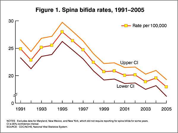
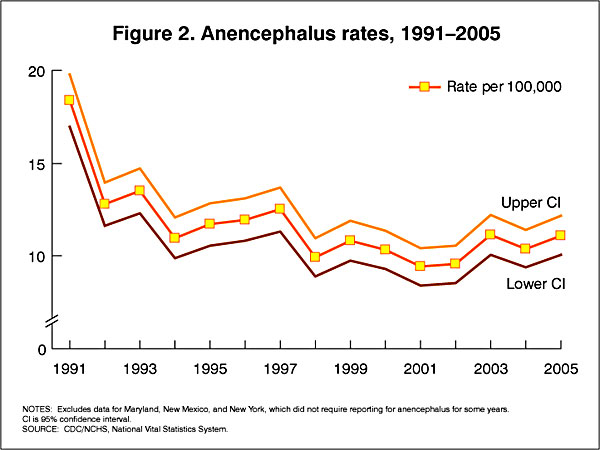
In the U.S.A. the Food and Drug Administration in 1996 authorized that all enriched cereal grain products be fortified with folic acid, with optional fortification beginning in March 1996 and mandatory fortification in January 1998. The data below shows the subsequent changes in anencephaly and spina bifida rate over that period.

USA spina bifida rates.jpg

USA anencephaly rates.jpg

Data CDC Report[12]

Latin America Statistics

A recent 2018 study of Latin American countries, has demonstrated a Female association and a substantial decrease in this ratio following folate fortification.[1] In contrast, an earlier 2015 USA study showed no sex association in their population.[13]


Neural tube defects sex ratio graph 01.png

Sex ratio changes for NTD cases and total births in Chile, Argentina, and Venezuela (1990–2013).[1]

NTD: neural tube defect; FAF: folic acid fortification; M/F: male/female; Sex ratio (male/female) for neural tube defect cases (full blue line), sex ratio for total births (dashed red line). Sex ratios estimated by multivariate regression models adjusted by hospital.

Potential Risk Factors

Neural Tube Defects - Potential risk factors
Maternal nutrition Alcohol use, Caffeine use, low folic acid, Low dietary quality, Elevated glycaemic load or index, Low methionine intake, Low serum choline level, Low serum vitamin B12 level, Low vitamin C level, Low zinc intake
Other maternal factors smoking, maternal hyperthermia, Low socio-economic status, Maternal infections and illnesses (TORCH, viral infection, bacterial infection, fungal infection), maternal diabetes, Pregestational obesity, Psychosocial stress, Valproic acid use
Environmental factors Ambient air pollution, Disinfectant by-products in drinking water, Indoor air pollution, Nitrate-related compounds, Organic solvents, Pesticides, Polycyclic aromatic hydrocarbons
Table data from review[14] (see original review for references)

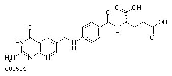
Folate Biosynthesis

Formula: C19H19N7O6

Alternate Names: Folic acid, Folate, Pteroylglutamic acid
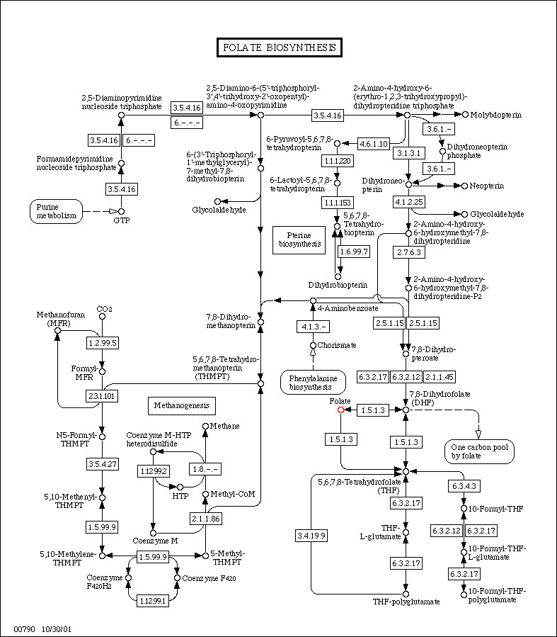

Folate Biosynthesis.jpg

(Data from KEGG)

Folate Biosynthesis(click image for full size or get original Map)
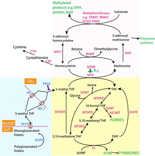
Folate one-carbon metabolism.jpg
Folate one-carbon metabolism[15]

Folate Supplementation and Other Abnormalities

A recent study of periconceptional folate supplementation using the Cochrane Pregnancy and Childbirth Group's Trials Register (July 2010) identified no statistically significant evidence of any effects on prevention of cleft palate, cleft lip, congenital cardiovascular defects, miscarriages or any other birth defects.[16]


Surgery for Spina Bifida

Fetal Surgery

The neural tube remaining open during fetal development can lead to neural damage through exposure to amniotic fluid and mechanical effects. There are experimental fetal surgical techniques in some countries that allow prenatal repair and perhaps prevent further neurological damage. There are though fetal and maternal risks associated with this surgical procedure.[17] A recent 2014 Cochrane Database study[18] identified that "insufficient evidence to recommend drawing firm conclusions on the benefits or harms of prenatal repair as an intervention for fetuses with spina bifida."

Neonatal Surgery

The surgical repair after birth is a more common procedure.


References

  1. 1.0 1.1 1.2 1.3 Poletta FA, Rittler M, Saleme C, Campaña H, Gili JA, Pawluk MS, Gimenez LG, Cosentino VR, Castilla EE & López-Camelo JS. (2018). Neural tube defects: Sex ratio changes after fortification with folic acid. PLoS ONE , 13, e0193127. PMID: 29538416 DOI.
  2. Kawakubo-Yasukochi T, Morioka M, Ohe K, Yasukochi A, Ozaki Y, Hazekawa M, Nishinakagawa T, Ono K, Nakamura S & Nakashima M. (2019). Maternal folic acid depletion during early pregnancy increases sensitivity to squamous tumor formation in the offspring in mice. J Dev Orig Health Dis , 10, 683-691. PMID: 31131784 DOI.
  3. Gao X, Finnell RH, Wang H & Zheng Y. (2018). Network correlation analysis revealed potential new mechanisms for neural tube defects beyond folic acid. Birth Defects Res , , . PMID: 29732722 DOI.
  4. Shi Z, Yang X, Li BB, Chen S, Yang L, Cheng L, Zhang T, Wang H & Zheng Y. (2018). Novel Mutation of LRP6 Identified in Chinese Han Population Links Canonical WNT Signaling to Neural Tube Defects. Birth Defects Res , 110, 63-71. PMID: 28960852 DOI.
  5. Li H, Zhang J, Chen S, Wang F, Zhang T & Niswander L. (2018). Genetic contribution of retinoid-related genes to neural tube defects. Hum. Mutat. , , . PMID: 29297599 DOI.
  6. 6.0 6.1 AIHW 2016. Monitoring the health impacts of mandatory folic acid and iodine fortification 2016. Cat. no. PHE 208. Canberra: AIHW. PDF
  7. Hollis ND, Allen EG, Oliver TR, Tinker SW, Druschel C, Hobbs CA, O'Leary LA, Romitti PA, Royle MH, Torfs CP, Freeman SB, Sherman SL & Bean LJ. (2013). Preconception folic acid supplementation and risk for chromosome 21 nondisjunction: a report from the National Down Syndrome Project. Am. J. Med. Genet. A , 161A, 438-44. PMID: 23401135 DOI.
  8. Surén P, Roth C, Bresnahan M, Haugen M, Hornig M, Hirtz D, Lie KK, Lipkin WI, Magnus P, Reichborn-Kjennerud T, Schjølberg S, Davey Smith G, Øyen AS, Susser E & Stoltenberg C. (2013). Association between maternal use of folic acid supplements and risk of autism spectrum disorders in children. JAMA , 309, 570-7. PMID: 23403681 DOI.
  9. Lee MS, Bonner JR, Bernard DJ, Sanchez EL, Sause ET, Prentice RR, Burgess SM & Brody LC. (2012). Disruption of the folate pathway in zebrafish causes developmental defects. BMC Dev. Biol. , 12, 12. PMID: 22480165 DOI.
  10. Mallard SR, Gray AR & Houghton LA. (2012). Periconceptional bread intakes indicate New Zealand's proposed mandatory folic acid fortification program may be outdated: results from a postpartum survey. BMC Pregnancy Childbirth , 12, 8. PMID: 22333513 DOI.
  11. 11.0 11.1 Macaldowie, A. and Hilder, L. (2011) Neural tube defects in Australia: prevalence before mandatory folic acid fortification. Cat. no. PER 53. Canberra: AIHW.
  12. CDC Trends in Spina Bifida and Anencephalus in the United States, 1991-2005
  13. Michalski AM, Richardson SD, Browne ML, Carmichael SL, Canfield MA, VanZutphen AR, Anderka MT, Marshall EG & Druschel CM. (2015). Sex ratios among infants with birth defects, National Birth Defects Prevention Study, 1997-2009. Am. J. Med. Genet. A , 167A, 1071-81. PMID: 25711982 DOI.
  14. Copp AJ, Adzick NS, Chitty LS, Fletcher JM, Holmbeck GN & Shaw GM. (2015). Spina bifida. Nat Rev Dis Primers , 1, 15007. PMID: 27189655 DOI.
  15. Greene ND, Stanier P & Copp AJ. (2009). Genetics of human neural tube defects. Hum. Mol. Genet. , 18, R113-29. PMID: 19808787 DOI.
  16. De-Regil LM, Fernández-Gaxiola AC, Dowswell T & Peña-Rosas JP. (2010). Effects and safety of periconceptional folate supplementation for preventing birth defects. Cochrane Database Syst Rev , , CD007950. PMID: 20927767 DOI.
  17. Adzick NS. (2013). Fetal surgery for spina bifida: past, present, future. Semin. Pediatr. Surg. , 22, 10-7. PMID: 23395140 DOI.
  18. Grivell RM, Andersen C & Dodd JM. (2014). Prenatal versus postnatal repair procedures for spina bifida for improving infant and maternal outcomes. Cochrane Database Syst Rev , , CD008825. PMID: 25348498 DOI.

Reviews

Veenboer PW, Bosch JL, van Asbeck FW & de Kort LM. (2012). Upper and lower urinary tract outcomes in adult myelomeningocele patients: a systematic review. PLoS ONE , 7, e48399. PMID: 23119003 DOI.

Eichholzer M, Tönz O & Zimmermann R. (2006). Folic acid: a public-health challenge. Lancet , 367, 1352-61. PMID: 16631914 DOI.

Tamura T & Picciano MF. (2006). Folate and human reproduction. Am. J. Clin. Nutr. , 83, 993-1016. PMID: 16685040

Wen SW & Walker M. (2005). An exploration of health effects of folic acid in pregnancy beyond reducing neural tube defects. J Obstet Gynaecol Can , 27, 13-9. PMID: 15937577

Articles

Giordan E, Bortolotti C, Lanzino G & Brinjikji W. (2018). Spinal Arteriovenous Vascular Malformations in Patients with Neural Tube Defects. AJNR Am J Neuroradiol , 39, 597-603. PMID: 29284599 DOI.

Molloy AM. (2002). Folate bioavailability and health. Int J Vitam Nutr Res , 72, 46-52. PMID: 11887752 DOI.

Daly LE, Kirke PN, Molloy A, Weir DG & Scott JM. (1995). Folate levels and neural tube defects. Implications for prevention. JAMA , 274, 1698-702. PMID: 7474275

Smithells RW, Sheppard S & Schorah CJ. (1976). Vitamin deficiencies and neural tube defects. Arch. Dis. Child. , 51, 944-50. PMID: 1015847

Leck I. (1977). Folates and the fetus. Lancet , 1, 1099-100. PMID: 68194


Search PubMed

Search Bookshelf: Folic Acid and Neural Tube Defects | Folic Acid


Search Pubmed: Folic Acid and Neural Tube Defects | Folic Acid

External Links

External Links Notice - The dynamic nature of the internet may mean that some of these listed links may no longer function. If the link no longer works search the web with the link text or name. Links to any external commercial sites are provided for information purposes only and should never be considered an endorsement. UNSW Embryology is provided as an educational resource with no clinical information or commercial affiliation.


Glossary Links

Glossary: A | B | C | D | E | F | G | H | I | J | K | L | M | N | O | P | Q | R | S | T | U | V | W | X | Y | Z | Numbers | Symbols | Term Link


Cite this page: Hill, M.A. (2024, March 19) Embryology Abnormal Development - Folic Acid and Neural Tube Defects. Retrieved from https://embryology.med.unsw.edu.au/embryology/index.php/Abnormal_Development_-_Folic_Acid_and_Neural_Tube_Defects

What Links Here?
© Dr Mark Hill 2024, UNSW Embryology ISBN: 978 0 7334 2609 4 - UNSW CRICOS Provider Code No. 00098G