Lecture - Neural Development: Difference between revisions
mNo edit summary |
mNo edit summary |
||
| Line 235: | Line 235: | ||
:'''Links:''' [[Endocrine - Thyroid Development]] | :'''Links:''' [[Endocrine - Thyroid Development]] | ||
==Postnatal== | |||
Revision as of 09:54, 12 October 2015
Introduction

- Understand early neural development.
- Understand the formation of the brain; grey and white matter from the neural tube.
- Understand the formation of spinal cord.
- Understand the role of migration of neurons during neural development.
- Detailed structure of the adult nervous system is provided in other Anatomy courses.
- History - Santiago Ramón y Cajal
Lecture Resources
| Movies | |||||||||||||||||||||||
|---|---|---|---|---|---|---|---|---|---|---|---|---|---|---|---|---|---|---|---|---|---|---|---|
|
|
|
|
|
| ||||||||||||||||||
|
|
|
|


Early Brain Structure
Primary Vesicles

- rostral neural tube forms 3 primary brain vesicles (week 4)
- 3 primary vesicles: prosencephalon (forebrain), mesencephalon (midbrain), rhombencephalon (hindbrain)
Brain Flexures
Rapid growth folds the neural tube forming 3 brain flexures
- cephalic flexure - pushes mesencephalon upwards
- cervical flexure - between brain stem and spinal cord
- pontine flexure - generates 4th ventricle

Secondary Vesicles

From the 3 primary vesicles developing to form 5 secondary vesicles
- prosencephalon- telencephalon (endbrain, forms cerebral hemispheres), diencephalon (betweenbrain, forms optic outgrowth)
- mesencephalon
- rhombencephalon- metencephalon (behindbrain), myelencephalon (medullabrain)
Carnegie stage 13 Embryo showing neural tube and brain flexures.
| Neural Tube | Primary Vesicles | Secondary Vesicles | Adult Structures |
|---|---|---|---|
| week 3 | week 4 | week 5 | adult |
| prosencephalon (forebrain) | telencephalon | Rhinencephalon, Amygdala, hippocampus, cerebrum (cortex), hypothalamus, pituitary | Basal Ganglia, lateral ventricles | |
| diencephalon | epithalamus, thalamus, Subthalamus, pineal, posterior commissure, pretectum, third ventricle | ||
| mesencephalon (midbrain) | mesencephalon | tectum, Cerebral peduncle, cerebral aqueduct, pons | |
| rhombencephalon (hindbrain) | metencephalon | cerebellum | |
| myelencephalon | medulla oblongata, isthmus | ||
| spinal cord, pyramidal decussation, central canal | |||

Historic figure showing the parts derived from the walls of the fore-brain. (After Wilhelm His (1831-1904))
Rhombomeres
- Hindbrain - Rhombomeres represent the crania-caudal segmentation of the neural tube at the levee lot the hindbrain.
- Historic - Identified morphologically as identifiable regions.
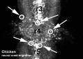
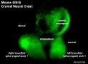
- Modern - Represent the different expression levels of Hox genes and levels of neural crest migration.

|

|
| Historic image of embryonic rhombomeres | Hindbrain neural crest migration |

Hox-proteins crania-caudal expression (species comparison)
Neural Layers
- Ventricular Germinal Zone (VGZ) - mitosis at the ventricular luminal surface, produces early-generated macroneurons
- Subventricular Zone (SVZ) - mitosis away from the ventricular surface, produces later-generated microneurons and glia
Brain

|

|
| Human Embryo developing head cross section (Week 8, Stage 22) | Detail of developing cortex (shown in blue box) |
- Neural progenitor cells migrate from the ventricular layer along radial glia.
- Cortex layers develops inside (first) outside (last)
- Glial progenitor cells develop later from the same ventricular stem cells.
Spinal Cord
- Similar processes to those described for brain.
- Remember notochord ventral patterning by SHH and dorsal ectoderm (dorsalisation).
- Identify the different regions within the neural tube (floor plate, basal plate, alar plate, roof plate)

|

|
| Stage 13 | Stage 22 |
| Half of a transverse section of the spinal cord | ||
|---|---|---|

|

|

|
| Human embryo of 18.5 mm (7.5 weeks). | Human embryo of 24 mm (8.5 weeks). | Human fetus of about 3 months. |
| Wilhelm His (1831-1904) |
Ventricular Development

- The ventricular system develops from the single cavity formed from the hollow neural tube.
- This fluid-filled space is separated from the amnion following fusion of the neural tube and closure of neuropores.
- At different regions sites within the wall (floor of lateral ventricle and roof of the third and fourth ventricles) differentiate to form choroid plexus a modified vascular structure which will produce Cerebrospinal fluid (CSF)
- choroid plexus is a modified vascular structure which will produce Cerebrospinal fluid (CSF)

(FYI - you do not need to know detailed stage development)
| Human Ventricular Development Timeline |
|---|
Data from O'Rahilly R, Müller F., 1990[1] |
Cranial Nerves

Historic diagram showing the relationship of the Cranial Nerves to the Primitive Segments of the Head.
Fetal Neural

Timeline of events in Human Neural Development

|
During the fetal period there is ongoing growth in size, weight and surface area of the brain and spinal cord. Microscopically there is ongoing: cell migration, extension of processes, cell death and glial cell development.
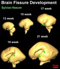
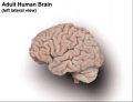
Cortical maturation (sulcation and gyration) and vascularization of the lateral surface of the brain starts with the insular cortex (insula, insulary cortex or insular lobe) region during the fetal period. This cerebral cortex region in the adult brain lies deep within the lateral sulcus between the temporal lobe and the parietal lobe.
Insular Gyral and Sulcal Development
(Data from[2])
(Text - modified from [3])
|


|

|

|
| Human brain at three months (median sagittal section) | Human brain at four months (inferior surface) | Human brain at five months (outer surface) |
Thyroid System and Neural Development

Timeline of human thyroid system and brain development from conception to birth.[4] (Estimation of neurogenesis adapted from Bayer et al.[5])
Postnatal
References
Historic Embryology
| Historic Disclaimer - information about historic embryology pages |
|---|
 |
- Chapter XV. The Brain and Spinal Cord Human Embryology and Morphology. Keith, A. (1902) London: Edward Arnold.
- Contributions to Embryology Carnegie Institution No.59 Relative Weight and Volume of the Component Parts of the Brain of the Human Embryo at Different Stages of Development. Jenkins, G.B. (1921). pp5-54.
Images
| Text-Book of Embryology - 1921 |
|---|
Bailey, F.R. and Miller, A.M. (1921). Text-Book of Embryology. New York: William Wood and Co.
|


| Anatomy of the Human Bod - 1918 |
|---|
- 2015 Course: Week 2 Lecture 1 Lecture 2 Lab 1 | Week 3 Lecture 3 Lecture 4 Lab 2 | Week 4 Lecture 5 Lecture 6 Lab 3 | Week 5 Lecture 7 Lecture 8 Lab 4 | Week 6 Lecture 9 Lecture 10 Lab 5 | Week 7 Lecture 11 Lecture 12 Lab 6 | Week 8 Lecture 13 Lecture 14 Lab 7 | Week 9 Lecture 15 Lecture 16 Lab 8 | Week 10 Lecture 17 Lecture 18 Lab 9 | Week 11 Lecture 19 Lecture 20 Lab 10 | Week 12 Lecture 21 Lecture 22 Lab 11 | Week 13 Lecture 23 Lecture 24 Lab 12 | 2015 Projects: Three Person Embryos | Ovarian Hyper-stimulation Syndrome | Polycystic Ovarian Syndrome | Male Infertility | Oncofertility | Preimplantation Genetic Diagnosis | Students | Student Designed Quiz Questions | Moodle page
Glossary Links
- Glossary: A | B | C | D | E | F | G | H | I | J | K | L | M | N | O | P | Q | R | S | T | U | V | W | X | Y | Z | Numbers | Symbols | Term Link
Cite this page: Hill, M.A. (2024, May 21) Embryology Lecture - Neural Development. Retrieved from https://embryology.med.unsw.edu.au/embryology/index.php/Lecture_-_Neural_Development
- © Dr Mark Hill 2024, UNSW Embryology ISBN: 978 0 7334 2609 4 - UNSW CRICOS Provider Code No. 00098G