File:Stage12 SEM3.jpg
From Embryology
No higher resolution available.
Stage12_SEM3.jpg (507 × 600 pixels, file size: 68 KB, MIME type: image/jpeg)
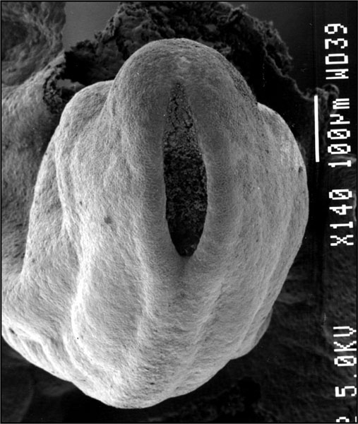
Human Embryo Caudal Neuropore (Carnegie stage 12)
This is an SEM image of the human embryo (Carnegie stage 12, week 4) caudal end.
The neural tube is shown still open at the caudal neuropore (posterior neuropore).
Neural
SEM Images
- Stage 12 SEM Images: Bright Field 1 | Bright Field 3 | Bright Field 3 | SEM1 | SEM2 | SEM3 | SEM4 dorsolateral head and arches | SEM5 lateral head and arches | SEM6 ventrolateral head and arches | SEM7 lateral | SEM8 ventrolateral | SEM9 cloacal membrane | SEM9 labeled | Carnegie stage 12
Stage 12
- Carnegie Stages: 1 | 2 | 3 | 4 | 5 | 6 | 7 | 8 | 9 | 10 | 11 | 12 | 13 | 14 | 15 | 16 | 17 | 18 | 19 | 20 | 21 | 22 | 23 | About Stages | Timeline
| Week: | 1 | 2 | 3 | 4 | 5 | 6 | 7 | 8 |
| Carnegie stage: | 1 2 3 4 | 5 6 | 7 8 9 | 10 11 12 13 | 14 15 | 16 17 | 18 19 | 20 21 22 23 |
Reference
Image Source: Scanning electron micrographs of the Carnegie stages of the early human embryos are reproduced with the permission of Prof Kathy Sulik, from embryos collected by Dr. Vekemans and Tania Attié-Bitach. Images are for educational purposes only and cannot be reproduced electronically or in writing without permission.
File history
Click on a date/time to view the file as it appeared at that time.
| Date/Time | Thumbnail | Dimensions | User | Comment | |
|---|---|---|---|---|---|
| current | 15:43, 10 August 2009 | | 507 × 600 (68 KB) | MarkHill (talk | contribs) | Original file name: Stage12semneuropore.jpg |
You cannot overwrite this file.
File usage
The following 22 pages use this file:
- 2009 Lecture 6
- 2010 BGD Lecture - Development of the Embryo/Fetus 1
- 2010 BGD Lecture - Development of the Embryo/Fetus 2
- 2010 BGD Practical 6 - Week 4
- 2010 Lab 3
- 2010 Lecture 6
- 2011 Lab 3 - Week 4
- ANAT2341 Lab 3 - Week 4
- Abnormal Development - Thalidomide
- BGDA Lecture - Development of the Embryo/Fetus 1
- BGDA Lecture - Development of the Embryo/Fetus 2
- BGDA Lecture - Development of the Nervous System
- BGDA Practical 7 - Week 4
- C
- Carnegie stage 12
- Ectoderm
- Human Embryo SEM
- K12 Thalidomide
- Neural System Development
- Talk:2011 Lab 3
- Talk:BGDA Lecture - Development of the Nervous System
- Template:Carnegie stage 11-14 image table