File:Heart outflow tract stage 14 03.jpg: Difference between revisions
(Z8600021 uploaded a new version of File:Heart outflow tract stage 14 03.jpg) |
mNo edit summary |
||
| Line 1: | Line 1: | ||
==Heart Outflow Tract (Carnegie Stage 14) EFIC== | |||
[[Episcopic_Fluorescence_Image_Capture|EFIC images]] are from an episcopic data set prepared from a human embryo at [[Carnegie stage 14|Carnegie stage 14]], representing the end of the [[Week 5|fifth week]] of development. | |||
'''B''' cut in frontal fashion at the junction of the outflow tract with the pharyngeal mesenchyme, shows the origins of the pharyngeal arch arteries from the aortic sac. The back wall of the sac, shown by the star, is the putative aortopulmonary septum. The two single-headed arrows show the extent of the pericardial cavity. | |||
:'''Links:''' [[:File:Heart outflow tract stage 14 01.jpg|Image EFIC outflow tract frontal]]] | [[:File:Heart outflow tract stage 14 03.jpg|Image EFIC - OFT RA RV]] | [[:File:Heart outflow tract stage 14 03.jpg|Image EFIC - OFT RA LA]] | [[Carnegie stage 14]] | [[Week 5]] | [[Episcopic_Fluorescence_Image_Capture]] | |||
{{Heart Links}} | |||
===Reference=== | |||
<pubmed>27587491</pubmed> | |||
https://www.ncbi.nlm.nih.gov/pmc/articles/PMC5011314/ | |||
http://journals.sagepub.com/doi/abs/10.1177/2150135116651114 | |||
PMID 27587491 | |||
====Copyright==== | |||
© The Author(s) 2016 | |||
https://creativecommons.org/licenses/by/3.0/ | |||
Figure 2. cropped and resized. | |||
{{Footer}} | |||
[[Category:Cardiovascular]][[Category:Heart]][[Category:Carnegie Stage 14]][[Category:Week 5]][[Category:EFIC]] | |||
Revision as of 12:05, 29 January 2017
Heart Outflow Tract (Carnegie Stage 14) EFIC
EFIC images are from an episcopic data set prepared from a human embryo at Carnegie stage 14, representing the end of the fifth week of development.
B cut in frontal fashion at the junction of the outflow tract with the pharyngeal mesenchyme, shows the origins of the pharyngeal arch arteries from the aortic sac. The back wall of the sac, shown by the star, is the putative aortopulmonary septum. The two single-headed arrows show the extent of the pericardial cavity.
- Links: Image EFIC outflow tract frontal] | Image EFIC - OFT RA RV | Image EFIC - OFT RA LA | Carnegie stage 14 | Week 5 | Episcopic_Fluorescence_Image_Capture
Reference
<pubmed>27587491</pubmed>
https://www.ncbi.nlm.nih.gov/pmc/articles/PMC5011314/
http://journals.sagepub.com/doi/abs/10.1177/2150135116651114
PMID 27587491
Copyright
© The Author(s) 2016
https://creativecommons.org/licenses/by/3.0/
Figure 2. cropped and resized.
Cite this page: Hill, M.A. (2024, May 21) Embryology Heart outflow tract stage 14 03.jpg. Retrieved from https://embryology.med.unsw.edu.au/embryology/index.php/File:Heart_outflow_tract_stage_14_03.jpg
- © Dr Mark Hill 2024, UNSW Embryology ISBN: 978 0 7334 2609 4 - UNSW CRICOS Provider Code No. 00098G
File history
Click on a date/time to view the file as it appeared at that time.
| Date/Time | Thumbnail | Dimensions | User | Comment | |
|---|---|---|---|---|---|
| current | 12:00, 29 January 2017 | 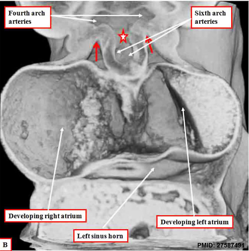 | 989 × 996 (134 KB) | Z8600021 (talk | contribs) | |
| 12:00, 29 January 2017 | 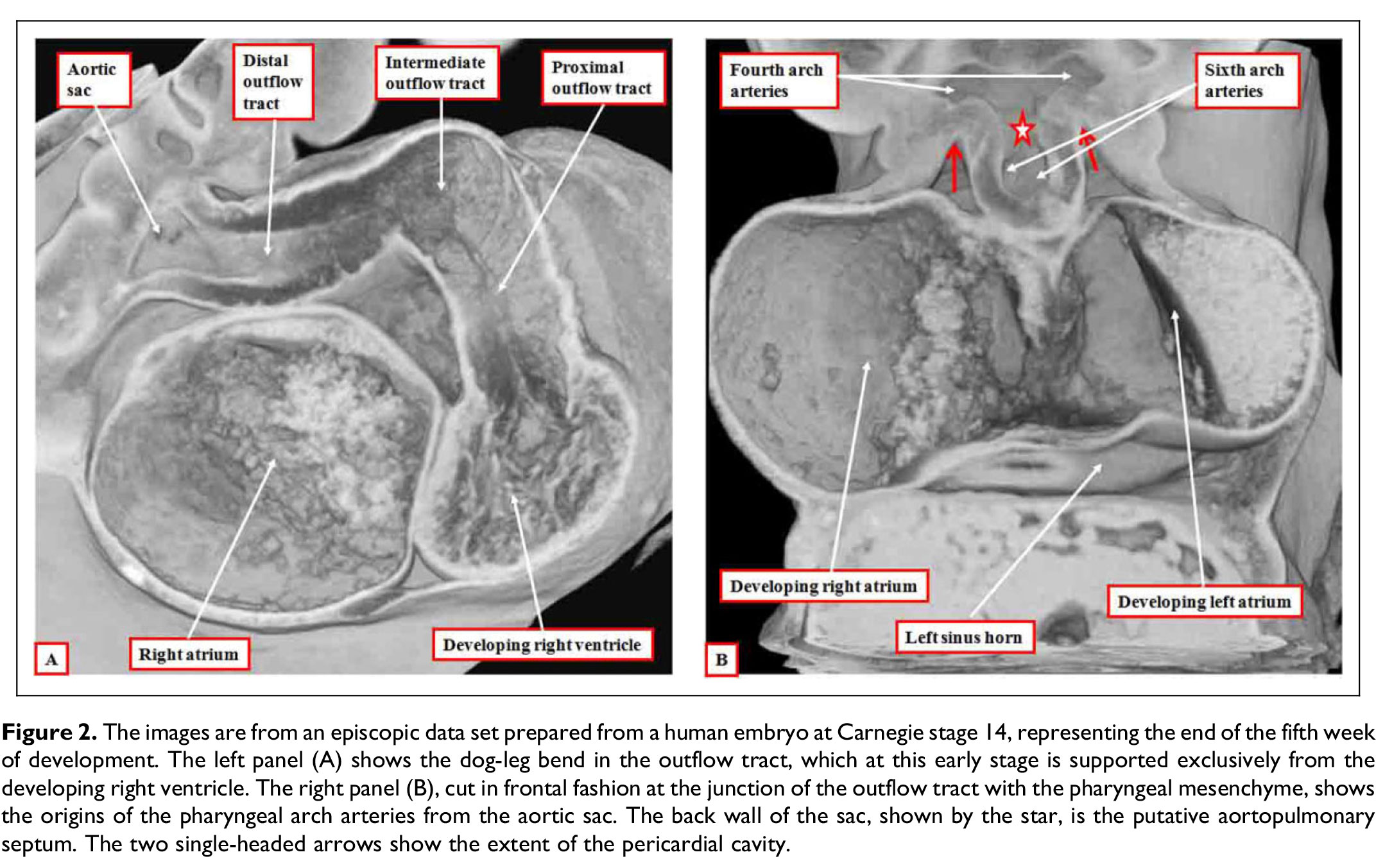 | 2,144 × 1,353 (421 KB) | Z8600021 (talk | contribs) |
You cannot overwrite this file.
File usage
The following page uses this file: