Developmental Signals - Hippo

From Embryology
Revision as of 09:28, 10 June 2015 by Z8600021 (talk | contribs) (Created page with "{{Header}} ==Introduction== The Hippo pathway, first identified in Drosophila, controls organ size by regulating cell proliferation and apoptosis. {{Factor Links}}| :Categ...")
(diff) ← Older revision | Latest revision (diff) | Newer revision → (diff)
Embryology - 27 Apr 2024    Facebook link Pinterest link Twitter link  Expand to Translate  
Google Translate - select your language from the list shown below (this will open a new external page)

العربية | català | 中文 | 中國傳統的 | français | Deutsche | עִברִית | हिंदी | bahasa Indonesia | italiano | 日本語 | 한국어 | မြန်မာ | Pilipino | Polskie | português | ਪੰਜਾਬੀ ਦੇ | Română | русский | Español | Swahili | Svensk | ไทย | Türkçe | اردو | ייִדיש | Tiếng Việt    These external translations are automated and may not be accurate. (More? About Translations)

Introduction

The Hippo pathway, first identified in Drosophila, controls organ size by regulating cell proliferation and apoptosis.


Factor Links: AMH | hCG | BMP | sonic hedgehog | bHLH | HOX | FGF | FOX | Hippo | LIM | Nanog | NGF | Nodal | Notch | PAX | retinoic acid | SIX | Slit2/Robo1 | SOX | TBX | TGF-beta | VEGF | WNT | Category:Molecular

| Category:Retinoic acid

Some Recent Findings

  • Retinoic acid signaling and neuronal differentiation[1] "The identification of neurological symptoms caused by vitamin A deficiency pointed to a critical, early developmental role of vitamin A and its metabolite, retinoic acid (RA). The ability of RA to induce post-mitotic, neural phenotypes in various stem cells, in vitro, served as early evidence that RA is involved in the switch between proliferation and differentiation. In vivo studies have expanded this "opposing signal" model, and the number of primary neurons an embryo develops is now known to depend critically on the levels and spatial distribution of RA. The proneural and neurogenic transcription factors that control the exit of neural progenitors from the cell cycle and allow primary neurons to develop are partly elucidated, but the downstream effectors of RA receptor (RAR) signaling (many of which are putative cell cycle regulators) remain largely unidentified. The molecular mechanisms underlying RA-induced primary neurogenesis in anamniote embryos are starting to be revealed; however, these data have been not been extended to amniote embryos. There is growing evidence that bona fide RARs are found in some mollusks and other invertebrates, but little is known about their necessity or functions in neurogenesis. One normal function of RA is to regulate the cell cycle to halt proliferation, and loss of RA signaling is associated with dedifferentiation and the development of cancer. Identifying the genes and pathways that mediate cell cycle exit downstream of RA will be critical for our understanding of how to target tumor differentiation. Overall, elucidating the molecular details of RAR-regulated neurogenesis will be decisive for developing and understanding neural proliferation-differentiation switches throughout development."
More recent papers
Mark Hill.jpg
PubMed logo.gif

This table allows an automated computer search of the external PubMed database using the listed "Search term" text link.

  • This search now requires a manual link as the original PubMed extension has been disabled.
  • The displayed list of references do not reflect any editorial selection of material based on content or relevance.
  • References also appear on this list based upon the date of the actual page viewing.


References listed on the rest of the content page and the associated discussion page (listed under the publication year sub-headings) do include some editorial selection based upon both relevance and availability.

More? References | Discussion Page | Journal Searches | 2019 References | 2020 References

Search term: Embryo Retinoic acid | Images

<pubmed limit=5>Embryo Retinoic acid</pubmed>

Organ Expression

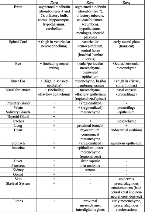
Rar gene expression.jpg

Summary of Rar gene expression patterns in mouse developing organ systems.[2]

Endoderm

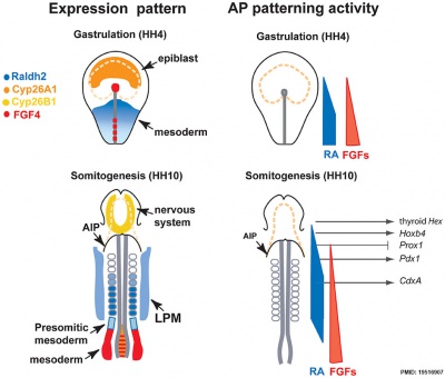
Chicken antero-posterior endoderm patterning cartoon

Chicken antero-posterior endoderm patterning[3]


Links: Endoderm | Chicken Development

Fetal Gonad

Human fetal gonad retinoid receptor expression.jpg

Immunohistochemical localisation of retinoid receptor expression in the human fetal gonad[4]


Neural

Model retinoic acid extracellular signal-regulated kinase and Wnt pathway interactions.jpg

Model retinoic acid extracellular signal-regulated kinase and Wnt pathway interactions[5]

Mouse embryo cortex radial expansion.jpg

RA Is Not Required for Radial Expansion of the Embryonic Cortex

References

  1. <pubmed>25558812</pubmed>
  2. <pubmed>19471585</pubmed>
  3. 19516907</pubmed>| PLoS One.
  4. Childs AJ, Cowan G, Kinnell HL, Anderson RA, Saunders PTK (2011) Retinoic Acid Signalling and the Control of Meiotic Entry in the Human Fetal Gonad. PLoS ONE 6(6): e20249. PMID 21674038 | PLoS One
  5. <pubmed>19642999</pubmed>


Articles

<pubmed>21673209</pubmed>| Can Fam Physician

Search Pubmed

Search Bookshelf Retinoic acid


Search Pubmed Now: Retinoic acid


Glossary Links

Glossary: A | B | C | D | E | F | G | H | I | J | K | L | M | N | O | P | Q | R | S | T | U | V | W | X | Y | Z | Numbers | Symbols | Term Link


Cite this page: Hill, M.A. (2024, April 27) Embryology Developmental Signals - Hippo. Retrieved from https://embryology.med.unsw.edu.au/embryology/index.php/Developmental_Signals_-_Hippo

What Links Here?
© Dr Mark Hill 2024, UNSW Embryology ISBN: 978 0 7334 2609 4 - UNSW CRICOS Provider Code No. 00098G