Book - The comparative anatomy of the nervous system of vertebrates including man 1
| Embryology - 27 Apr 2024 |
|---|
| Google Translate - select your language from the list shown below (this will open a new external page) |
|
العربية | català | 中文 | 中國傳統的 | français | Deutsche | עִברִית | हिंदी | bahasa Indonesia | italiano | 日本語 | 한국어 | မြန်မာ | Pilipino | Polskie | português | ਪੰਜਾਬੀ ਦੇ | Română | русский | Español | Swahili | Svensk | ไทย | Türkçe | اردو | ייִדיש | Tiếng Việt These external translations are automated and may not be accurate. (More? About Translations) |
Kappers CUA. Huber GC. and Elizabeth C. Crosby EC. The comparative anatomy of the nervous system of vertebrates including man Volume I. (1936) The Macmillan Company, New York.
| Historic Disclaimer - information about historic embryology pages |
|---|
 |
The Comparative Anatomy of the Nervous System of Vertebrates including Man - Volume I
By
C. U. Ariens Kappers, M.D., Sc.D., Ll.D.
Director of The Central Institute Of Brain Research, Amsterdam, And Professor Op Comparative Neurology In The University Op Amsterdam
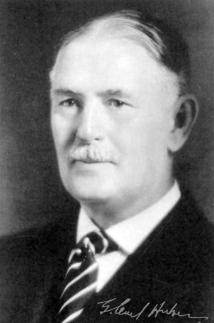
G. Carl Huber, M.D., Sc.D.
Late Dean Of The Qhaddate School, Director Op The Anatomical Laboratories, And Professor Of Anatomy In The University Op Michigan
And
Elizabeth Caroline Crosby, Ph.D.
Associate Professor Of Anatomy In The University Op Michigan
Template page only.
Dedicated to Charles Judson Herrick, Great Pioneer In American Comparative Neurology
Preface
“ I do the very best I know how; the very best I can ; and I mean to keep doing so until the end. If the end brings me out all right, what is said against me won’t amount to anything. If the end brings me out wrong, ten angels swearing I was right would make no difference.”
Abraham Lincoln
The quotation given above hung over the desk of Dean G. Carl Huber, and was to him undoubtedly an inspiration for that intellectual independence which was so characteristic of him and which was so inspiring to those who collaborated with him in this book as well as in other fields. Without his wide range of knowledge in the sphere of neurological research, accumulated over many years from his early work on nerve repair in 1892 to the time of his death, and his great editorial skill, the result of many years of editorship of scientific journals and articles, it would have been quite impossible to prepare for publication and to have accepted, under present conditions, a text so technical and so large as is the present book. Through the years of preparation of this book it was his enthusiasm, his industry, his unfailing optimism, which carried the task on to completion. It is a matter of the greatest regret to both his collaborators and to the publishers that, while he saw the manuscript completed, he should not have lived to see, in published form, this work, which is so largely a product of his scholarship and inspiration.
This book is an outgrowth of “Die vergleichende Anatomie des Nervensystems,” which was written by C. U. Ariens Kappers in 1920-1921. When, in 1926, the present text was begun, the original plan on the part of both Dutch and American collaborators was to translate the German text into English and to make such revision as should be deemed necessary in the fight of later literature. However, so great additions had been made to the knowledge of the comparative anatomy of the nervous system during the years following its publication that it soon became evident that, in order to do justice to the situation, much of the text must be entirely rewritten, and additions made to the figures. The idea, then, of presenting a translation was abandoned. Thus, with the exception of certain portions of the text, such, for example, as the introduction and the pages on gyri and sulci, the present book offers a new presentation of the material available in comparative neurology, based on the available literature and on results of the research programs of the Institute of Brain Research at Amsterdam and the Laboratory of Comparative Neurology at the University of Michigan, both of which have collaborated in the preparation of the present text.
This collaboration has been so complete that it would be difficult to assign exclusive credit to either laboratory for any special portion. Mention should be made, however, of certain fields which have received attention from one laboratory or the other.
Foremost among the contributions from the Institute of Brain Research at .\msterdam are the framework of plan and material provided by the German edition and the thorough revision of the presentation of the theory of neurobiotaxis, written by the Dutch collaborator. Certain facts and figures with regard to the newer work on efferent cranial nerve nuclei and their roots were made available through the work of Dr. J. L. Addens of the Institute. The authors wsh here to express their thanks to Dr. Addens for his kindness in permitting the use of his figures and for suggestions with regard to this portion of the text. Some new figures have been added and cliches provided for the figures used from the German text by the laboratory at Amsterdam.
The chapters on diencephalon and telencephalon, particularly those portions dealing with these regions in reptiles, birds, and mammals, have received particular attention from the Laboratory of Comparative Neurology of the Anatomy Department at Michigan. The material presented concerning these regions includes the results of published and, to some extent, unpublished work along these lines, derived from the research programs of the American authors and of others associated in this laboratory, now or formerly, as staff or students. Also, from the work of staff members at Michigan, additions have been made to the accounts of the vestibular centers of reptiles, to trigeminal and facial centers of various vertebrates, and to the spinal cord of birds. The sources of any such information used, whether published or unpublished, are credited in the text, but especial mention is made here of the account of the marginal nuclei of birds, which was written by Dr. J. F. Huber. To these associates and colleagues who have so generously permitted the use of the results of their work, the authors wish to offer their most sincere thanks. Various figures, partly from published contributions but in many cases drawn especially for this text, have been contributed by the laboratory at Michigan. Other figures have been redrawn or relabeled to meet the requirements of American publishers, most of thus latter work having been done by Dean Huber.
Very greatly appreciated permission to use figures which had appeared in WLtar publications was granted by the Wistar Institute. Through the kindne.'S of Dr. Woollard, Editor of the Journal of Anatomy, a similar permission was granted for the use of certain figures from this English journal. The authors arc also indebted to various investigators in other laboratories for the use of figures or, in a few ca-ses, of unpublished manuscript. It is not possible to mention here all of those to whom thanks arc due, but the sources of all such material u.-cd are credited in each case in the text. However, the authors wish to thank particularly Profe.'ssor C. Judson Herrick for the privilege of examining unpubli.'hed manuscript and figures and Dr. Jeannette Obenchain for certain timely suggestions.
To publish such large a book under present conditions is very difficult. The rights of publication held by the Dutch firm of de Erven F. Bohn were mo.st kindly turned over by this firm to the American publishers. It is quite impt. 'ible for the authors to express adequately their sense of deep obligation to The Macmillan Company for the unbounded courtesy, help, and encouragement which have made possible the completion of this book under conditions which have been percularly trying. No effort has been spared in any particular on the part of the representatives of this company to make this book all that it should be.
The correcting of the proof and the preparation of the subject index and author index were, of necessity, handled at the University of Michigan. In these fields the younger members of the Department offered greatly needed and greatly appreciated help. Dr. Tryphena Humphrey, Dr. Jean Weston, Dr. Russell Woodburne, and Dr. John Barnard, members of the staff of the Anatomy Department, and the secretaries. Miss Catherine Brook and Miss Elizabeth Switzer, all helped in making the prehminary drafts for the indexes. Dr. Weston, Dr. Humphrey, and Miss Switzer helped not only in typing the manuscript but in checking for error in the bibhographies and proof and in the final preparation of these indexes. Those who have attempted to edit manuscript on the scale of that presented in this book can appreciate how heartfelt the gratitude is to these younger people.
Almost ten years have passed since the book was begun, and now that the task is completed it represents only in small part the ideal that its authors were trying to attain. If, however, it in any way aids some of those who are working in this field to a better comprehension of the problems involved in the study of the nervous system and the possibilities offered by such study, the authors will feel that their labor has been well repaid.
C. U. Ariens Kappers Elizabeth C. Crosby
Introduction
The nervous system associates impulses arising within the body with those due to surface stimulations and prqvides appropriate effectory paths. It associates and integrates impulses from neuromuscular and neurotendinous endings and the semicircular canals (proprioceptive impulses of Sherrington, ’06) yet in such fashion that within certain limits the various components remain recognizable, each retaining its specific character. In similar ways it associates various stimuli from the outside world (exteroceptive impulses) and interrelates those received from various organs (interoceptive or visceral afferent impulses). Finally, it coordinates and integrates exteroceptive, proprioceptive, and interoceptive impulses into a dynamic, effective whole.
It is customary and correct to divide the constituents of the nervous system into nervous and non-nervous elements. To the former belong those elements, usually termed neurons, which serve the functions of reception (sensitivity), conduction, and integration of stimuli, all of which, but most particularly the last, give the nervous system its special significance. These various functions are characteristic of living substance in general and the protoplasm of cells, other than neurons, does not lack such properties. The protoplasm of unicellular forms has been shown to be capable not only of receiving stimuli, but also of conducting such stimuli to remote portions of the cell body and there instigating movements. In these unicellular animals, different impulses occurring at the same time may reenforce or inhibit each other. Also, in such simple multicellular forms as the sponges, which as yet have not developed a nervous system, conductivity is evident, in addition to the sensitivity of the body cells. This does not confine itself to the body of the stimulated cell but is intercellular, passing from one cell to another over protoplasmic intercellular bridges which make possible, not only the diffusion of single impulses but the correlation of various impulses occurring at the same time. Such intercellular conductivity, by means of intercellular protoplasmic bridges instead of through a nervous system, has been found in certain amphibian embryos (Schaper, ’98 ; Goldstein, ’04; Wintrebert, ’04) and is present also in smooth muscle tissue. It is only the enhancement of these previously mentioned functions which characterizes a neuron.
Another general characteristic of living substance {Hering, ’70; Butler, ’80; Laycock, ’75; Semon, ’ll), which is particularly outstanding in relation to the nervous system, is an internal power for setting up stimuli (engram formation). This is the ability to continue for a longer time than the impulse itself persists, the conditions produced by it. That such an ability is more characteristic of neurons than of other cells remains to be substantiated ; indeed the facts suggest the contrary to be true. This inherent power for the retention of the results of stimulation is merely more evident in the nerve cells because in them these results are frequently a part of conscious processes. This inherent ability for retention is the organic basis for memory (better ekphoria), becoming functional through the conduction and correlation of impulses. Under the term memory is implied that attribute by which sensations which are latent come to expression again through impulses which arouse the earlier sensation, either directly through new perceptions or indirectly through associations with previous sensations. This ability to remember is more particularly characteristic of the nervous system than of other parts of the body and its ekphoria has the further characteristic that it may disappear from consciousness without losing the engram.
Not only memory, but also attention and association, so characteristic of nervous functions, may be considered as characteristic of protoplasm as well. Attention is the tendency to concentrate on one function, a process evident in the specific development of other than nervous tissues, while association or correlation is observed in the functional relations of tissues such as tendon, bone, or muscle, or of organs and organ systems such as the gastrointestinal and respiratory systems.
In the nervous system, as in all living substance, there is an active striving, an inherent tendency, to supplement the activities and to elaborate the various impulses, which is recognizable chiefly through the end results, but which may be felt consciously as an indeterminate realization of the force of thought and will. The results of this entelechic* tendency (see p. 5a) are seen in the progressive development of the brain in accordance with a general plan, in the progressive differentiation and adjustment of its constituents, and in their mutual general relations. The entrance of more or less heterogeneous sensations and their correlation and adequate expression through efferent responses leads to a continually increasing and finely adapted consolidation in which the correlation of the various exteroceptive, proprioceptive, and even interoceptive impulses plays a large part. The development of the cortex in particular presents a continually increasing and finely adapted association and control of proprioceptive and exteroceptive impulses.
This development of a central nervous system is preceded by the formation of regions of particular sensitivity (the sense organs), which further the perception of those stimuli which relate the individual to the outside world. Among these, the perceptions of the so-called"" vital ” or protopathic impulses develop somewhat earlier than those of the more objective “gnostic” or epicritic sensations. This is to be expected, since the former are more .subjective and are largely protective, being concerned with impulses harmful or useful to the body, while the more objective gnostic or epicritic sensibility increases the knowledge of the outside world.
The impulses coming into the nervous system become effective through efferent nerve paths which provide an outlet for them. Yet not all afferent impulses are followed by motor responses. The interplay of the various incoming impulses may result merely in a condition of central equilibrium or correlation, so that there is no discharge over efferent paths, or a discharge may be initiated which is inhibitory in its effects, or merely gnostic in character in that it adds to the knowledge of the environment.
No teleological explanation of the development of the nervous system was intended in the reference to the inner urge or development and expression characteristic of living matter. The nervous system has not developed during phylogeny with the brain of man as its fixed pattern or goal. Evolution as a whole has occurred without molding itself to a form established in anticipation of the progressive development. The ape-like progenitor of man did not have in mind the characteristic human form as a pattern wliich he should imitate. This may be expressed by saying that the development here is entelechic rather than teleologic, for each stage is an end within itself — not a step toward a well known, predetermined goal.
- The expression " entelpphy ” is not used here as a property apart from the organism, superimposed upon it but a« an inherent organic property.
Contents
- The Evolution And Morphology Of Nervous Elements
- The Structure of Neurons and Their Processes
- Synaptic Relations between Neurons
- The Connections of Nerves with Other Tissues
- Sensory Endings
- Motor and Other Effectory Endings
- The Ectodersial Supporting Tissue of the Central Nervous System
- The Mesodermal Supporting Tissue of the Central Nervous System
- Supporting Elements of the Peripheral Nervous System
- The Ectodermal Supporting Tissue of the Peripheral Nerves
- The Mesodermal Supporting Tissue of the Peripheral Nerves
- Structural Laws of the Nervous System
- The Principles of Neurobiotaxis
- Monoaxonism and Polydendritism
- Selectivity in the Connections of Neurons
- Review and Conclusions
- BIBLIOGRAPHY
- The Comparative Anatomy Of The Spinal Cord
- The Brain and Spinal Cord of Amphioxus
- The Spinal Cord of Cyclostojies
- The Spinal Cord of Plagiostomes
- The Spinal Cord of Ganoids and Teleosts
- The Spinal Cord of Amphibians
- The Spinal Cord of Reptiles
- The Spinal Cord of Birds
- The Spinal Cord of Mammals
- Extent and Gross Morphology of the Spinal Cord
- Dorsal and Ventral Root Fibers
- Somatic Efferent Centers : Their Arrangement and the Distribution of Their Fibers
- The Visceral Efferent Tenters
- The Sympathetic System
- The Central Location of Preganglionic Neurons
- The Relations and Certain Functions of the Sympathetic System. Muscle Tonus
- The Primary Neurons of the Visceral Afferent System
- The Primary Neurons of the Somatic Afferent System
- The Spinal Ganglia 243
- The Distribution of the Spinal Ganglion Cell Dendrites to the Skin
- The Secondary Centers within the Cord
- Secondary Ascending Paths
- Paths within the Cord for Exteroceptive Pain and Temperature
- Paths within the Cord for Tactile Impulses and Proprioceptive Paths of the Dorsal Funiculi
- The Bulbar Terminations of the Tactile and Proprioceptive Paths of the Dorsal Funiculi
- The Proprioceptive Paths in the Lateral Funiculus of the Cord
- Paths within the Cord for Visceral Afferent Impulses
- Paths Descending into the Cord
- Size, Growth, and Chemical Differentiation of the Spinal Cord
- Review of the Organization and Progressive Development of the Spinal Cord
- BIBLIOGRAPHY
- The Medulla Oblongata
- Taste and the General Afferent Systems
- The Structure, Distribution, and Number of Taste Buds in Vertebrates
- Chemical Sense and Taste
- The Peripheral and Central Relations of the Afferent Components of the Vagus, Glossopharyngeal, and Facial Nerves
- Discussion of the Peripheral Taste Paths in Relation to the Trigeminus
- The Sensory Components of the Trigeminal Nerve
- Review of the Development of the Branchial Nerves and of Their Afferent Connections 406
- BIBLIOGRAPHY 412
- The Lateral Line And Acoustic Systems
- The Acoustico-lateral Systems of Cyclostomes
- The Acoustico-lateral Systems of Plagiostombs
- The Acoustico-lateral Systems of Ganoids and Teleosts
- The Lateral Line and Acoustic Systems op Amphibians
- The Vestibular and Cochlear Systems of Reptiles
- The Vestibular and Cochlear Systems of Birds
- The Vestibular and Cochlear Systems of Mammals
- Review of the Lateral Line and Acoustic Systems
- BIBLIOGRAPHY
- The Effectory System Of The Midbrain And The Medulla Oblongata
- Phylogeny of Cranial Efferents
- The Effectory System of Cyclostomes
- The Effectory System of Plagiostomes
- The Effectory System of the Ganoids and Teleosts
- The Effectory System of Amphibians
- The Effectory System of Reptiles
- The Effectory System in Birds
- The Effectory System of Mammals
- Resume of the Effectory System of the Medulla Oblongata and Midbrain
- BIBLIOGRAPHY
- The Coordinating Apparatus
- The Reticular Centers of the Medulla Oblongata and the Midbrain
- Resume of the Relations of the Reticular Cells op the Medulla Oblongata AND Midbrain
- Certain Other Coordinating Systems of the Medulla Oblongata
- The Inferior Ouvary Nucleus
- Review of the Phylogenetic Development of the Inferior Olive
- BIBLIOGRAPHY
Cite this page: Hill, M.A. (2024, April 27) Embryology Book - The comparative anatomy of the nervous system of vertebrates including man 1. Retrieved from https://embryology.med.unsw.edu.au/embryology/index.php/Book_-_The_comparative_anatomy_of_the_nervous_system_of_vertebrates_including_man_1
- © Dr Mark Hill 2024, UNSW Embryology ISBN: 978 0 7334 2609 4 - UNSW CRICOS Provider Code No. 00098G