Category:Cardiovascular: Difference between revisions
From Embryology
No edit summary |
mNo edit summary |
||
| Line 2: | Line 2: | ||
See also the narrower categories [[:Category:Heart]] and [[:Category:Blood]]. | See also the narrower categories [[:Category:Heart]] and [[:Category:Blood]]. | ||
{{Heart Links}} | |||
[[Category:System Development]] | [[Category:System Development]] | ||
Revision as of 12:39, 13 February 2017
This is a direct link to pages, images and media related to cardiovascular system development.
See also the narrower categories Category:Heart and Category:Blood.
Subcategories
This category has the following 13 subcategories, out of 13 total.
Pages in category 'Cardiovascular'
The following 200 pages are in this category, out of 519 total.
(previous page) (next page)H
- HM Practical - Blood Vessel Histology
- HM Practical - Cardiac Histology
- Template:Hofbauer cell
- Template:Hofbauer cells
- Template:Human Embryology 2 18-6 Dorsal segmental artery table1
- Template:Human Embryology 2 18-6 Dorsal segmental artery table2
- Template:Human Embryology 2 18-6 Mesonephric artery table3
- Human System Development
- Template:Hypoplastic left heart
- Hypoplastic Left Heart Syndrome Movie
I
- Template:ICD-10-circulatory system Q20-Q28 table
- Template:Immune
- Intermediate - Atrial Ventricular Septation
- Intermediate - Cardiac Abnormalities
- Intermediate - Heart Tube Looping
- Intermediate - Heart Tube Looping 2
- Intermediate - Heart Tube Looping 3
- Intermediate - Heart Valves
- Intermediate - Outflow Tract
- Intermediate - Primordial Heart Tube
- Intermediate - Vascular Overview
- Intermediate Cardiac Embryology
- Template:Intermediate Cardiac menu
L
M
P
- Paper - A case of early ectopic gestation
- Paper - A congenital anomaly of the heart - truncus arteriosus communis (1927)
- Paper - A contribution to the early development of the heart in mammalia, with special reference to the guinea pig
- Paper - A Contribution to the Embryology of the Liver and Vascular System in Man
- Paper - A rare vascular anomaly-opening of the upper left pulmonary vein into a persistent left superior vena cava (1915)
- Paper - A specimen showing complete remains of the left superior vena cava (1915)
- Paper - A study of wandering mesenchymal cells on the living yolk-sac and their developmental products (1915)
- Paper - Coarctation of the aorta 1942
- Paper - Complete situs inversus of the vena cava superior (1930)
- Paper - Congenital deficiency of the pericardium (1916)
- Paper - Cor biloculare, with a note on the development of the pulmonary veins (1937)
- Paper - Development and vascularization of the testis (1906)
- Paper - Development of postcava and tributaries in the domestic cat (1907)
- Paper - Development of the great anterior veins in man and mammalia
- Paper - Development of the human heart from its earliest appearance to the stage found in embryos of twenty paired somites (1927)
- Paper - Development of the inferior vena cava (1929)
- Paper - development of the postcaval vein in birds (1903)
- Paper - Development of the vascular system in five to twenty-one somite dog embryos
- Paper - Developmental Changes in the Pericardium, the Mesocardia, and the Pleural Sacs in the Human Embryo
- Paper - Developmental defects at the foramen ovale (1938)
- Paper - Equivalence of different hematopoietic anlages 1 (1916)
- Paper - First contractions of the heart without cytological differentiation
- Paper - Four cases of anomalous inferior vena cava with an explanation of their developmental origin (1928)
- Paper - Functional limitations of the foramen ovale in the human foetal heart
- Paper - Further evidence on the origin of the lymphatic endothelium from the endothelium of the blood-vascular system
- Paper - General observations on early superficial lymphatics in living chick embryos (1912)
- Paper - Growth allometry of the myocardium in human embryos from stages 15 to 23
- Paper - Hematopoiesis in young human embryos
- Paper - Histogenesis of the heart muscle of the pig in relation to the appearance and development of the intercalated discs (1919)
- Paper - Histogenesis of the human aorta
- Paper - Initiation and early changes in the character of the heart beat in vertebrate embryos
- Paper - Injection and reconstruction of the jugular lymph sac in the chick (1912)
- Paper - Migration processes during ontogeny with reference to the venous development in the dorsal body wall (1946)
- Paper - Normal haemopoiesis in intra-uterine and neonatal life (1941)
- Paper - Observations on the development of the earliest lymphatics in the region of the posterior lymph heart in living chick embryos (1912)
- Paper - Observations upon the occurrence, structure and function of the giant cells of the marrow (1890)
- Paper - On the cervical veins and lymphatics in four human embryos, with an interpretation of anomalies on the subclavian and jugular veins in the adult (1909)
- Paper - On the development of the aortae cardinal and umbilical veins and the other blood vessels of vertebrate embryos from capillaries (1909)
- Paper - On the development of the atrial septum and the valvular apparatus in the right atrium of the pig embryo (1916)
- Paper - On the development of the blood-vessels of the brain in the human embryo (1905)
- Paper - On the Development of the Human Heart
- Paper - On the earliest blood-vessels in the anterior limb-buds of birds and their relation to the primary subclavian artery
- Paper - On the fate of the posterior cardinal veins and their relation to the development of the vena cava and azygos in the embryo pig (1915)
- Paper - On the origin of the abdominal lymphatics in mammals from the vena cava and the renal veins (1912)
- Paper - On the origin of the lymphatic system from the veins and the development of the lymph hearts and thoracic duct in the pig (1902)
- Paper - On the origin of the pulmonary arteries in mammals
- Paper - On the origin of the pulmonary arteries in mammals 2
- Paper - On the placentation of the macaque (Macaca mulatta), from the time of implantation until the formation of the definitive placenta
- Paper - On the position of the vitelline arteries in human embryos
- Paper - On the time of the post-natal obliteration of the fetal blood-passages (1918)
- Paper - Origin and development of the primitive vessels of the chick and of the pig (1917)
- Paper - Origin of the pulmonary vessels in the chick (1922)
- Paper - Origin, development and degeneration of the blood vessels of the ovary (1899)
- Paper - Persistence of the left posterior cardinal vein (1911)
- Paper - Persistent left superior vena cava, left duct of cuvier and left horn of the sinus venosus
- Paper - Persistent left superior vena cava, left duct of cuvier and left horn of the sinus venosus (1930)
- Paper - Preliminary note on the differentiation of angioblasts in the living chick (1917)
- Paper - Significant superficial anastomoses in the arterial blood supply to the human brain (1959)
- Paper - Six specimens of abnormal heart (1912)
- Paper - Some abnormal developments in the vascular system of the frog (rana temporaria) (1915)
- Paper - Some observations on the cardio-vascular system in the viable foetal lamb (1940)
- Paper - Studies on the area vasculosa of the embryo chick 2 (1937)
- Paper - Teratogenecity in the setting of cardiac development and maldevelopment
- Paper - The aortic arch derivatives in human adult (1951)
- Paper - The circle of Willis - An examination of 700 specimens (1905)
- Paper - The course of the blood flow through the fetal mammalian heart
- Paper - The course of the blood through the heart of the fetal mammal, with a note on the reptilian and amphibian circulations (1909)
- Paper - The development of the aorta and aortic arches in rabbits
- Paper - The development of the arteries of the human lower extremity
- Paper - The Development of the Atrio-Ventricular Valves in Man
- Paper - The development of the cardiac loop in the rabbit with especial reference to the bulboventricular groove and origin of the interventricular septum (1919)
- Paper - The development of the cardiac-coronary circulatory system
- Paper - The development of the cranial arteries in the human embryo
- Paper - The development of the cranial venous system in man, from the viewpoint of comparative anatomy
- Paper - The development of the heart in man
- Paper - The development of the mammalian spleen, with special reference to its hematopoietic activity (1921)
- Paper - The Development of the Pars Membranacea Septi in the Human Heart
- Paper - The development of the principal arterial stems in the forelimb of the pig (1922)
- Paper - The development of the pulmonary vein in the domestic cat (1913)
- Paper - The development of the subcutaneous vascular plexus in the head of the human embryo (1923)
- Paper - The development of the vascular system in the human embryo prior to the establishment of the heart
- Paper - The development of the veins in the limbs of rabbit embryos
- Paper - The development of the vena cava inferior (1902)
- Paper - The development of the vena cava inferior in man (1925)
- Paper - The development of the venous sinuses of the dura mater in the human embryo
- Paper - The developmental alterations in the vascular system of the brain of the human embryo (1921)
- Paper - The ductus arteriosus in the human fetus and newborn infant
- Paper - The ductus venosus in the fetus and in the adult (1923)
- Paper - The Earliest Blood-Vessels in Man
- Paper - The earliest stages of development of the blood-vessels and of the heart in ferret embryos
- Paper - The earliest stages of development of the blood-vessels and of the heart in ferret embryos 2
- Paper - The early development of the sheep heart (1946)
- Paper - The early stages of the development of the pericardium
- Paper - The effect of the heart-beat upon the development of the vascular system in the chick (1918)
- Paper - The equivalence of different homatopoietic anlages by method of stimulation of the different stem cells 1
- Paper - The equivalence of different homatopoietic anlages by method of stimulation of the different stem cells 2
- Paper - The fifth aortic arch of mammalian embryos; the nature of the last pharyngeal evagination
- Paper - The first contractions of the heart in rat embryos
- Paper - The form and the functions of the uterine blood vessels in the rhesus monkey
- Paper - The formation of the cardiac loop in the chick
- Paper - The Formation of the Pars Membranacea Septi (1916)
- Paper - The formation of the venous valves, the foramen secundum and the septum secundum in the human heart
- Paper - The frequency of an opening between the right and left auricles at the seat of the foetal foramen ovale (1900)
- Paper - The fusion of the cardiac anlages and the formation of the cardiac loop in the cat (1916)
- Paper - The genesis, development, and adult anatomy of the nasofrontal region in man
- Paper - The genetic interpretation of the development of the mammalian lymphatic system (1908)
- Paper - The genetic principles of the development of the systemic lymphatic vessels in the mammalian embryo (1910)
- Paper - The human embryonic heart in the ninth week
- Paper - The human embryonic heart in the ninth week (1954)
- Paper - The human embryonic heart in the seventh week (1962)
- Paper - The life-history of the formed elements of the blood, especially the red blood corpuscles (1890)
- Paper - The morphology of human uteroplacental vasculature
- Paper - The nerve supply of the mammalian ductus arteriosus (1941)
- Paper - The origin and development of the carotid body (1924)
- Paper - The origin and early development of the posterior lymph heart in the chick (1915)
- Paper - The origin and occurrence of the single umbilical artery in normal and abnormal human fetuses (1922)
- Paper - The origin of blood and vascular endothelium in embryos without a circulation of the blood and in the normal embryo (1915)
- Paper - The origin of blood cells (1916)
- Paper - The origin of the heart and blood vessels in felis domestica (1924)
- Paper - The origin, development and function of the blood cells in certain marine teleosts 1 (1939)
- Paper - The partitioning of the truncus and conus and the formation of the membranous portion of the interventricular septum in the human heart (1942)
- Paper - The phylogenetic relations of the lymphatic and bloodvascular systems in vertebrates (1910)
- Paper - The physiology of the embryonic mammalian heart before circulation
- Paper - The relation between the size of the artery and the capillary bed in the embryo (1937)
- Paper - The relative role played by the embryonic veins in the development of the mammalian vena cava posterior
- Paper - Three examples of a right aortic arch
- Paper - Time and rate of loss of nuclei by the red blood cells of human embryos
- Paper - Transformation of the aortic-arch system during the development of the human embryo (1922)
- Paper - Transposition of the ventricles and the arterial stems (1931)
- Paper - True congenital diverticulum of the trachea in a subject showing also right aortic arch (1929)
- Paper - Two cases considered from the developmental standpoint in which the right subclavian artery arose from the arch of the aorta (1915)
- Paper - Variations and anomalies of the venous valves of the right atrium of the human heart (1929)
- Paper - Wilhelm His - His relation to the institution of learning
- Paper The development of the subcutaneous vascular plexus in the head of the human embryo (1923)
- Paper- The primary divisions of the myocardium in the human embryo
- Template:Patent ductus arteriosus
- Patent Ductus Venosus Movie
- Template:PDGF
- Template:Persistent right umbilical vein
- Template:Pia mater
- Template:Placenta vascular
- Template:Placenta vascular bed
- Template:Placental cord
- Template:Placental villi
- Template:Pre-eclampsia
Media in category 'Cardiovascular'
The following 200 files are in this category, out of 695 total.
(previous page) (next page) Abbott 16-18.jpg 771 × 1,000; 164 KB
 Abbott 19.jpg 880 × 791; 171 KB
 Abbott 1915.jpg 554 × 776; 38 KB
 Abbott 20.jpg 854 × 800; 129 KB
 Abbott 21.jpg 619 × 1,000; 151 KB
 Abbott 211.jpg 600 × 600; 51 KB
 Abbott 212.jpg 600 × 600; 55 KB
 Abbott 213.jpg 600 × 600; 51 KB
 Abbott 214.jpg 600 × 600; 54 KB
 Abbott 215.jpg 600 × 600; 50 KB
 Abbott 216.jpg 600 × 600; 78 KB
 Abbott 22.jpg 947 × 800; 220 KB
 Abbott 23.jpg 884 × 800; 167 KB
 Abbott 231.jpg 914 × 800; 163 KB
 Abbott 24.jpg 588 × 800; 129 KB
 Abbott 241.jpg 827 × 800; 148 KB
 Abbott 25.jpg 585 × 810; 126 KB
 Abbott 251.jpg 908 × 1,000; 167 KB
 Abbott 26.jpg 933 × 728; 140 KB
 Abbott 261.jpg 953 × 823; 162 KB
 Abbott 27.jpg 628 × 796; 132 KB
 Abbott 271.jpg 979 × 901; 161 KB
 Abbott 28.jpg 607 × 754; 118 KB
 Abbott 281.jpg 681 × 1,000; 151 KB
 Abbott 29.jpg 606 × 597; 111 KB
 Abbott 291.jpg 1,030 × 800; 188 KB
 Abbott 30.jpg 500 × 674; 85 KB
 Abbott 301.jpg 779 × 800; 118 KB
 Abbott 31.jpg 684 × 535; 81 KB
 Abbott 311.jpg 1,134 × 800; 162 KB
 Abbott 32-34.jpg 846 × 800; 84 KB
 Abbott 32.jpg 504 × 374; 21 KB
 Abbott 33.jpg 504 × 374; 19 KB
 Abbott 34.jpg 504 × 374; 22 KB
 Abbott plate 05.jpg 671 × 1,000; 132 KB
 Abbott plate 51.jpg 618 × 800; 108 KB
 Abnormal81-92-heart.png 481 × 344; 6 KB
 Accessory renal artery.jpg 800 × 798; 103 KB
 Adhesion regulation blood or vessel differentiation.jpg 600 × 788; 42 KB
 Adult heart CT01.jpg 957 × 951; 212 KB
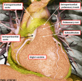
 Adult heart outflow tract CT01.jpg 747 × 747; 58 KB
 Adult heart outflow tract CT02.jpg 747 × 747; 65 KB
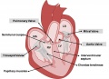 Adult Heart Valves.jpg 1,475 × 1,070; 113 KB
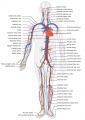 Adult human cardiovascular system.jpg 707 × 1,000; 151 KB
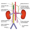 Adult renal venous cartoon.jpg 600 × 600; 62 KB
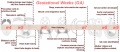 Advanced Heart Development Timeline GA.jpg 1,000 × 434; 97 KB
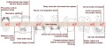 Advanced Heart Development Timeline.jpg 1,772 × 769; 158 KB
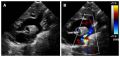 Aorta coarctation echocardiogram.jpg 601 × 283; 28 KB
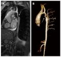 Aorta coarctation MRI.jpg 455 × 423; 25 KB
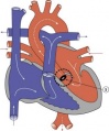
 Aortic arch and ductus arteriosus.jpg 600 × 720; 68 KB
 Aortic Stenosis.jpg 290 × 350; 16 KB
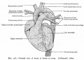
 Arey1924 fig193.jpg 1,200 × 962; 126 KB
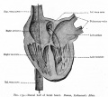
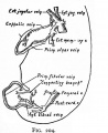
 Arey1924 fig194.jpg 1,200 × 710; 122 KB
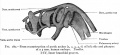
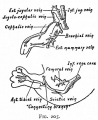
 Arey1924 fig195.jpg 1,583 × 950; 267 KB
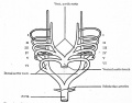
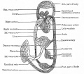
 Arey1924 fig196.jpg 1,200 × 881; 196 KB
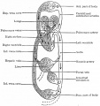
 Arey1924 fig197.jpg 1,200 × 1,615; 227 KB
 Arey1924 fig198.jpg 1,200 × 1,102; 187 KB
 Arey1924 fig199.jpg 1,200 × 956; 161 KB
 Arey1924 fig200.jpg 1,774 × 1,484; 209 KB
 Atrial & Ventricular Septation 1.jpg 1,482 × 960; 97 KB
 Atrial & Ventricular Septation 2.jpg 1,482 × 1,075; 113 KB
 Atrial Septal Defect.jpg 287 × 350; 16 KB
 Atrial Septation.jpg 1,482 × 960; 90 KB
 AV Canal Division (Superior View).jpg 1,487 × 489; 69 KB
 AV Canal Division.jpg 1,482 × 970; 93 KB
 AV Valves.jpg 1,183 × 1,085; 129 KB
 Bailey156.jpg 921 × 617; 150 KB
 Bailey157.jpg 725 × 562; 93 KB
 Bailey158.jpg 898 × 509; 101 KB
 Bailey159.jpg 933 × 896; 238 KB
 Bailey160.jpg 916 × 715; 202 KB
 Bailey161.jpg 645 × 629; 101 KB
 Bailey162.jpg 799 × 642; 102 KB
 Bailey163.jpg 757 × 691; 107 KB
 Bailey164.jpg 928 × 862; 149 KB
 Bailey165.jpg 613 × 1,045; 127 KB
 Bailey166.jpg 787 × 656; 90 KB
 Bailey167.jpg 610 × 458; 58 KB
 Bailey168.jpg 657 × 314; 43 KB
 Bailey169.jpg 504 × 264; 27 KB
 Bailey170.jpg 709 × 457; 67 KB
 Bailey171.jpg 954 × 507; 81 KB
 Bailey173.jpg 888 × 620; 113 KB
 Bailey174.jpg 955 × 542; 88 KB
 Bailey175.jpg 885 × 306; 51 KB
 Bailey176.jpg 918 × 352; 62 KB
 Bailey177.jpg 943 × 873; 199 KB
 Bailey178.jpg 913 × 653; 125 KB
 Bailey179.jpg 892 × 794; 114 KB
 Bailey180.jpg 938 × 431; 70 KB
 Bailey181.jpg 839 × 658; 67 KB
 Bailey182.jpg 913 × 718; 103 KB
 Bailey183.jpg 847 × 448; 57 KB
 Bailey184.jpg 625 × 462; 48 KB
 Bailey185.jpg 944 × 499; 98 KB
 Bailey187.jpg 817 × 732; 70 KB
 Bailey188.jpg 906 × 538; 64 KB
 Bailey189.jpg 810 × 632; 63 KB
 Bailey190.jpg 801 × 584; 69 KB
 Bailey191.jpg 863 × 509; 109 KB
 Bailey192.jpg 960 × 806; 133 KB
 Bailey193.jpg 747 × 848; 94 KB
 Bailey194.jpg 841 × 638; 80 KB
 Bailey195.jpg 924 × 781; 79 KB
 Bailey196.jpg 760 × 510; 47 KB
 Bailey197.jpg 878 × 705; 122 KB
 Bailey198.jpg 931 × 623; 83 KB
 Bailey199.jpg 924 × 451; 62 KB
 Bailey200.jpg 898 × 671; 149 KB
 Bailey201.jpg 1,059 × 1,033; 259 KB
 Bailey202.jpg 906 × 848; 138 KB
 Bailey203.jpg 406 × 614; 48 KB
 Bailey204.jpg 534 × 653; 62 KB
 Bailey205.jpg 534 × 653; 68 KB
 Bailey206.jpg 973 × 854; 162 KB
 Bailey207.jpg 896 × 951; 139 KB
 Bailey208.jpg 1,179 × 952; 165 KB
 Bailey209.jpg 1,248 × 988; 319 KB
 Bailey210.jpg 565 × 558; 63 KB
 Bailey211.jpg 767 × 820; 182 KB
 Bailey212.jpg 914 × 938; 218 KB
 Bailey213.jpg 800 × 851; 101 KB
 Bailey214.jpg 439 × 781; 81 KB
 Bailey215.jpg 944 × 958; 361 KB
 Bailey216.jpg 905 × 734; 261 KB
 Bailey217.jpg 767 × 694; 62 KB
 Bailey218.jpg 872 × 542; 84 KB
 Bailey219.jpg 911 × 575; 97 KB
 Bailey220.jpg 833 × 555; 105 KB
 Bailey221.jpg 904 × 774; 181 KB
 Bailey222.jpg 928 × 583; 145 KB
 Bailey245.jpg 499 × 733; 47 KB
 Bailey247.jpg 877 × 592; 113 KB
 Bailey248.jpg 814 × 521; 111 KB
 Baileytable03.jpg 821 × 667; 73 KB
 Bast1931 plate01.jpg 1,280 × 871; 113 KB
 Blood capillary EM 01.jpg 1,107 × 714; 260 KB
 Blood capillary EM 02.jpg 600 × 600; 99 KB
 Blood capillary EM 03.jpg 1,560 × 1,230; 441 KB
 Blood capillary EM 04.jpg 1,560 × 1,230; 462 KB
 Blood capillary EM 05.jpg 1,015 × 800; 205 KB
 Blood capillary EM 06.jpg 1,015 × 800; 216 KB
 Blood vessel wall cartoon.jpg 450 × 600; 71 KB
 Blood-brain barrier cartoon.jpg 765 × 1,000; 75 KB
 Blood-brain barrier EM01.jpg 1,656 × 810; 250 KB
 Bremer1914 plate01.jpg 684 × 1,000; 102 KB
 Bremer1914 plate02.jpg 716 × 1,000; 147 KB
 Bremer1914 plate03.jpg 706 × 1,000; 127 KB
 Bremer1914 plate04.jpg 643 × 1,000; 127 KB
 Bremer1914 plate05.jpg 643 × 1,000; 144 KB
 Buell-plate01.jpg 1,221 × 1,500; 236 KB
 Buell-plate02.jpg 1,124 × 1,500; 287 KB
 Buell01.jpg 713 × 800; 70 KB
 Buell02.jpg 977 × 800; 86 KB
 Buell03.jpg 790 × 800; 73 KB
 Buell04.jpg 913 × 800; 69 KB
 Buell05.jpg 1,176 × 800; 96 KB
 Buell06.jpg 1,037 × 800; 76 KB
 Buell07.jpg 1,200 × 878; 195 KB
 Buell08.jpg 1,037 × 1,000; 176 KB
 Buell09.jpg 724 × 1,000; 83 KB
 Buell10.jpg 665 × 1,000; 113 KB
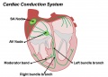 Cardiac Conduction System.jpg 1,201 × 862; 81 KB
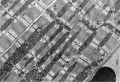 Cardiac muscle EM01.jpg 1,072 × 735; 231 KB
 Cardiac muscle EM02.jpg 1,072 × 735; 224 KB
 Cardiac muscle EM03.jpg 849 × 615; 135 KB
 Cardiac muscle EM04.jpg 1,000 × 680; 191 KB
 Cardiac Muscle EM05.jpg 992 × 733; 158 KB
 Cardiac Neural Crest Migration.jpg 1,517 × 1,116; 122 KB
 Cephalic plexus.png 600 × 557; 559 KB
 Cerebral blood supply development 01.jpg 1,200 × 460; 67 KB
 Cerebral brain artery development 01.jpg 845 × 600; 74 KB
 Cerebral brain artery development 02.jpg 996 × 400; 57 KB
 Cervical intersomitic vessels.png 600 × 462; 308 KB
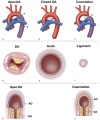
 Coarctation of the Aorta.jpg 289 × 350; 16 KB
 Complete atrioventricular canal.jpg 320 × 240; 22 KB
 Congdon-table01.jpg 764 × 1,000; 167 KB
 Congdon1922-1-16.jpg 980 × 1,000; 157 KB
 Congdon1922-17.jpg 1,000 × 411; 55 KB
 Congdon1922-18-25.jpg 1,200 × 795; 179 KB
 Congdon1922-18.jpg 494 × 506; 29 KB
 Congdon1922-19.jpg 653 × 471; 31 KB
 Congdon1922-20.jpg 794 × 446; 43 KB
 Congdon1922-21.jpg 578 × 407; 27 KB
 Congdon1922-22.jpg 511 × 489; 27 KB
 Congdon1922-23.jpg 519 × 412; 26 KB
 Congdon1922-24.jpg 790 × 482; 40 KB
 Congdon1922-25.jpg 509 × 358; 25 KB
 Congdon1922-26.jpg 746 × 726; 54 KB
 Congdon1922-27-28.jpg 997 × 612; 68 KB
 Congdon1922-29.jpg 976 × 1,000; 81 KB
 Congdon1922-30.jpg 1,133 × 1,000; 176 KB
 Congdon1922-31.jpg 1,063 × 1,000; 93 KB
 Congdon1922-32.jpg 1,133 × 1,000; 132 KB
 Congdon1922-33.jpg 920 × 1,000; 107 KB
 Congdon1922-34.jpg 920 × 1,000; 122 KB
 Congdon1922-35.jpg 920 × 1,000; 97 KB
 Congdon1922-36.jpg 920 × 1,000; 113 KB
 Congdon1922-37.jpg 1,200 × 838; 163 KB
 Congdon1922-38.jpg 1,187 × 1,000; 165 KB


.jpg)