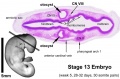
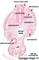
Carnegie stage 13
| Embryology - 19 May 2024 |
|---|
| Google Translate - select your language from the list shown below (this will open a new external page) |
|
العربية | català | 中文 | 中國傳統的 | français | Deutsche | עִברִית | हिंदी | bahasa Indonesia | italiano | 日本語 | 한국어 | မြန်မာ | Pilipino | Polskie | português | ਪੰਜਾਬੀ ਦੇ | Română | русский | Español | Swahili | Svensk | ไทย | Türkçe | اردو | ייִדיש | Tiếng Việt These external translations are automated and may not be accurate. (More? About Translations) |
Introduction
 |
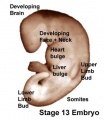
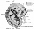
Factsweek 4 to week 5, 28 - 32 days. The embryos have a crown rump length (CRL) of 4 - 6 mm and somite number 30 pairs. Gestational Age GA - end week 6 beginning week 7 Summary
See also Events Features
| |||
|
Carnegie Stage 13 Movies: Gastrointestinal Tract | Cardiovascular | Central Nervous System | Carnegie Stage 13 | Stage 22 Movies | Movies
|

| Week: | 1 | 2 | 3 | 4 | 5 | 6 | 7 | 8 |
| Carnegie stage: | 1 2 3 4 | 5 6 | 7 8 9 | 10 11 12 13 | 14 15 | 16 17 | 18 19 | 20 21 22 23 |
- Carnegie Stages: 1 | 2 | 3 | 4 | 5 | 6 | 7 | 8 | 9 | 10 | 11 | 12 | 13 | 14 | 15 | 16 | 17 | 18 | 19 | 20 | 21 | 22 | 23 | About Stages | Timeline
Bright Field


- Embryo Links: Embryo and Placenta | Embryo | Embryo (label) | Embryo (animated) | Embryo (animated large) | Head region | Head region (label) | Body region | Body region (label) | Carnegie stage 13
Embryo Virtual Slide
| Stage 13 - Left Ventrolateral View
|
| Stage 13 | Embryo Slides |
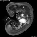
Scanning EM


Kyoto Collection


|

| |||
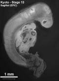
| Upper half of embryo shown in median sagittal plane. Scale bar 1 mm. | ||||

|

| |||
| Upper half of embryo shown in ventral view. Scale bar 0.5 mm. | Oral cavity floor. Scale bar 0.5 mm. | |||

|

|
Image source: The Kyoto Collection images are reproduced with the permission of Prof. Kohei Shiota and Prof. Shigehito Yamada, Anatomy and Developmental Biology, Kyoto University Graduate School of Medicine, Kyoto, Japan for educational purposes only and cannot be reproduced electronically or in writing without permission.
Carnegie Collection


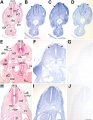
- Carnegie stage 13: 6473 left | 6473 dorsal | 6473 right | 6469 right | 8066 dorsal | 8066 left | 8119 left | 7433 right | 7433 ventral
| Carnegie Collection Embryos - Stage 13 | ||||||||||
|---|---|---|---|---|---|---|---|---|---|---|
| Serial No. | Size (mm) | Grade | Fixative | Embedding Medium | Plane | Thinness (µm) | Stain | Year | Notes | |
| 1 | E.,4.5 Ch., 30x30 | Poor | Salicylic acid | P | Transverse | 10 | Hemat. | 1887 | Obtained by Mall while student | |
| 19 | E., 5.5 Ch., 18x14 | Poor | p | ? | Transverse | 20 | Al. coch. | 1895 | ||
| 98 | E., 4 Ch., 24x16x9 | Poor | p | P | Transverse | 20 | Al. coch. | 1896 | ||
| 76 | E., 4.5 Ch., 22x20 | Poor | Alc. | P | Transverse | 20 | Al. coch. | 1897 | ||
| 112 | E., 4 | Poor | p | P | Sagittal | 10 | Al. coch. | p | ||
| 116 | E., 5 | Poor | p | ? | Sagittal | 10 | Al. coch. | 1898 | ||
| 148 | E.,4.3 Ch., 17x14x10 | Poor | Alc. | P | Coronal | 10 | (Stain - Haematoxylin Eosin) | 1899 | Abnormal. Nasal discs fused | |
| 186 | E.,3.5 Ch., 25x20x15 | Poor | Alc. | P | Transverse | 20 | Al. coch. | 1901 | ||
| 239 | E., 3.0 | Poor | Formalin | P | Transverse | 10 | (Stain - Haematoxylin Eosin) | 1903 | ||
| 248 | E., 4.5 Ch., 30x23x15 | Poor | p | ? | Coronal | 50 | Al. coch. | 1904 | ||
| 407 | E.,4 Ch., 14x13X7 | Poor | Formalin | ? | Transverse | 40 | Al. coch. | 1907 | ||
| 463 | E., 3.9 Ch., 17x12x7 | Good | Formalin | P | Coronal | 10 | Al. coch. | 1910 | ||
| 523 | E., 5 Ch., 25x25x15 | Fair | Formalin | P | Transverse | p | Al. coch. | 1911 | ||
| 588 | E., 4.0 Ch., 19x15x8 | Good | Corros. acetic | P | Coronal | 15 | (Stain - Haematoxylin Eosin) | 1912 | Advanced | |
| 786 | E., 4.5 Ch., 19x10x10 | Poor | Alc. | P | Sagittal | 15 | Al. coch. | 1913 | ||
| 800 | E., 6.0 | Good | Corros. acetic | P | Transverse | 10 | H | 1913 | Curettage. Anencephaly | |
| 808 | E.,4.0 | Poor | Corros. acetic | P | Transverse | 15 | Al. coch. | 1914 | Tubal Incomplete | |
| 826 | E., 5.0 Ch., 13x13x9 | Good | Formalin | P | Transverse | 20 | Al. coch. | 1914 | Shrunken | |
| 836 | E.,4.0 Ch., 22x18x11 | Exc. | Corros. acetic | P | Transverse | 15 | Al. coch. | 1914 | Less advanced | |
| 963 | E.,4.0 Ch., 23x18x16 | Good | Formalin | P | Coronal | 20 | Al. coch. | 1914 | ||
| 1075 | E.,6.0 Ch., 46x32x20 | Exc. | Corros. acetic | P | Coronal | 20 | (Stain - Haematoxylin Eosin) or. G. | 1915 | Most advanced in group | |
| 3956 | E., 4.0 | Poor | Formalin | P | Transverse | 20 | Al. coch. | 1922 | Tubal Incomplete | |
| 4046 | E.,5 Ch., 22x20x20 | Poor | Formalin | P | Transverse | 50 | Al. coch. | 1922 | ||
| 5541 | E., 6.0 Ch., 35x30x20 | Good | Formalin | P | Transverse | 10 | Al. coch., eosin | 1927 | ||
| 5682 | E., 5.3 Ch., 29x25x13 | Poor | Formalin | P | Coronal | 20 | Al. coch. | 1928 | ||
| 5874 | E., 4.8 | Exc. | Bouin | P | Transverse | 10 | (Stain - Haematoxylin Eosin) | 1929 | Hysterotomy. Bromides only | |
| 6032 | E., 5.8 Ch., 30x24x13 | Poor | Formalin | P | P | ? | p | 1929 | Not good enough to cut | |
| 6469 | E., 5.0 Ch., 25x18x18 | Poor | Formalin | P | P | P | P | 1932 | Fragmented on cutting. Not saved | |
| 6473 | E., 5.0 Ch, 30x30x15 | Exc. | Formalin | C-P | Coronal | 6 | Al. coch. | 1932 | Less advanced. Ag added | |
| 7433 | E., 5.2 Ch., 15x13x13 | Exc. | Formalin | C-P | Coronal | 8 | (Stain - Haematoxylin Eosin) | 1937 | Tubal | |
| 7618 | E, 48 Ch, 18x15x15 | Exc. | Bouin | C-P | Coronal | 10 | (Stain - Haematoxylin Eosin) | 1939 | Hystereaomy. Advanced. Ag added | |
| 7669 | E, 5.0 Ch., 23x16x14 | Good | Formalin | C-P | Coronal | 6 | (Stain - Haematoxylin Eosin) | 1939 | Hysterectomy. Least advanced in group, Ag added | |
| 7889 | E, 4.2 | Exc. | Bouin | C-P | Coronal | 6 | (Stain - Haematoxylin Eosin) | 1941 | Hysterectomy | |
| 8066 | E,53 Ch , 20x18xI8 | Exc. | Bouin | C-P | Transverse | 8 | (Stain - Haematoxylin Eosin) | 1942 | Hysterectomy. Ag added to slide 2 | |
| 8119 | E., 5.3 Ch., 32x28x6.5 | Exc. | Bouin | C-P | Transverse | 8 | (Stain - Haematoxylin Eosin) | 1943 | Hysterectomy | |
| 8147 | E., 5.2 Ch., 27x21x19 | Poor | Formalin | ? | ? | p | p | 1943 | Tubal Not cut | |
| 8239 | E., 4.3 | Exc. | Bouin | C-P | Sagittal | 8 | H. phlox. | 1944 | ||
| 8372 | E., 5.6 | Exc. | Alc.& Bouin | P | Transverse | 10 | Azan | 1946 | ||
| 8581 | E., 4.8 | Good | Kaiserling | C-P | Sagittal | 8 | Azan | 1948 | Most-advanced third | |
| 8967 | E., 5.7 | Exc. | Acetic Zenker | C-P | Transverse | 6 | (Stain - Haematoxylin Eosin) | 1931 | Head injured. Univ. Chicago No. H1426 | |
| 9296 | E,4.5 | Exc. | C-P | Coronal | 8 | Azan | 1955 | |||
| 9297 | E., 4.5 | Exc. | C-P | Sagittal | 8 | Azan | 1955 | |||
| 9697 | E., 5.5 | Bouin | 1956 | not cut | ||||||
Abbreviations
| ||||||||||
| iBook - Carnegie Embryos | |
|---|---|

|
|
Hill Collection
| Hill HH145 | |
|---|---|

|

|

|

|

|

|

|

|
- Links: Hill Collection
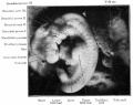
Stage 13 Serial Section Images
These are the original serial images prepared from embryo sections for Embryology practical classes and transferred online in 1996. This embryo shows some features present on the later Stage 14 embryo.
- Links: Carnegie stage 13 - serial sections | Carnegie stage 22 - serial sections | Carnegie stage 22
Labeled Sections

|

|

|

|

| ||
| A1L | A2L | A3L | A4L | A5L | A6L | A7L |

|

|

|

|

|

|

|
| B1L | B2L | B3L | B4L | B5L | B6L | B7L |

|

|

|

|

|

|

|
| C1L | C2L | C3L | C4L | C5L | C6L | C7L |

|

|

|

|

|

|

|
| D1L | D2L | D3L | D4L | D5L | D6L | D7L |

|

|

|

|

|

| |
| E1L | E2L | E3L | E4L | E5L | E6L | E7L |

|

|

|

|

|

|

|
| F1L | F2L | F3L | F4L | F5L | F6L | F7L |

|

|

|

|

|

|

|
| G1L | G2L | G3L | G4L | G5L | G6L | G7L |
Unlabeled Sections

|

|

|

| |||
| A1 | A2 | A3 | A4 | A5 | A6 | A7 |

|

|

|

|

|

|

|
| B1 | B2 | B3 | B4 | B5 | B6 | B7 |

|

|

|

|

|

|

|
| C1 | C2 | C3 | C4 | C5 | C6 | C7 |

|

|

|

|

|

|

|
| D1 | D2 | D3 | D4 | D5 | D6 | D7 |

|

|

|

|

|

| |
| E1 | E2 | E3 | E4 | E5 | E6 | E7 |

|

|

|

|

|

|

|
| F1 | F2 | F3 | F4 | F5 | F6 | F7 |

|

|

|

|

|

|

|
| G1 | G2 | G3 | G4 | G5 | G6 | G7 |
Stage 13 Movies
|
| Sagittal EFIC |
| Page | Play |
|
|
|
|
Events
- hearing - otic vesicle closes from surface and endolymphatic appendage apparent. A capillary network forms around the otic vesicle with esoderm becomes condensed as the otic capsule.[1] Vestibulocochlear ganglion (vestibular part) and vestibular nerve fires present.
- Vision - By the end of the fourth week the optic vesicle lies close to the surface ectoderm. Optic evagination differentiation allows identification of optic part of retina, future pigmented layer of retina, and optic stalk. The surface ectoderm overlying the optic vesicle, in response to this contact, has thickened to form the lense placode.[2]
References
- ↑ Streeter GL. Developmental horizons in human embryos. Description of age group XIII, embryos about 4 or 5 millimeters long, and age group XIV, period of indentation of the lens vesicle. (1945) Carnegie Instn. Wash. Publ. 557, Contrib. Embryol., Carnegie Inst. Wash., 31: 27-63.
- ↑ <pubmed>7364662</pubmed>
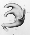
Additional Images
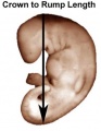
Stage 13 crown rump length
Stage 13 surface bulges
Stage 13 Optical Projection Tomography
Stage 13 otocyst
Stage 13 caudal trunk
Cross-section showing neural tube, notochord, limb bud
Selected image of above cross-section showing neural tube, notochord, limb bud


Historic
| Historic Disclaimer - information about historic embryology pages |
|---|
 |
Historic - Human embryo with twenty-seven primitive segments (7 mm., 26 days)
Historic - Human embryo with 28 primitive segments (7.5 mm)
1911 Cloaca model Carnegie embryo 186


- Carnegie Stages: 1 | 2 | 3 | 4 | 5 | 6 | 7 | 8 | 9 | 10 | 11 | 12 | 13 | 14 | 15 | 16 | 17 | 18 | 19 | 20 | 21 | 22 | 23 | About Stages | Timeline
Image Source: Scanning electron micrographs of the Carnegie stages of the early human embryos are reproduced with the permission of Prof Kathy Sulik, from embryos collected by Dr. Vekemans and Tania Attié-Bitach. Images are for educational purposes only and cannot be reproduced electronically or in writing without permission.
Cite this page: Hill, M.A. (2024, May 19) Embryology Carnegie stage 13. Retrieved from https://embryology.med.unsw.edu.au/embryology/index.php/Carnegie_stage_13
- © Dr Mark Hill 2024, UNSW Embryology ISBN: 978 0 7334 2609 4 - UNSW CRICOS Provider Code No. 00098G