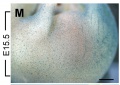
Category:Mouse E15.5
The Embryology pages and media listed below relate to E15.5 mouse development.
- Mouse Stages: E1 | E2.5 | E3.0 | E3.5 | E4.5 | E5.0 | E5.5 | E6.0 | E7.0 | E7.5 | E8.0 | E8.5 | E9.0 | E9.5 | E10 | E10.5 | E11 | E11.5 | E12 | E12.5 | E13 | E13.5 | E14 | E14.5 | E15 | E15.5 | E16 | E16.5 | E17 | E17.5 | E18 | E18.5 | E19 | E20 | Timeline | About timed pregnancy
| Carnegie | Stage | |||||||||||||||||||||||
| Human | Days | 1 | 2-3 | 4-5 | 5-6 | 7-12 | 13-15 | 15-17 | 17-19 | 20 | 22 | 24 | 28 | 30 | 33 | 36 | 40 | 42 | 44 | 48 | 52 | 54 | 55 | 58 |
| Mouse | Days | 1 | 2 | 3 | E4.5 | E5.0 | E6.0 | E7.0 | E8.0 | E9.0 | E9.5 | E10 | E10.5 | E11 | E11.5 | E12 | E12.5 | E13 | E13.5 | E14 | E14.5 | E15 | E15.5 | E16 |
| Rat | Days | 1 | 3.5 | 4-5 | 5 | 6 | 7.5 | 8.5 | 9 | 10.5 | 11 | 11.5 | 12 | 12.5 | 13 | 13.5 | 14 | 14.5 | 15 | 15.5 | 16 | 16.5 | 17 | 17.5 |
| Note these Carnegie stages are only approximate day timings for average of embryos. Links: Carnegie Stage Comparison | ||||||||||||||||||||||||
| ||||||||||||||||||||||||
| Timeline Links: human timeline | mouse timeline | mouse detailed timeline | chicken timeline | rat timeline | Medaka | Category:Timeline |
Events
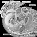
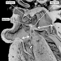
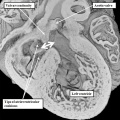
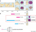
- heart - E15.5 (TS24) - E18.5 (TS27) Definitive external prenatal configuration achieved, Atrioventricular valve leaflets are being modified, Coronary arteries are being modified.[1]
- musculoskeletal - Abdominal wall myotubes were abundant in all five of the muscle layers with the unidirectional orientation in all layers.[2]
- palate - secondary palatal shelf fusion is complete, mesenchymal condensation followed by osteogenic differentiation occurs.
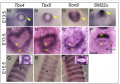
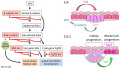
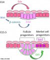
- tongue - mesenchyme alters tongue shape and size, Hippo signalling and cell proliferation in a region- and stage-specific manner - Nf2cKO mutants had a decreased level of Hippo signalling transcription factor Yes-associated protein (Yap). At E15.5, loss of Nf2 in the NC lineage resulted in distinct changes in cell proliferation in different regions, that is, high in epithelium and mesenchyme subjacent to the epithelium, and lower in deeper layers of the mesenchyme.[3]
References
- ↑ Savolainen SM, Foley JF & Elmore SA. (2009). Histology atlas of the developing mouse heart with emphasis on E11.5 to E18.5. Toxicol Pathol , 37, 395-414. PMID: 19359541 DOI.
- ↑ Nichol PF, Corliss RF, Yamada S, Shiota K & Saijoh Y. (2012). Muscle patterning in mouse and human abdominal wall development and omphalocele specimens of humans. Anat Rec (Hoboken) , 295, 2129-40. PMID: 22976993 DOI.
- ↑ Ishan M, Chen G, Yu W, Wang Z, Giovannini M, Cao X & Liu HX. (2021). Deletion of Nf2 in neural crest-derived tongue mesenchyme alters tongue shape and size, Hippo signalling and cell proliferation in a region- and stage-specific manner. Cell Prolif , 54, e13144. PMID: 34697858 DOI.
Search Pubmed: Mouse E15.5
Pages in category 'Mouse E15.5'
The following 2 pages are in this category, out of 2 total.
Media in category 'Mouse E15.5'
The following 21 files are in this category, out of 21 total.
 Anderson2016-fig28a.jpg 800 × 800; 117 KB
 Anderson2016-fig28b.jpg 800 × 800; 110 KB
 Anderson2016-fig34a.jpg 800 × 800; 105 KB
 Embryonic Trachea.png 468 × 326; 205 KB
 Integumentary touch dome model 01.jpg 1,280 × 724; 144 KB
 Integumentary touch dome model 03.jpg 603 × 724; 73 KB
 Mouse - palate MMP-25 expression.jpg 1,000 × 818; 243 KB
 Mouse cochlea gene expression.jpg 1,000 × 346; 75 KB
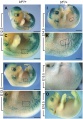
 Mouse external genital development.jpg 800 × 701; 75 KB
 Mouse gonad Gcnf expression 01.jpg 1,947 × 843; 304 KB
 Mouse gonad Gcnf expression E15.5.jpg 338 × 782; 53 KB
 Mouse interdigit apoptosis 01.jpg 800 × 800; 81 KB
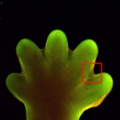 Mouse interdigit apoptosis 02.jpg 764 × 764; 61 KB
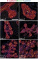
 Mouse melanoblast distribution 01.jpg 697 × 1,000; 192 KB
 Mouse melanoblast distribution 05.jpg 761 × 540; 94 KB
 Mouse pancreas development.jpg 600 × 939; 261 KB
 Mouse pax7 limb 01.jpg 1,320 × 549; 112 KB
 Mouse- respiratory development 01.jpg 1,000 × 571; 125 KB
 Renal - early glomerulus.jpg 1,155 × 432; 52 KB
 Renal - podocyte development 01.jpg 1,200 × 908; 155 KB
 Renal - S-shaped body stage.jpg 1,155 × 432; 99 KB


