Neural System - Abnormalities
| Embryology - 27 Jun 2024 |
|---|
| Google Translate - select your language from the list shown below (this will open a new external page) |
|
العربية | català | 中文 | 中國傳統的 | français | Deutsche | עִברִית | हिंदी | bahasa Indonesia | italiano | 日本語 | 한국어 | မြန်မာ | Pilipino | Polskie | português | ਪੰਜਾਬੀ ਦੇ | Română | русский | Español | Swahili | Svensk | ไทย | Türkçe | اردو | ייִדיש | Tiếng Việt These external translations are automated and may not be accurate. (More? About Translations) |
Introduction


There are many different congenital and environmentally derived abnormalities associated with the nervous system. There are potentially 1000's of neurological abnormalities that could be listed on this page, therefore this current page is only a very brief introduction to some of these neural abnormalities. For additional information see links at the bottom of each sub-heading.
As the nervous system continues to develop fetally and postnatally, environmental issues both before and after birth are relevant to neural development. Embryonic and fetal nutrition folate (required early for neural tube closure) and iodine (required later for neural growth and differentiation). The developmental environment can also impact upon neural growth; maternal drugs such as alcohol and heavy metals such as lead (mining, historically both petrol and paint). Finally, postnatally other sensory abnormalities (hearing) can also impact on achieving developmental milestones.
Some Recent Findings
|
| More recent papers |
|---|
 This table allows an automated computer search of the external PubMed database using the listed "Search term" text link.
More? References | Discussion Page | Journal Searches | 2019 References | 2020 References Search term: Abnormal Neural Development <pubmed limit=5>Abnormal Neural Development</pubmed> |
Neural Tube Closure

Dysraphism is the term often used to describe the defective fusion of the neural folds. The position and degree of failure of fusion will result in either embryonic death or a range of different neural defects. The way (mode) in which the human neural tube fuses has been a source of contention. In humans, fusion appears to initiate at multiple sites along the neural groove[6][7], this mode of closure may differ from that found in many animal species used in developmental studies.
Human Embryonic Death:
- 5 weeks with total failure of fusion.
- 6.5 weeks with opening over the rhombencephalon.
- survive beyond 7 weeks with a defect at the frontal and parietal regions.

Mouse posterior neuropore Axd mutant[8]
Spina Bifida (Meningomyelocele)


Double meningomyelocele[9]

|

|
| Meningomyelocele | Spina Bifida Rates (USA Data) |
There are potentially many different causes of spina bifida and neural tube defects. The basis of the abnormality is a failure of the neural tube to close caudally. At least one known cause is a low maternal diet of folic acid (folate) containing foods. (see Neural Tube Defects and Low Folic Acid or Folic Acid below)
Neural tube defects from failure to close can be screened by amniocentesis or ultrasound.
Alpha-fetoproetin normally present in the CSF, leaks and can be detected in amniotic fluid.
| International Classification of Diseases |
|---|
Q05 Spina bifidaIncl.: hydromeningocele (spinal), meningocele (spinal), meningomyelocele, myelocele, myelomeningocele, rachischisis, spina bifida (aperta)(cystica), syringomyelocele Excl.: Arnold-Chiari syndrome (Q07.0), spina bifida occulta (Q76.0)
|
Cephalic Disorders
Cephalic (Greek, kephale = head) are a large group of abnormalities that relate to both skeletal (skull) and neural (brain) associated defects including: anencephaly, hydrocephalus, encephalocele, colpocephaly (occipital horn enlargement), lissencephaly (smooth brain), porencephaly (cyst or cavity in cerebral hemisphere), acephaly (absence of head), exencephaly (brain outside skull), macrocephaly (large head), micrencephaly (small brain), otocephaly (absence of lower jaw), brachycephaly (premature fusion of coronal suture), oxycephaly (premature fusion of coronal suture + other), plagiocephaly (premature unilateral fusion of coronal or lambdoid sutures), scaphocephaly (premature fusion of sagittal suture), trigonocephaly.
Microcephaly

A recent Spanish study[10] has shown a concordance between a head circumference growth function and intellectual disability in relation with the cause of microcephaly.
- "3,269 head circumference (HC) charts of patients from a tertiary neuropediatric unit were reviewed and 136 microcephalic participants selected. Using the Z-scores of registered HC measurements we defined the variables: HC Minimum, HC Drop and HC Catch-up. We classified patients according to the presence or absence of intellectual disability (IQ below 71) and according to the cause of microcephaly (idiopathic, familial, syndromic, symptomatic and mixed). Using Discriminant Analysis a C-function was defined as C=HC Minimum + HC Drop with a cut-off level of C=-4.32 Z-score. In our sample 95% of patients scoring below this level, severe microcephaly, were classified in the disabled group while the overall concordance was 66%. In the symptomatic-mixed group the concordance between HC function and outcome reached 82% in contrast to only 54% in the idiopathic-syndromic group (P-value=0.0002)."

Microlissencephaly (MRI)[11] - Zika virus associated lishencephaly (smooth brain) in combination with severe congenital microcephaly (small head).
Anencephaly

This is a neural tube defect, the basis of the abnormality is a failure of the neural tube to close cranially. Also called exencephaly or craniorachischisis.
| International Classification of Diseases |
|---|
Q00 Anencephaly and similar malformations
|
| Fetal Anencephaly | |
|---|---|

|

|
| ventral view | lateral view |

Anencephaly in a fetus (GA week 18) from a diabetic mother. Ultrasound images (coronal) show a complete absence of the cranial vault and brain and enlarged orbits.[13]
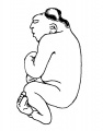
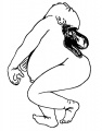
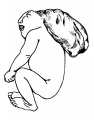
Anencephaly Historic Drawings
These drawings are from a 1921 study of a single embryonic anencephaly.[14]
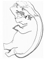
Fig. 1. Reconstruction of anencephalic embryo.
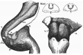
Fig. 2. Hind-brain 28 mm embryo (left view).


These drawings are from a 1925 study of 57 cases of anencephaly.[15]
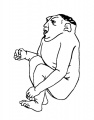
anencephalic acranius
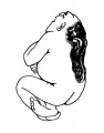
anencephalic craniorhachischisis
microcephalic acrauius
microcephalic craniorhachischisis
exocephalic acranius


- Links: Maternal Diabetes
Holoprosencephaly
International Classification of Diseases - Q04 Other congenital malformations of brain - Q04.2 Holoprosencephaly
Below are shown images of fetal holoprosencephaly and associated cyclopia.[16]

|

|
| Human holoprosencephaly cyclopean dissection | Proboscis histology |
Hydrocephalus
Hydrocephalus (historic image from Hess, 1922)
This is a defect of cerebrospinal fliud (CSF) flow, excess fluid production or impaired fluid absorption and can be congenital or acquired. Estimated incidence of 1 in 1000 live births the condition leads to enlarged ventricles and head, separated skull cranial sutures and fontanelles. Obstruction of CSF flow can occur at any time (prenatally or postnatally) and leads to accumulation of within the ventricles. The time of onset will have different effects and should be compared to the equilivant neurological events that are occuring.
Ventricular obstruction usually occurs at the level of the cerebral aqueduct (narrowest site), but can occur elsewhere, and can be caused by viral infection or zoonotic disease.
- Links: Congenital Hydrocephalus
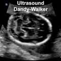
Dandy Walker malformation
| Dandy Walker malformation (MRI)[17] is another form of ventricular abnormality that affects cerebellar development.
The vermis of the cerebellum can be small or absent, the fourth ventricle enlarges due to cyst formation. An ultrasound study[18] of fetuses with Dandy-Walker malformation 13 to 16 weeks (GA 15-18 weeks) identified the fourth ventricle widely open posteriorly, even in the standard transcerebellar view, and the brainstem-vermis (BV) angle was > 45°, significantly increased compared to that in normal fetuses (P < 0.0001). Note that at this age, an open fourth ventricle can also found in about 10% of normal fetuses. Named after Walter Dandy (1886 – 1946) and Arthur Earl Walker (1907 – 1995), two USA neurosurgeons.
|

|
- Links: Congenital Hydrocephalus
Encephalocele
This defect is generally mesodermal in origin, leading to protrusion of brain and meninges outside the crainal cavity. The severity of the disorder can vary dependent upon the degree of mesodermal abnormality.
Encephalitis
Normally a postnatal clinical syndrome of the central nervous system resulting in inflammation of the brain parenchyma and caused by a range of pathogens (viral, bacterial and protozoal infections). This infectious disease is due mainly to viral pathogens: Herpes simplex encephalitis 10%–20% of cases, Murray Valley encephalitis virus, Japanese encephalitis virus, Australian bat lyssavirus, West Nile virus, Hendra virus and Nipah virus. The pathogen is generally included in the specific encephalitis naming; Varicella encephalitis and Toxoplasma meningoencephalitis.
- Links: Neural System - Abnormalities | TORCH Infections | PMC2700670)
Fragile X Syndrome
Fragile X Syndrome (FXS) is the most common form of inherited mental retardation and autism. The condition is caused by a loss of the functional fragile X mental retardation protein (FMRP) an RNA-binding protein that can regulate the translation of specific mRNAs. There are several suggested additional roles for this protein including synaptic development and function[19] and in adult neurogenesis.[20]
ICD: Q99.2 Fragile X chromosome Fragile X syndrome
- Links: Fragile X Syndrome
Autism
Autism (autism spectrum disorder, ASD) is a behaviourally defined brain disorder in children. Features include: impoverished verbal and non-verbal communication skills, reduced social interactions (bias their attention towards objects rather than the surrounding social situation), behavioural impairments in attention engagement/disengagement, poor emotional discrimination and facial recognition, and fail to response to their own names. There exist many different and unproven claims as to the origins of autism.
Developmentally associated with neural maturation changes in cortical thickness and organization, and particularly affecting pyramidal neurons. A rat model shows structural and behavioural features of autism as a result of altering the trajectory of early postnatal cortical development.[21]
- Links: Neural Exam Movies
Rett Syndrome
(RTT) A severe neurodevelopment disorder, with intellectual disability and abnormalities of movement, mainly caused by mutations in the X-linked (Xq28) Methyl-CpG-binding protein 2 (MECP2) gene and therefore almost exclusively in females. The congenital variant of Rett syndrome is caused by heterozygous mutation in the FOXG1 gene on chromosome 14q13.
Cerebral Palsy
| ICD-10 G80 Cerebral palsy |
|---|
|
Cerebral palsy is a group of disorders in motor impairment that limits activity, and is attributed to non-progressive disturbances during brain development in fetuses or infants (3-4 / 1,000).[22]
Represented by one or more of these features:
- impaired cognition
- impaired communication
- impaired sensory perception
- behavioural abnormalities
- seizure disorders
- Links: Neural Exam Movies | Medline Plus | NINDS - Info | CDC Screening and Diagnosis | Cerebral Palsy Alliance
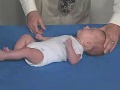
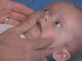
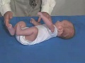
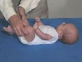
Newborn Neural Exam
Neural - The collapsed tables below link to a number of short videos that demonstrate simple assessments of the postnatal developing nervous system.

|
| ||||||||||||||||||||||||||||||||||||||||||||||||||||||||||||||||||||||||||
|
|
International Classification of Diseases
The International Classification of Diseases (ICD) World Health Organization's classification used worldwide as the standard diagnostic tool for epidemiology, health management and clinical purposes. This includes the analysis of the general health situation of population groups. It is used to monitor the incidence and prevalence of diseases and other health problems. Within this classification "congenital malformations, deformations and chromosomal abnormalities" are (Q00-Q99) but excludes "inborn errors of metabolism" (E70-E90).
Note that the current ICD10 classification system is being updated to ICD11 that is currently in beta testing status. The data on this page, other than the information in the collapsed table below, is from ICD10 classification.
| International Classification of Diseases ICD-11 20 Developmental anomalies (beta draft) |
|---|
| ICD-11 Beta Draft - NOT FINAL, updated on a daily basis, It is not approved by WHO, NOT TO BE USED for CODING except for agreed FIELD TRIALS.
Chapter 20 Developmental anomalies, only a few examples of the draft ICD-11 Beta coding and tree structure for "structural developmental anomalies" within this section are shown in the table below. |
| Mortality and Morbidity Statistics - 20 Developmental Anomalies |
Structural Developmental Anomalies
|
| Multiple developmental anomalies or syndromes |
| Chromosomal anomalies, excluding gene mutations |
| Conditions with disorders of intellectual development as a relevant clinical feature |
| LD6Y Other specified developmental anomalies
LD6Z Developmental anomalies, unspecified |
| CD-11 Beta Draft - NOT FINAL, updated on a daily basis, It is not approved by WHO, NOT TO BE USED for CODING except for agreed FIELD TRIALS. |
Congenital malformations of the nervous system (Q00-Q07)
Q00 Anencephaly and similar malformations
- Q00.0 Anencephaly, Acephaly, Acrania, Amyelencephaly, Hemianencephaly, Hemicephaly
- Q00.1 Craniorachischisis
- Q00.2 Iniencephaly
Q01 Encephalocele
Incl.: encephalomyelocele, hydroencephalocele, hydromeningocele, cranial meningocele, cerebral meningoencephalocele
Excl.: Meckel-Gruber syndrome (Q61.9)
- Q01.0 Frontal encephalocele
- Q01.1 Nasofrontal encephalocele
- Q01.2 Occipital encephalocele
- Q01.8 Encephalocele of other sites
- Q01.9 Encephalocele, unspecified
Q02 Microcephaly
Incl.: Hydromicrocephaly Micrencephalon Excl.: Meckel-Gruber syndrome (Q61.9)
Q03 Congenital hydrocephalus
Q03 Congenital hydrocephalus Incl.: hydrocephalus in newborn Excl.: Arnold-Chiari syndrome (Q07.0) hydrocephalus: acquired (G91.-) due to congenital toxoplasmosis (P37.1) with spina bifida (Q05.0-Q05.4)
- Q03.0 Malformations of aqueduct of Sylvius Aqueduct of Sylvius: anomaly obstruction, congenital stenosis
- Q03.1 Atresia of foramina of Magendie and Luschka Dandy-Walker syndrome
- Q03.8 Other congenital hydrocephalus
- Q03.9 Congenital hydrocephalus, unspecified
Q04 Other congenital malformations of brain
Excl.: cyclopia (Q87.0) macrocephaly (Q75.3)
- Q04.0 Congenital malformations of corpus callosum, Agenesis of corpus callosum
- Q04.1 Arhinencephaly
- Q04.2 Holoprosencephaly
- Q04.3 Other reduction deformities of brain, Absence, Agenesis, Aplasia, Hypoplasia of part of brain, Agyria, Hydranencephaly, Lissencephaly, Microgyria, Pachygyria Excl.: congenital malformations of corpus callosum (Q04.0)
- Q04.4 Septo-optic dysplasia
- Q04.5 Megalencephaly
- Q04.6 Congenital cerebral cysts, Porencephaly, Schizencephaly, Excl.: acquired porencephalic cyst (G93.0)
- Q04.8 Other specified congenital malformations of brain, Macrogyria
- Q04.9 Congenital malformation of brain, unspecified Congenital: anomaly, deformity, disease or lesion, multiple anomalies NOS of brain
Q05 Spina bifida
Incl.: hydromeningocele (spinal), meningocele (spinal), meningomyelocele, myelocele, myelomeningocele, rachischisis, spina bifida (aperta)(cystica), syringomyelocele Excl.: Arnold-Chiari syndrome (Q07.0), spina bifida occulta (Q76.0)
- Q05.0 Cervical spina bifida with hydrocephalus
- Q05.1 Thoracic spina bifida with hydrocephalus Spina bifida: dorsal thoracolumbar with hydrocephalus
- Q05.2 Lumbar spina bifida with hydrocephalus, Lumbosacral spina bifida with hydrocephalus
- Q05.3 Sacral spina bifida with hydrocephalus
- Q05.4 Unspecified spina bifida with hydrocephalus
- Q05.5 Cervical spina bifida without hydrocephalus
- Q05.6 Thoracic spina bifida without hydrocephalus Spina bifida: dorsal NOS, thoracolumbar NOS
- Q05.7 Lumbar spina bifida without hydrocephalus, Lumbosacral spina bifida NOS
- Q05.8 Sacral spina bifida without hydrocephalus
- Q05.9 Spina bifida, unspecified
Q06 Other congenital malformations of spinal cord
- Q06.0 Amyelia
- Q06.1 Hypoplasia and dysplasia of spinal cord, Atelomyelia, Myelatelia, Myelodysplasia of spinal cord
- Q06.2 Diastematomyelia
- Q06.3 Other congenital cauda equina malformations
- Q06.4 Hydromyelia Hydrorachis
- Q06.8 Other specified congenital malformations of spinal cord
- Q06.9 Congenital malformation of spinal cord, unspecified Congenital: anomaly, deformity, disease or lesion, NOS of spinal cord or meninges
Q07 Other congenital malformations of nervous system
Excl.: familial dysautonomia [Riley-Day] (G90.1), neurofibromatosis (nonmalignant) (Q85.0)
- Q07.0 Arnold-Chiari syndrome
- Q07.8 Other specified congenital malformations of nervous system Agenesis of nerve, Displacement of brachial plexus, Jaw-winking syndrome, Marcus Gunn's syndrome
- Q07.9 Congenital malformation of nervous system, unspecified Congenital: anomaly, deformity, disease or lesion, NOS of nervous system
References
- ↑ Im K, Guimaraes A, Kim Y, Cottrill E, Gagoski B, Rollins C, Ortinau C, Yang E & Grant PE. (2017). Quantitative Folding Pattern Analysis of Early Primary Sulci in Human Fetuses with Brain Abnormalities. AJNR Am J Neuroradiol , 38, 1449-1455. PMID: 28522661 DOI.
- ↑ Goldstein IS, Erickson DJ, Sleeper LA, Haynes RL & Kinney HC. (2017). The Lateral Temporal Lobe in Early Human Life. J. Neuropathol. Exp. Neurol. , 76, 424-438. PMID: 28498956 DOI.
- ↑ 3.0 3.1 Narisawa A, Komatsuzaki S, Kikuchi A, Niihori T, Aoki Y, Fujiwara K, Tanemura M, Hata A, Suzuki Y, Relton CL, Grinham J, Leung KY, Partridge D, Robinson A, Stone V, Gustavsson P, Stanier P, Copp AJ, Greene ND, Tominaga T, Matsubara Y & Kure S. (2012). Mutations in genes encoding the glycine cleavage system predispose to neural tube defects in mice and humans. Hum. Mol. Genet. , 21, 1496-503. PMID: 22171071 DOI.
- ↑ Adzick NS, Thom EA, Spong CY, Brock JW, Burrows PK, Johnson MP, Howell LJ, Farrell JA, Dabrowiak ME, Sutton LN, Gupta N, Tulipan NB, D'Alton ME & Farmer DL. (2011). A randomized trial of prenatal versus postnatal repair of myelomeningocele. N. Engl. J. Med. , 364, 993-1004. PMID: 21306277 DOI.
- ↑ Abeywardana S & Sullivan EA 2008. Neural tube defects in Australia. An epidemiological report. Cat. no. PER 45. Sydney: AIHW National Perinatal Statistics Unit | PDF.
- ↑ Van Allen MI, Kalousek DK, Chernoff GF, Juriloff D, Harris M, McGillivray BC, Yong SL, Langlois S, MacLeod PM & Chitayat D. (1993). Evidence for multi-site closure of the neural tube in humans. Am. J. Med. Genet. , 47, 723-43. PMID: 8267004 DOI.
- ↑ Nakatsu T, Uwabe C & Shiota K. (2000). Neural tube closure in humans initiates at multiple sites: evidence from human embryos and implications for the pathogenesis of neural tube defects. Anat. Embryol. , 201, 455-66. PMID: 10909899
- ↑ <pubmed>21262862</pubmed>| PMC3063985
- ↑ Sarda D, Kothari P, Laddha A, Kulkarni B. Double meningomyelocele: Embryogenesis. J Pediatr Neurosci [serial online] 2007 [cited 2013 Mar 22];2:26-7. Available from: http://www.pediatricneurosciences.com/text.asp?2007/2/1/26/32004
- ↑ Coronado R, Macaya Ruíz A, Giraldo Arjonilla J & Roig-Quilis M. (2015). [Concordance between a head circumference growth function and intellectual disability in relation with the cause of microcephaly]. An Pediatr (Barc) , 83, 109-16. PMID: 25534043 DOI.
- ↑ de Fatima Vasco Aragao M, van der Linden V, Brainer-Lima AM, Coeli RR, Rocha MA, Sobral da Silva P, Durce Costa Gomes de Carvalho M, van der Linden A, Cesario de Holanda A & Valenca MM. (2016). Clinical features and neuroimaging (CT and MRI) findings in presumed Zika virus related congenital infection and microcephaly: retrospective case series study. BMJ , 353, i1901. PMID: 27075009
- ↑ Mathews TJ. Trends in spina bifida and anencephalus in the United States, 1991-2005, National Vital Statistics System.
- ↑ Alorainy IA, Barlas NB & Al-Boukai AA. (2010). Pictorial Essay: Infants of diabetic mothers. Indian J Radiol Imaging , 20, 174-81. PMID: 21042439 DOI.
- ↑ Frazer JE. Report on an anencephalic embryo. (1921) J Anat. 56(1): 12-9. PMID 17103933
- ↑ Nañagas JC. A comparison of the growth of the body dimensions of anencephalic human fetuses with normal fetal growth as determined by graphic analysis and empirical formulae. (1925) American J. Anatomy. 455-494.
- ↑ Arnold WH & Meiselbach V. (2009). 3-D reconstruction of a human fetus with combined holoprosencephaly and cyclopia. Head Face Med , 5, 14. PMID: 19563629 DOI.
- ↑ Saleem SN. (2014). Fetal MRI: An approach to practice: A review. J Adv Res , 5, 507-23. PMID: 25685519 DOI.
- ↑ Contro E, Volpe P, De Musso F, Muto B, Ghi T, De Robertis V & Pilu G. (2014). Open fourth ventricle prior to 20 weeks' gestation: a benign finding?. Ultrasound Obstet Gynecol , 43, 154-8. PMID: 24151160 DOI.
- ↑ Bassell GJ & Warren ST. (2008). Fragile X syndrome: loss of local mRNA regulation alters synaptic development and function. Neuron , 60, 201-14. PMID: 18957214 DOI.
- ↑ Luo Y, Shan G, Guo W, Smrt RD, Johnson EB, Li X, Pfeiffer RL, Szulwach KE, Duan R, Barkho BZ, Li W, Liu C, Jin P & Zhao X. (2010). Fragile x mental retardation protein regulates proliferation and differentiation of adult neural stem/progenitor cells. PLoS Genet. , 6, e1000898. PMID: 20386739 DOI.
- ↑ Chomiak T, Karnik V, Block E & Hu B. (2010). Altering the trajectory of early postnatal cortical development can lead to structural and behavioural features of autism. BMC Neurosci , 11, 102. PMID: 20723245 DOI.
- ↑ Aisen ML, Kerkovich D, Mast J, Mulroy S, Wren TA, Kay RM & Rethlefsen SA. (2011). Cerebral palsy: clinical care and neurological rehabilitation. Lancet Neurol , 10, 844-52. PMID: 21849165 DOI.
Journals
Journal of Pediatric Neurosciences - is official publication of the Indian Society for Pediatric Neurosurgery.
Search PubMed
Search term: Neural Development Abnormalities | Anencephaly | Hydrocephalus | Encephalocele | Holoprosencephaly | Autism
Glossary Links
- Glossary: A | B | C | D | E | F | G | H | I | J | K | L | M | N | O | P | Q | R | S | T | U | V | W | X | Y | Z | Numbers | Symbols | Term Link
Cite this page: Hill, M.A. (2024, June 27) Embryology Neural System - Abnormalities. Retrieved from https://embryology.med.unsw.edu.au/embryology/index.php/Neural_System_-_Abnormalities
- © Dr Mark Hill 2024, UNSW Embryology ISBN: 978 0 7334 2609 4 - UNSW CRICOS Provider Code No. 00098G