Carnegie stage 5: Difference between revisions
mNo edit summary |
mNo edit summary |
||
| Line 133: | Line 133: | ||
===Stage 5a=== | ===Stage 5a=== | ||
* [[:Category:Carnegie Embryo 8225|Carnegie No. 8225]] - Described briefly by Hertig and Rock (1945b). Hysterectomy (bicornuate uterus). Anterior wall of uterus. Chorion, 0.33 x 0.306 mm. Chorionic cavity, 0.228 x 0.2 mm. Embryonic disc, 0.09 x 0.078 mm. Perhaps more advanced than No. 8020 (Mazanec, 1959; Harris and Ramsey, 1966), but has also been interpreted as less advanced | * [[:Category:Carnegie Embryo 8225|Carnegie No. 8225]] - Described briefly by Hertig and Rock (1945b). Hysterectomy (bicornuate uterus). Anterior wall of uterus. Chorion, 0.33 x 0.306 mm. Chorionic cavity, 0.228 x 0.2 mm. Embryonic disc, 0.09 x 0.078 mm. Perhaps more advanced than No. 8020 (Mazanec, 1959; Harris and Ramsey, 1966), but has also been interpreted as less advanced.<ref name=Hertig1956>{{Ref-Hertig1956}}</ref> Photomicrograph in Hertig, Rock, and Adams (1956, [[:File:Hertig1956 fig09.jpg|fig. 9]]). Presumed age, 7 days. | ||
* [[:Category:Carnegie Embryo 8020|Carnegie No. 8020]] - (figs. 5-3 to 5-5). Described by Hertig and Rock (1945a). Hysterectomy. Posterior wall of uterus. Chorion, 0.45 x 0.3 mm. Chorionic cavity, 0.288 x 0.186 mm. Embryonic disc, 0.126 x 0.092 mm. New model of blood vessels at implantation site has been prepared (Harris and Ramsey, 1966). Presumed age, 7 days. | * [[:Category:Carnegie Embryo 8020|Carnegie No. 8020]] - (figs. 5-3 to 5-5). Described by Hertig and Rock (1945a).<ref name=Hertig1945a>{{Ref-1945a}}</ref> Hysterectomy. Posterior wall of uterus. Chorion, 0.45 x 0.3 mm. Chorionic cavity, 0.288 x 0.186 mm. Embryonic disc, 0.126 x 0.092 mm. New model of blood vessels at implantation site has been prepared (Harris and Ramsey, 1966). Presumed age, 7 days. | ||
* Fruhling, Ginglinger, and Gandar (1954) described briefly a specimen of about 8 days. Curettage. Early implantation. Few trophoblastic digitations. Beginning amniotic cavity. Most sections through embryonic disc lost. | * Fruhling, Ginglinger, and Gandar (1954) described briefly a specimen of about 8 days. Curettage. Early implantation. Few trophoblastic digitations. Beginning amniotic cavity. Most sections through embryonic disc lost. | ||
* [[:Category:Carnegie Embryo 8155|Carnegie No. 8155]] - (fig. 14). Described by Hertig and Rock (1949). Hysterectomy. Anterior wall of uterus. Chorion, 0.306 x 0.210 mm. Chorionic cavity, 0.168 x 0.082 mm. Embryonic disc, 0.09 x 0.05 mm. “Tropho-epiblastic cavity” (Luckett, 1975). | * [[:Category:Carnegie Embryo 8155|Carnegie No. 8155]] - (fig. 14). Described by Hertig and Rock (1949).<ref name=Hertig1949>{{Ref-1949}}</ref> Hysterectomy. Anterior wall of uterus. Chorion, 0.306 x 0.210 mm. Chorionic cavity, 0.168 x 0.082 mm. Embryonic disc, 0.09 x 0.05 mm. “Tropho-epiblastic cavity” (Luckett, 1975). | ||
===Stage 5b=== | ===Stage 5b=== | ||
| Line 157: | Line 157: | ||
* Kleinhans. Described by Grosser (1922). Autopsy. Only one section. Embryonic disc not seen. Chorion, 0.8 x 0.65 mm. Chorionic cavity, 0.35 x 0.15 mm. | * Kleinhans. Described by Grosser (1922). Autopsy. Only one section. Embryonic disc not seen. Chorion, 0.8 x 0.65 mm. Chorionic cavity, 0.35 x 0.15 mm. | ||
* Barnes. Described by Hamilton, Barnes, and Dodds (1943), and further by Hamilton and Boyd (1960). Hysterectomy. Posterior wall of uterus. Pathological edema in endometrium (Davies, 1944). Chorion, 0.931 x 0.77 x 0.737 mm. Primary umbilical vesicle and perhaps early differentiation of secondary vesicle (Luckett, 1978). Presumed age, 10-11 days or perhaps 12 days (Davies, 1944). | * Barnes. Described by Hamilton, Barnes, and Dodds (1943), and further by Hamilton and Boyd (1960). Hysterectomy. Posterior wall of uterus. Pathological edema in endometrium (Davies, 1944). Chorion, 0.931 x 0.77 x 0.737 mm. Primary umbilical vesicle and perhaps early differentiation of secondary vesicle (Luckett, 1978). Presumed age, 10-11 days or perhaps 12 days (Davies, 1944). | ||
* Werner (Prof. Werner Gerlach). Described by Stieve (1936), with graphic reconstruction by Florian. Autopsy. Chorion, 0.78 x 1.36 x 0.72 mm. Embryonic disc, 0.18 x 0.12 mm. Some large, round cells in the epiblast are mentioned as possible primordial germ cells. Primary umbilical vesicle and perhaps early differentiation of secondary vesicle (Luckett, 1978). Primordia (mesoblastic crests) of chorionic villi (Hertig and Rock, 1941). ‘Connecting stalk’ very indistinct. Rostrocaudal axis but no primitive streak; epiblast fused with extra-embryonic mesoblast caudally and perhaps is origin of latter (Florian, 1933 and 1945). Perhaps 12 days. | |||
* Knoth and Larsen (1972) studied an implantation site by electron microscopy. No villi, but “beginning” to form. Primary umbilical vesicle “not so easily seen.” Probably 11 days. | * Knoth and Larsen (1972) studied an implantation site by electron microscopy. No villi, but “beginning” to form. Primary umbilical vesicle “not so easily seen.” Probably 11 days. | ||
* Dankmeijer and Wielenga (1968) described a specimen of stage 5. Curettage. Incomplete. | * Dankmeijer and Wielenga (1968) described a specimen of stage 5. Curettage. Incomplete. | ||
| Line 170: | Line 170: | ||
* Keller and Keller (1954) found a pathological specimen embedded in the stroma of the ostium uteri. | * Keller and Keller (1954) found a pathological specimen embedded in the stroma of the ostium uteri. | ||
Several pathological specimens of stage 5c are in the Carnegie Collection. Nos. 8370, 7770, 8299 (malpositioned embryonic disc, Hertig, 1968, fig. 132), 8329, 8000 (superficial implantation), and 7771 (no embryo) were measured and illustrated by Hertig, Rock, and Adams (1956). | Several pathological specimens of stage 5c are in the Carnegie Collection. Nos. 8370, 7770, 8299 (malpositioned embryonic disc, Hertig, 1968, fig. 132), 8329, 8000 (superficial implantation), and 7771 (no embryo) were measured and illustrated by Hertig, Rock, and Adams (1956).<ref name=Hertig1956>{{Ref-Hertig1956}}</ref> | ||
==Events== | ==Events== | ||
Revision as of 12:17, 24 February 2017
| Embryology - 16 Jun 2024 |
|---|
| Google Translate - select your language from the list shown below (this will open a new external page) |
|
العربية | català | 中文 | 中國傳統的 | français | Deutsche | עִברִית | हिंदी | bahasa Indonesia | italiano | 日本語 | 한국어 | မြန်မာ | Pilipino | Polskie | português | ਪੰਜਾਬੀ ਦੇ | Română | русский | Español | Swahili | Svensk | ไทย | Türkçe | اردو | ייִדיש | Tiếng Việt These external translations are automated and may not be accurate. (More? About Translations) |
Introduction

|
Facts: Week 1 - 2, size 0.1 - 0.2 mm
SummaryImplantation completed, inner cell mass, bilaminar embryo, trophoblast development. Historically, this stage was subdivided again into three separate sub-stages (a, b, and c), currently these are discussed as a single stage.
|

| Week: | 1 | 2 | 3 | 4 | 5 | 6 | 7 | 8 |
| Carnegie stage: | 1 2 3 4 | 5 6 | 7 8 9 | 10 11 12 13 | 14 15 | 16 17 | 18 19 | 20 21 22 23 |
- Carnegie Stages: 1 | 2 | 3 | 4 | 5 | 6 | 7 | 8 | 9 | 10 | 11 | 12 | 13 | 14 | 15 | 16 | 17 | 18 | 19 | 20 | 21 | 22 | 23 | About Stages | Timeline
| <html5media height="300" width="250">File:Chorion 001.mp4</html5media> |
Animation (left) shows the events following implantation and focuses on changes in the the spaces surrounding the embryonic disc, the extraembryonic coelom. The blastoceol cavity is converted into two separate spaces: the yolk sac and the chorionic cavity. The third space lies above the epiblast, the amniotic cavity.
|
|
| Chorionic Cavity Movie | MP4 movie |
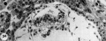
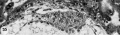
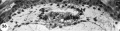
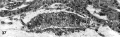
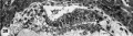
Surface View
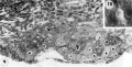
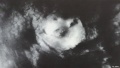
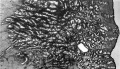
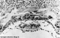
This is a uterine surface view showing the site of implantation (pale region in centre of image). The conceptus can be identified located at the dark central region within the pale region.

Carnegie Collection
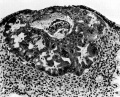
Stage 5a
- Streeter Horizon Va
Carnegie 8225 7.5 day
Carnegie 8020 7.5 day
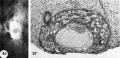
Carnegie 8155 8 day


- Carnegie 8155
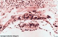
Embryo No.8155
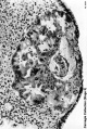
Embryo No.8155
Embryo No.8155


Stage 5b
- Streeter Horizon Vb - 9 day
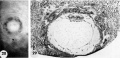
Carnegie 8215 embryo, chorionic cavity and trophoblast
Carnegie 8171 predecidual reaction
Carnegie 8171 embryo and trophoblast
Carnegie 8004 endometrium and intact specimen
Carnegie 8004 embryo, chorionic cavity and trophoblast


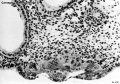
- Embryo No. 8004
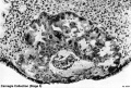


Stage 5c
- Streeter Horizon Vc - Surface and Cross-section view
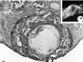
Carnegie 7699 11 day
Carnegie 7950 12 day
Carnegie 7700 12 day
- Hertig1956 fig31-32.jpg
Carnegie 855812-day
Carnegie 8330 12-day


- Streeter Horizon Vc - Embryonic Disc
Carnegie 7699 11 day
Carnegie 7950 12 day
Carnegie 7700 12 day
Carnegie 8558 12 day
Carnegie 8330 12 day
Historic terminology is "germ disc"
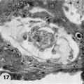
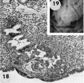
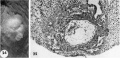
- Carnegie Embryo 7700
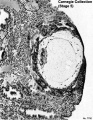
Embryo No.7700
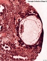
Embryo No.7700
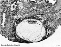
Embryo No.7700
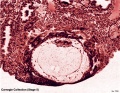
Embryo No.7700
Embryo No.7700
Embryo No.7700
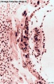
Embryo No.7700
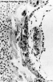
Embryo No.7700


| Carnegie Collection - Stage 5 | ||||||||||
|---|---|---|---|---|---|---|---|---|---|---|
| Serial No. | Stage | Grade | Fixative | Embedding Medium | Thinness (µm) | Stain | Year | Notes | ||
| 8020 | 5a | Exc. | Alc. & Bouin | C-P | 6 | (Stain - Haematoxylin Eosin) | 1942 | Hertig and Rock (1945a)[1] | ||
| 8155 | 5a | Exc. | Bouin | C-P | 6 | (Stain - Haematoxylin Eosin) | 1943 | Hertig and Rock (1949)[2] | ||
| 8225 | 5a | Exc. | Alc. & Bouin | C-P | 6 | (Stain - Haematoxylin Eosin) | 1944 | Hertig and Rock (1945b)[3] | ||
| 8004 | 5b | Exc | Alc. & Bouin | C-P | 6 | (Stain - Haematoxylin Eosin) | 1942 | Hertig and Rock (1945a)[1] | ||
| 8171 | 5b | Exc | Alc. | C-P | 6 | (Stain - Haematoxylin Eosin) | 1943 | Hertig and Rock (1949)[2] | ||
| 8215 | 5b | Exc | Alc. & Bouin | C-P | 6 | (Stain - Haematoxylin Eosin) | 1944 | Hertig and Rock (1945c)[4] | ||
| 9350 | 5b | Exc | Bouin | ? | ? | (Stain - Haematoxylin Eosin) | 1955 | Heuser (1956)[5] | ||
| 4900 | 5c | Poor | p | P | 10 | p | 1925 | Incomplete. Streeter (1926)[6] | ||
| 7699 | 5c | Exc. | Bouin | C-P | 6 | (Stain - Haematoxylin Eosin) | 1939 | Hertig and Rock (1941)[7] | ||
| 7700 | 5c | Exc. | Bouin | C-P | 6 | (Stain - Haematoxylin Eosin) | 1938 | Hertig and Rock (1941)[7] | ||
| 7771 | 5c | Exc. | Bouin | C-P | 10 | (Stain - Haematoxylin Eosin) | 1940 | Abnormal | ||
| 7950 | 5c | Exc. | Alc. & Bouin | C-P | 6 | (Stain - Haematoxylin Eosin) | 1941 | Hertig and Rock (1944)[8] | ||
| 8000 | 5c | Poor | Alc. & Bouin | C-P | 8 | (Stain - Haematoxylin Eosin) | 1942 | Abnormal | ||
| 8139 | 5c | Exc. | ? | C-P | 6 | (Stain - Haematoxylin Eosin) | 1943 | Incomplete. Marchetti (1945)[9] | ||
| 8299 | 5c | Exc. | Alc. & Bouin | C-P | 6 | (Stain - Haematoxylin Eosin), phlox. | 1945 | Abnormal | ||
| 8329 | 5c | Exc. | Alc. & Bouin | C-P | 6 | (Stain - Haematoxylin Eosin), phlox. | 1945 | Abnormal | ||
| 8330 | 5c | Exc. | Alc. & Bouin | C-P | 6 | (Stain - Haematoxylin Eosin), phlox. | 1945 | |||
| 8370 | 5c | Poor | Alc. & Bouin | C-P | 6 | (Stain - Haematoxylin Eosin), phlox. | 1946 | Abnormal | ||
| 8558 | 5c | Exc. | Alc. & Bouin | C-P | 6 | (Stain - Haematoxylin Eosin) | 1947 | |||
| Stage 5 was originally subdivided into 3 sequential parts a, b, c.
Abbreviations
| ||||||||||
References
| ||||||||||
| iBook - Carnegie Embryos | |
|---|---|

|
|
Historic Stage 5 Embryos
Stage 5a
- Carnegie No. 8225 - Described briefly by Hertig and Rock (1945b). Hysterectomy (bicornuate uterus). Anterior wall of uterus. Chorion, 0.33 x 0.306 mm. Chorionic cavity, 0.228 x 0.2 mm. Embryonic disc, 0.09 x 0.078 mm. Perhaps more advanced than No. 8020 (Mazanec, 1959; Harris and Ramsey, 1966), but has also been interpreted as less advanced.[1] Photomicrograph in Hertig, Rock, and Adams (1956, fig. 9). Presumed age, 7 days.
- Carnegie No. 8020 - (figs. 5-3 to 5-5). Described by Hertig and Rock (1945a).[2] Hysterectomy. Posterior wall of uterus. Chorion, 0.45 x 0.3 mm. Chorionic cavity, 0.288 x 0.186 mm. Embryonic disc, 0.126 x 0.092 mm. New model of blood vessels at implantation site has been prepared (Harris and Ramsey, 1966). Presumed age, 7 days.
- Fruhling, Ginglinger, and Gandar (1954) described briefly a specimen of about 8 days. Curettage. Early implantation. Few trophoblastic digitations. Beginning amniotic cavity. Most sections through embryonic disc lost.
- Carnegie No. 8155 - (fig. 14). Described by Hertig and Rock (1949).[3] Hysterectomy. Anterior wall of uterus. Chorion, 0.306 x 0.210 mm. Chorionic cavity, 0.168 x 0.082 mm. Embryonic disc, 0.09 x 0.05 mm. “Tropho-epiblastic cavity” (Luckett, 1975).
Stage 5b
- Carnegie No. 8171 - Described by Hertig and Rock (1949). Hysterectomy. Posterior wall of uterus. Abnormal leucocytic infiltration of endometrium. Chorion, 0.422 x 0.404 mm. Chorionic cavity, 0.164 X 0.138 mm. Embryonic disc, 0.114 x 0.088 mm. A cellular remnant within the umbilical vesicle, because it is probably derived from the endoderm, “may, in a sense, be regarded as an abnormal form of twin embryo” (ibid.). Presumed age, 9 days.
- Carnegie No. 8215 - Described briefly by Hertig and Rock (1945c). Hysterectomy. Posterior wall of uterus. Chorion, 0.525 X 0.498 mm. Chorionic cavity, 0.228 X 0.21 mm. Embryonic disc, 0.084 x 0.052 mm. Lacunae perhaps further developed than in No. 8171 (Mazanec, 1959), but specimen has been “considered to be slightly younger because the decidual reaction is not yet apparent” (Hertig, Rock, and Adams, 1956). Photomicrographs in Hertig, Rock, and Adams (1956, figs. 15 and 17). Presumed age, 9 days.
- Carnegie No. 8004 - (figs. 5-7 to 5-9). Described by Hertig and Rock (1945a). Hysterectomy. Posterior wall of uterus. Chorion, 0.582 x 0.45 mm. Chorionic cavity, 0.312 X 0.185 mm. Embryonic disc, 0.132 x 0.1 mm.
- Carnegie No. 9350 - Described briefly by Heuser (1956). Hysterectomy. At junction of posterior and anterior walls. Chorion, 0.59 x 0.58 mm. Chorionic cavity, 0.3 X 0.1 mm. Embryonic disc, 0.132 x 0.09 mm. Presumed age, 9 days.
Stage 5c
- Davies-Harding. Described by Davies (1944). Hysterectomy. Anterior wall of uterus. Incomplete (almost one-half of embryonic disc missing). “No true villi.” Primary umbilical vesicle present. Extensive extra-embryonic meshwork. Slightly later stage of development than No. 8004 (Davies, 1944, Addendum), which belongs to 5b. “Possibly pathological” (Boyd and Hamilton, 1970). Chorion, 1.18 x 0.55 mm. Chorionic cavity, 0.409 x 0.238 mm. Embryonic disc, 0.117 mm. May be regarded as transitional between 5b and 5c. Presumed age, 9-10 days.
- Carnegie No. 7699 - Described by Hertig and Rock (1941). Hysterectomy. Posterior wall of uterus. Chorion, 1.026 x 0.713 mm. Chorionic cavity, 0.48 x 0.336 mm. Embryonic disc, 0.138 x 0.138 mm. New model of blood vessels at implantation site has been prepared (Harris and Ramsey, 1966). Presumed age, 11 days.
- Carnegie No. 4900 - Miller. Described by Streeter (1926). Curettage. Angiogenesis described by Hertig (1935). Incomplete (some sections missing). Primary umbilical vesicle present. New graphic reconstruction made by Streeter (1939a,b). Chorion, 0.9 mm. Chorionic cavity, 0.4 mm. Presumed age, 10-11 days or perhaps even 12 days (Krafka,1941).
- Dible-West. Described by Dible and West (1941). Autopsy. Posterior wall of uterus. Incomplete. Chorionic cavity, 0.47 x 0.28 mm. Embryonic disc, 0.1 x 0.02 mm. Presumed age, 11-13 days.
- Müller (1930) described an autopsy specimen. Only one section was near the embryonic disc.
- Wilson (1954) found a specimen by endometrial biopsy. Chorion, 0.5 x 0.6 mm. Chorionic cavity, 0.47 x 0.24 mm. Between the amnion and the cytotrophoblast, “a small accumulation of fibroblastic cells probably represents the earliest stage of the Bauchstiel.” The endoderm is described as being “delaminated from the embryonic disc.” Presumed age, 11 days.
- Carnegie No. 7950 - Described briefly by Hertig and Rock (1944). Chorion, 0.75 x 0.45 mm. Chorionic cavity, 0.4 x 0.26 mm. Embryonic disc, 0.16 x 0.07 mm. Slightly more developed than No. 7699, although embryo is a little less differentiated. Photomicrographs in Hertig, Rock, and Adams (1956; figs. 27 and 35). Presumed age, 12 days.
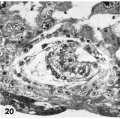
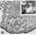
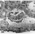
- Carnegie No. 7700 (figs. 18-22). Described by Hertig and Rock (1941). Hysterectomy. Posterior wall of uterus. Chorion, 0.948 x 0.835 mm. Chorionic cavity, 0.55 x 0.498 mm. Embryonic disc, 0.204 x 0.165 mm. Presumed age, 12 days.
- Carnegie No. 8558 - Measurements and photomicrographs in Hertig, Rock, and Adams (1956; figs. 30 and 37). Chorion, 0.96 x 0.52 mm. Chorionic cavity, 0.58 x 0.36 mm. Embryonic disc, 0.22 x 0.08 mm. Presumed age, 12 days.
- Carnegie No. 8330 - Measurements and photomicrographs in Hertig, Rock, and Adams (1956; figs. 30 and 37). Chorion, 0.85 x 0.65 mm. Chorionic cavity, 0.46 x 0.4 mm. Embryonic disc, 0.216 x 0.063 mm. Condensation of extra-embryonic mesoblast perhaps indicates “the beginning of axis formation” (ibid.). Presumed age, 12 days.
- Kleinhans. Described by Grosser (1922). Autopsy. Only one section. Embryonic disc not seen. Chorion, 0.8 x 0.65 mm. Chorionic cavity, 0.35 x 0.15 mm.
- Barnes. Described by Hamilton, Barnes, and Dodds (1943), and further by Hamilton and Boyd (1960). Hysterectomy. Posterior wall of uterus. Pathological edema in endometrium (Davies, 1944). Chorion, 0.931 x 0.77 x 0.737 mm. Primary umbilical vesicle and perhaps early differentiation of secondary vesicle (Luckett, 1978). Presumed age, 10-11 days or perhaps 12 days (Davies, 1944).
- Werner (Prof. Werner Gerlach). Described by Stieve (1936), with graphic reconstruction by Florian. Autopsy. Chorion, 0.78 x 1.36 x 0.72 mm. Embryonic disc, 0.18 x 0.12 mm. Some large, round cells in the epiblast are mentioned as possible primordial germ cells. Primary umbilical vesicle and perhaps early differentiation of secondary vesicle (Luckett, 1978). Primordia (mesoblastic crests) of chorionic villi (Hertig and Rock, 1941). ‘Connecting stalk’ very indistinct. Rostrocaudal axis but no primitive streak; epiblast fused with extra-embryonic mesoblast caudally and perhaps is origin of latter (Florian, 1933 and 1945). Perhaps 12 days.
- Knoth and Larsen (1972) studied an implantation site by electron microscopy. No villi, but “beginning” to form. Primary umbilical vesicle “not so easily seen.” Probably 11 days.
- Dankmeijer and Wielenga (1968) described a specimen of stage 5. Curettage. Incomplete.
- Carnegie No. 8139. Described by Marchetti (1945), who admitted that it is “not entirely normal.” Curettage. Chorion, 0.706 x 1.2 mm. Chorionic cavity, 0.635 x 0.582 mm. Embryonic disc, 0.126 x 0.048 x 0.116 mm. Embryo located centrally in chorionic cavity, which contains a meshwork. Embryonic disc, slightly ovoid (i.e., presents a longitudinal axis). Described originally as previllous, although “primitive villi” (stage 6) are mentioned by Boyd and Hamilton (1970): folds of undulating contour of cytotrophoblast are not yet “primitive unbranching villi, although they may be the primordial of them” (Marchetti, 1945; fig. 5). Primary umbilical vesicle present. May be regarded as transitional between stages 5 and 6. Presumed age, 13 days.
- Bandler (1912) found a specimen embedded in tubal mucosa and which showed “as yet absolutely no suggestion of chorionic villi.” No details available.
- Pommerenke (1958) described a specimen of stage 5. Curettage. Incomplete. Embryo not included.
- Macafee. Described by Morton (1949). Curettage. Probably belongs to stage 5. Embryonic disc not found.
- Scipiades (1938) described a specimen that belongs either to stage 5 or to stage 6, probably the former, “but its preservation is so poor that accurate conclusions are impossible” (Hertig and Rock, 1941). Curettage. Chorion, 1.498 x 0.49 mm. Chorionic cavity, 0.99 mm. Embryonic disc (only two sections available), 0.18 x 0.048 mm. Presumed age, 11-12 days.
- Thomas and van Campenhout (1963) found a specimen that probably belongs to stage 5, although some trophoblastic thickenings of a villous character were said to be present.
- Teacher-Bryce I. A pathological specimen of stage 5 described by Bryce (1924).
- Sch. (Schönig). A pathological specimen of stage 5 described by von Möllendorff (1921a).
- Keller and Keller (1954) found a pathological specimen embedded in the stroma of the ostium uteri.
Several pathological specimens of stage 5c are in the Carnegie Collection. Nos. 8370, 7770, 8299 (malpositioned embryonic disc, Hertig, 1968, fig. 132), 8329, 8000 (superficial implantation), and 7771 (no embryo) were measured and illustrated by Hertig, Rock, and Adams (1956).[1]
Events
- Mitosis - cells on surface of blastocyst (trophoblast cells) divide more rapidly than the inner cells (inner cell mass, embryoblast)
- Implantation - adhesion to endometrium epithelium and implantation through this layer into the underlying uterine storm.
- Endocrine signalling - trophoblast cells secrete hCG (human Chorionic Gonadotropin/Gonadotrophin) the signal of pregnancy.
- Cell invasion and fusion - trophoblast cells spread through the maternal uterine wall and also fuse together to form specialised multi-nucleated cells.
- Corpus luteum - signal from trophoblasts maintain this endocrine tissue in the maternal ovary that secretes progesterone.
References
- ↑ 1.0 1.1 Hertig AT. Rock J. and Adams EC. A description of 34 human ova within the first 17 days of development. (1956) Amer. J Anat., 98:435-493.
- ↑ Template:Ref-1945a
- ↑ Template:Ref-1949
- ↑ <pubmed></pubmed>
- Carnegie Stages: 1 | 2 | 3 | 4 | 5 | 6 | 7 | 8 | 9 | 10 | 11 | 12 | 13 | 14 | 15 | 16 | 17 | 18 | 19 | 20 | 21 | 22 | 23 | About Stages | Timeline
Glossary Links
- Glossary: A | B | C | D | E | F | G | H | I | J | K | L | M | N | O | P | Q | R | S | T | U | V | W | X | Y | Z | Numbers | Symbols | Term Link
Cite this page: Hill, M.A. (2024, June 16) Embryology Carnegie stage 5. Retrieved from https://embryology.med.unsw.edu.au/embryology/index.php/Carnegie_stage_5
- © Dr Mark Hill 2024, UNSW Embryology ISBN: 978 0 7334 2609 4 - UNSW CRICOS Provider Code No. 00098G