Category:Cardiovascular: Difference between revisions
From Embryology
mNo edit summary |
mNo edit summary |
||
| Line 1: | Line 1: | ||
This | This {{Embryology}} category shows pages, images and media related to cardiovascular system development. | ||
See also the narrower categories [[:Category:Heart]] and [[:Category:Blood]]. | See also the narrower categories [[:Category:Heart]] and [[:Category:Blood]]. | ||
{{Heart Links}} | {{Heart Links}} | ||
Latest revision as of 12:39, 13 February 2017
This Embryology category shows pages, images and media related to cardiovascular system development.
See also the narrower categories Category:Heart and Category:Blood.
Subcategories
This category has the following 13 subcategories, out of 13 total.
Pages in category 'Cardiovascular'
The following 200 pages are in this category, out of 519 total.
(previous page) (next page)H
- HM Practical - Blood Vessel Histology
- HM Practical - Cardiac Histology
- Template:Hofbauer cell
- Template:Hofbauer cells
- Template:Human Embryology 2 18-6 Dorsal segmental artery table1
- Template:Human Embryology 2 18-6 Dorsal segmental artery table2
- Template:Human Embryology 2 18-6 Mesonephric artery table3
- Human System Development
- Template:Hypoplastic left heart
- Hypoplastic Left Heart Syndrome Movie
I
- Template:ICD-10-circulatory system Q20-Q28 table
- Template:Immune
- Intermediate - Atrial Ventricular Septation
- Intermediate - Cardiac Abnormalities
- Intermediate - Heart Tube Looping
- Intermediate - Heart Tube Looping 2
- Intermediate - Heart Tube Looping 3
- Intermediate - Heart Valves
- Intermediate - Outflow Tract
- Intermediate - Primordial Heart Tube
- Intermediate - Vascular Overview
- Intermediate Cardiac Embryology
- Template:Intermediate Cardiac menu
L
M
P
- Paper - A case of early ectopic gestation
- Paper - A congenital anomaly of the heart - truncus arteriosus communis (1927)
- Paper - A contribution to the early development of the heart in mammalia, with special reference to the guinea pig
- Paper - A Contribution to the Embryology of the Liver and Vascular System in Man
- Paper - A rare vascular anomaly-opening of the upper left pulmonary vein into a persistent left superior vena cava (1915)
- Paper - A specimen showing complete remains of the left superior vena cava (1915)
- Paper - A study of wandering mesenchymal cells on the living yolk-sac and their developmental products (1915)
- Paper - Coarctation of the aorta 1942
- Paper - Complete situs inversus of the vena cava superior (1930)
- Paper - Congenital deficiency of the pericardium (1916)
- Paper - Cor biloculare, with a note on the development of the pulmonary veins (1937)
- Paper - Development and vascularization of the testis (1906)
- Paper - Development of postcava and tributaries in the domestic cat (1907)
- Paper - Development of the great anterior veins in man and mammalia
- Paper - Development of the human heart from its earliest appearance to the stage found in embryos of twenty paired somites (1927)
- Paper - Development of the inferior vena cava (1929)
- Paper - development of the postcaval vein in birds (1903)
- Paper - Development of the vascular system in five to twenty-one somite dog embryos
- Paper - Developmental Changes in the Pericardium, the Mesocardia, and the Pleural Sacs in the Human Embryo
- Paper - Developmental defects at the foramen ovale (1938)
- Paper - Equivalence of different hematopoietic anlages 1 (1916)
- Paper - First contractions of the heart without cytological differentiation
- Paper - Four cases of anomalous inferior vena cava with an explanation of their developmental origin (1928)
- Paper - Functional limitations of the foramen ovale in the human foetal heart
- Paper - Further evidence on the origin of the lymphatic endothelium from the endothelium of the blood-vascular system
- Paper - General observations on early superficial lymphatics in living chick embryos (1912)
- Paper - Growth allometry of the myocardium in human embryos from stages 15 to 23
- Paper - Hematopoiesis in young human embryos
- Paper - Histogenesis of the heart muscle of the pig in relation to the appearance and development of the intercalated discs (1919)
- Paper - Histogenesis of the human aorta
- Paper - Initiation and early changes in the character of the heart beat in vertebrate embryos
- Paper - Injection and reconstruction of the jugular lymph sac in the chick (1912)
- Paper - Migration processes during ontogeny with reference to the venous development in the dorsal body wall (1946)
- Paper - Normal haemopoiesis in intra-uterine and neonatal life (1941)
- Paper - Observations on the development of the earliest lymphatics in the region of the posterior lymph heart in living chick embryos (1912)
- Paper - Observations upon the occurrence, structure and function of the giant cells of the marrow (1890)
- Paper - On the cervical veins and lymphatics in four human embryos, with an interpretation of anomalies on the subclavian and jugular veins in the adult (1909)
- Paper - On the development of the aortae cardinal and umbilical veins and the other blood vessels of vertebrate embryos from capillaries (1909)
- Paper - On the development of the atrial septum and the valvular apparatus in the right atrium of the pig embryo (1916)
- Paper - On the development of the blood-vessels of the brain in the human embryo (1905)
- Paper - On the Development of the Human Heart
- Paper - On the earliest blood-vessels in the anterior limb-buds of birds and their relation to the primary subclavian artery
- Paper - On the fate of the posterior cardinal veins and their relation to the development of the vena cava and azygos in the embryo pig (1915)
- Paper - On the origin of the abdominal lymphatics in mammals from the vena cava and the renal veins (1912)
- Paper - On the origin of the lymphatic system from the veins and the development of the lymph hearts and thoracic duct in the pig (1902)
- Paper - On the origin of the pulmonary arteries in mammals
- Paper - On the origin of the pulmonary arteries in mammals 2
- Paper - On the placentation of the macaque (Macaca mulatta), from the time of implantation until the formation of the definitive placenta
- Paper - On the position of the vitelline arteries in human embryos
- Paper - On the time of the post-natal obliteration of the fetal blood-passages (1918)
- Paper - Origin and development of the primitive vessels of the chick and of the pig (1917)
- Paper - Origin of the pulmonary vessels in the chick (1922)
- Paper - Origin, development and degeneration of the blood vessels of the ovary (1899)
- Paper - Persistence of the left posterior cardinal vein (1911)
- Paper - Persistent left superior vena cava, left duct of cuvier and left horn of the sinus venosus
- Paper - Persistent left superior vena cava, left duct of cuvier and left horn of the sinus venosus (1930)
- Paper - Preliminary note on the differentiation of angioblasts in the living chick (1917)
- Paper - Significant superficial anastomoses in the arterial blood supply to the human brain (1959)
- Paper - Six specimens of abnormal heart (1912)
- Paper - Some abnormal developments in the vascular system of the frog (rana temporaria) (1915)
- Paper - Some observations on the cardio-vascular system in the viable foetal lamb (1940)
- Paper - Studies on the area vasculosa of the embryo chick 2 (1937)
- Paper - Teratogenecity in the setting of cardiac development and maldevelopment
- Paper - The aortic arch derivatives in human adult (1951)
- Paper - The circle of Willis - An examination of 700 specimens (1905)
- Paper - The course of the blood flow through the fetal mammalian heart
- Paper - The course of the blood through the heart of the fetal mammal, with a note on the reptilian and amphibian circulations (1909)
- Paper - The development of the aorta and aortic arches in rabbits
- Paper - The development of the arteries of the human lower extremity
- Paper - The Development of the Atrio-Ventricular Valves in Man
- Paper - The development of the cardiac loop in the rabbit with especial reference to the bulboventricular groove and origin of the interventricular septum (1919)
- Paper - The development of the cardiac-coronary circulatory system
- Paper - The development of the cranial arteries in the human embryo
- Paper - The development of the cranial venous system in man, from the viewpoint of comparative anatomy
- Paper - The development of the heart in man
- Paper - The development of the mammalian spleen, with special reference to its hematopoietic activity (1921)
- Paper - The Development of the Pars Membranacea Septi in the Human Heart
- Paper - The development of the principal arterial stems in the forelimb of the pig (1922)
- Paper - The development of the pulmonary vein in the domestic cat (1913)
- Paper - The development of the subcutaneous vascular plexus in the head of the human embryo (1923)
- Paper - The development of the vascular system in the human embryo prior to the establishment of the heart
- Paper - The development of the veins in the limbs of rabbit embryos
- Paper - The development of the vena cava inferior (1902)
- Paper - The development of the vena cava inferior in man (1925)
- Paper - The development of the venous sinuses of the dura mater in the human embryo
- Paper - The developmental alterations in the vascular system of the brain of the human embryo (1921)
- Paper - The ductus arteriosus in the human fetus and newborn infant
- Paper - The ductus venosus in the fetus and in the adult (1923)
- Paper - The Earliest Blood-Vessels in Man
- Paper - The earliest stages of development of the blood-vessels and of the heart in ferret embryos
- Paper - The earliest stages of development of the blood-vessels and of the heart in ferret embryos 2
- Paper - The early development of the sheep heart (1946)
- Paper - The early stages of the development of the pericardium
- Paper - The effect of the heart-beat upon the development of the vascular system in the chick (1918)
- Paper - The equivalence of different homatopoietic anlages by method of stimulation of the different stem cells 1
- Paper - The equivalence of different homatopoietic anlages by method of stimulation of the different stem cells 2
- Paper - The fifth aortic arch of mammalian embryos; the nature of the last pharyngeal evagination
- Paper - The first contractions of the heart in rat embryos
- Paper - The form and the functions of the uterine blood vessels in the rhesus monkey
- Paper - The formation of the cardiac loop in the chick
- Paper - The Formation of the Pars Membranacea Septi (1916)
- Paper - The formation of the venous valves, the foramen secundum and the septum secundum in the human heart
- Paper - The frequency of an opening between the right and left auricles at the seat of the foetal foramen ovale (1900)
- Paper - The fusion of the cardiac anlages and the formation of the cardiac loop in the cat (1916)
- Paper - The genesis, development, and adult anatomy of the nasofrontal region in man
- Paper - The genetic interpretation of the development of the mammalian lymphatic system (1908)
- Paper - The genetic principles of the development of the systemic lymphatic vessels in the mammalian embryo (1910)
- Paper - The human embryonic heart in the ninth week
- Paper - The human embryonic heart in the ninth week (1954)
- Paper - The human embryonic heart in the seventh week (1962)
- Paper - The life-history of the formed elements of the blood, especially the red blood corpuscles (1890)
- Paper - The morphology of human uteroplacental vasculature
- Paper - The nerve supply of the mammalian ductus arteriosus (1941)
- Paper - The origin and development of the carotid body (1924)
- Paper - The origin and early development of the posterior lymph heart in the chick (1915)
- Paper - The origin and occurrence of the single umbilical artery in normal and abnormal human fetuses (1922)
- Paper - The origin of blood and vascular endothelium in embryos without a circulation of the blood and in the normal embryo (1915)
- Paper - The origin of blood cells (1916)
- Paper - The origin of the heart and blood vessels in felis domestica (1924)
- Paper - The origin, development and function of the blood cells in certain marine teleosts 1 (1939)
- Paper - The partitioning of the truncus and conus and the formation of the membranous portion of the interventricular septum in the human heart (1942)
- Paper - The phylogenetic relations of the lymphatic and bloodvascular systems in vertebrates (1910)
- Paper - The physiology of the embryonic mammalian heart before circulation
- Paper - The relation between the size of the artery and the capillary bed in the embryo (1937)
- Paper - The relative role played by the embryonic veins in the development of the mammalian vena cava posterior
- Paper - Three examples of a right aortic arch
- Paper - Time and rate of loss of nuclei by the red blood cells of human embryos
- Paper - Transformation of the aortic-arch system during the development of the human embryo (1922)
- Paper - Transposition of the ventricles and the arterial stems (1931)
- Paper - True congenital diverticulum of the trachea in a subject showing also right aortic arch (1929)
- Paper - Two cases considered from the developmental standpoint in which the right subclavian artery arose from the arch of the aorta (1915)
- Paper - Variations and anomalies of the venous valves of the right atrium of the human heart (1929)
- Paper - Wilhelm His - His relation to the institution of learning
- Paper The development of the subcutaneous vascular plexus in the head of the human embryo (1923)
- Paper- The primary divisions of the myocardium in the human embryo
- Template:Patent ductus arteriosus
- Patent Ductus Venosus Movie
- Template:PDGF
- Template:Persistent right umbilical vein
- Template:Pia mater
- Template:Placenta vascular
- Template:Placenta vascular bed
- Template:Placental cord
- Template:Placental villi
- Template:Pre-eclampsia
Media in category 'Cardiovascular'
The following 200 files are in this category, out of 695 total.
(previous page) (next page) Keith1902 fig204.jpg 918 × 700; 0 bytes
 Keith1902 fig205.jpg 1,079 × 800; 164 KB
 Keith1902 fig206.jpg 896 × 700; 105 KB
 Keith1902 fig207.jpg 703 × 550; 44 KB
 Keith1902 fig208.jpg 556 × 550; 45 KB
 Keith1902 fig209.jpg 926 × 650; 67 KB
 Keith1902 fig210.jpg 808 × 750; 65 KB
 Keith1921 fig026.jpg 995 × 346; 71 KB
 Keith1921 fig129.jpg 827 × 683; 96 KB
 Kollmann512.jpg 737 × 842; 100 KB
 Kollmann528.jpg 744 × 587; 91 KB
 Kollmann529.jpg 737 × 593; 94 KB
 Kollmann530.jpg 733 × 524; 83 KB
 Kollmann531.jpg 688 × 570; 88 KB
 Kollmann532.jpg 702 × 409; 59 KB
 Kollmann533.jpg 717 × 594; 76 KB
 Kollmann534.jpg 684 × 599; 74 KB
 Kollmann535.jpg 646 × 577; 81 KB
 Kollmann536.jpg 655 × 577; 64 KB
 Kollmann537.jpg 481 × 489; 45 KB
 Kollmann538.jpg 519 × 472; 42 KB
 Kollmann539.jpg 736 × 696; 132 KB
 Kollmann540.jpg 746 × 376; 62 KB
 Kollmann541.jpg 757 × 535; 74 KB
 Kollmann542.jpg 732 × 520; 74 KB
 Kollmann543.jpg 739 × 445; 63 KB
 Kollmann544.jpg 731 × 379; 74 KB
 Kollmann545.jpg 731 × 568; 107 KB
 Kollmann546.jpg 718 × 491; 99 KB
 Kollmann547.jpg 519 × 475; 57 KB
 Kollmann548.jpg 738 × 525; 72 KB
 Kollmann549.jpg 689 × 573; 75 KB
 Kollmann550.jpg 852 × 667; 156 KB
 Kollmann551.jpg 776 × 688; 126 KB
 Kollmann552.jpg 634 × 469; 47 KB
 Kollmann553.jpg 724 × 734; 83 KB
 Kollmann554.jpg 625 × 568; 54 KB
 Kollmann555.jpg 605 × 585; 61 KB
 Kollmann556.jpg 659 × 588; 86 KB
 Kollmann557.jpg 568 × 570; 82 KB
 Kollmann558.jpg 743 × 852; 170 KB
 Kollmann559.jpg 720 × 505; 76 KB
 Kollmann560.jpg 705 × 457; 62 KB
 Kollmann561.jpg 739 × 591; 82 KB
 Kollmann562.jpg 670 × 581; 68 KB
 Kollmann563.jpg 754 × 848; 143 KB
 Lewis1906 fig425.jpg 1,000 × 1,191; 200 KB
 Lewis1909 fig01.jpg 600 × 710; 60 KB
 Lewis1909 fig02.jpg 500 × 733; 67 KB
 Lewis1909 fig03.jpg 1,329 × 1,132; 189 KB
 Lewis1909 fig04.jpg 1,000 × 1,027; 180 KB
 Lewis1909 fig05.jpg 1,280 × 1,013; 374 KB
 Lewis1909 fig06.jpg 1,000 × 991; 110 KB
 Lymphatic vessel formation model.jpg 600 × 692; 179 KB
 Mall1912-fig01.jpg 969 × 1,100; 365 KB
 Mall1912-fig02.jpg 797 × 730; 180 KB
 Mall1912-fig03.jpg 627 × 600; 95 KB
 Mall1912-fig04.jpg 832 × 650; 151 KB
 Mall1912-fig05.jpg 900 × 836; 177 KB
 Mall1912-fig06.jpg 1,000 × 991; 184 KB
 Mall1912-fig07.jpg 652 × 600; 93 KB
 Mall1912-fig08.jpg 800 × 471; 82 KB
 Mall1912-fig09.jpg 700 × 800; 158 KB
 Mall1912-fig10.jpg 800 × 550; 136 KB
 Mall1912-fig11.jpg 1,000 × 725; 124 KB
 Mall1912-fig12.jpg 816 × 800; 212 KB
 Mall1912-fig13.jpg 800 × 631; 173 KB
 Mall1912-fig14.jpg 538 × 600; 66 KB
 Mall1912-fig15.jpg 600 × 385; 33 KB
 Mall1912-fig16.jpg 735 × 650; 59 KB
 Mall1912-fig17.jpg 574 × 500; 39 KB
 Mall1912-fig18.jpg 600 × 425; 38 KB
 Mall1912-fig19.jpg 900 × 629; 77 KB
 Mall1912-fig20.jpg 400 × 494; 56 KB
 Mall1912-fig21.jpg 760 × 582; 141 KB
 Mall1912-fig22.jpg 800 × 600; 116 KB
 Mall1912-fig23.jpg 800 × 594; 159 KB
 Mall1912-fig24-25.jpg 850 × 1,000; 241 KB
 Mall1912-fig26.jpg 800 × 769; 150 KB
 Mall1912-fig27.jpg 542 × 600; 41 KB
 Mall1912-fig28.jpg 731 × 1,000; 396 KB
 Mall1912-fig29.jpg 472 × 580; 47 KB
 Mall1912-fig30.jpg 438 × 500; 58 KB
 Mall1912-fig31-33.jpg 963 × 1,000; 94 KB
 Mall1912-fig34-37.jpg 1,000 × 774; 96 KB
 McClure1925 fig16.jpg 1,000 × 919; 212 KB
 Model coupling hematopoiesis with osteopoiesis.jpg 486 × 600; 44 KB
 Model for granulocytic nuclear lobulation.jpg 600 × 930; 85 KB
 Molecular & Genetic Cardiac Development Factors.jpg 1,475 × 1,541; 269 KB
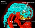
 Mouse E9 cervical intersomitic vessels A.jpg 1,714 × 1,142; 209 KB
 Mouse E9 cervical intersomitic vessels B.jpg 1,714 × 1,142; 175 KB
 Mouse E9 cervical intersomitic vessels C.jpg 1,714 × 1,142; 245 KB
 Mouse E9 cervical intersomitic vessels D.jpg 1,714 × 1,142; 147 KB
 Mouse E9 cervical intersomitic vessels.jpg 2,989 × 2,300; 777 KB
 Mouse embryo vascular.png 600 × 576; 518 KB
 Mouse heart E9.5.jpg 600 × 558; 31 KB
 Mouse hematopoietic stem cell.gif 600 × 595; 40 KB
 Mouse osteoblast 01.jpg 599 × 449; 118 KB
 Mouse osteoclast 01.jpg 600 × 453; 153 KB
 Mouse placenta blood vessel EM01.jpg 1,000 × 739; 132 KB
 Mouse-Cephalic-plexus-11somite 01.jpg 758 × 598; 106 KB
 Mouse-coronary vessel formation.jpg 800 × 282; 44 KB
 Mouse-heart E17.5.jpg 353 × 1,000; 145 KB
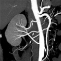 Multiple renal arteries 01.jpg 496 × 496; 40 KB
 Notch and yolk sac blood vessels model.jpg 600 × 775; 97 KB
 NOTCH-endothelial-cartoon.jpg 1,280 × 888; 92 KB
 ORahilly1987 fig20-2.jpg 600 × 549; 46 KB
 Outflow tract 001 icon.jpg 720 × 540; 33 KB
 Partial Anomalous Pulmonary Venous Drainage.jpg 290 × 350; 16 KB
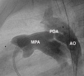 Patent ductus arteriosus angiogram.jpg 1,000 × 897; 91 KB
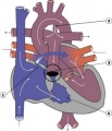
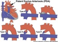 Patent ductus arteriosus classification.jpg 1,200 × 880; 138 KB
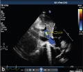
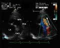 Patent ductus arteriosus echocardiogram.jpg 1,200 × 960; 133 KB
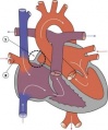
 Patent Ductus Arteriosus.jpg 294 × 350; 16 KB
 Patten024.jpg 1,047 × 451; 75 KB
 Patten047.jpg 785 × 765; 165 KB
 Patten048.jpg 797 × 857; 174 KB
 Patten049.jpg 807 × 864; 119 KB
 Patten050.jpg 800 × 890; 109 KB
 Patten054.jpg 764 × 1,060; 179 KB
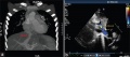
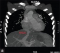
 Persistent fifth aortic arch and patent ductus arteriosus CT01.jpg 589 × 800; 69 KB
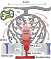 Placenta lobule blood flow cartoon.jpg 692 × 800; 134 KB
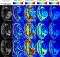 Placenta MRI01.jpg 1,280 × 1,227; 317 KB
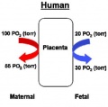 Placenta oxygen exchange levels.jpg 404 × 401; 18 KB
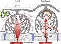 Placenta spiral artery conversion.jpg 592 × 423; 65 KB
 Placental cord vessels 01.jpg 650 × 488; 51 KB
 Placental cord vessels 02.jpg 800 × 477; 65 KB
 Platypus right auricle 01.jpg 800 × 640; 124 KB
 Platypus ventricular septum 01.jpg 683 × 800; 94 KB
 Postnatal persistant ductus venosus ultrasound 01.jpg 1,370 × 600; 96 KB
 Postnatal persistant ductus venosus ultrasound 02.jpg 671 × 600; 44 KB
 Postnatal persistant ductus venosus ultrasound 03.jpg 694 × 600; 54 KB
 Prentiss 286.jpg 799 × 933; 121 KB
 Pulmonary Atresia.jpg 301 × 350; 17 KB
 Pulmonary circulation cartoon.jpg 800 × 385; 65 KB
 Pulmonary Stenosis.jpg 289 × 350; 16 KB
 Quain594.jpg 592 × 1,000; 87 KB
 Quain595.jpg 1,200 × 803; 123 KB
 Quain596.jpg 869 × 819; 97 KB
 Quain597.jpg 811 × 1,200; 204 KB
 Robert Anderson.jpg 307 × 400; 16 KB
 Rugh 156.jpg 700 × 655; 86 KB
 Rugh 157.jpg 993 × 1,000; 183 KB
 Rugh 158.jpg 1,000 × 623; 158 KB
 Rugh 159.jpg 768 × 800; 87 KB
 Rugh 160.jpg 1,000 × 568; 97 KB
 Rugh 161.jpg 1,000 × 568; 104 KB
 Rugh 162.jpg 759 × 800; 75 KB
 Rugh 163.jpg 651 × 800; 75 KB
 Rugh 164.jpg 941 × 800; 108 KB
 Rugh 165.jpg 598 × 800; 132 KB
 Rugh 166.jpg 638 × 1,000; 175 KB
 Sabin1909 fig01-02.jpg 636 × 262; 19 KB
 Sabin1909 fig13.jpg 640 × 547; 114 KB
 Sabin1909 fig16.jpg 512 × 438; 120 KB
 Sabin1909 fig17.jpg 645 × 545; 80 KB
 Sabin1915 plate01.jpg 2,561 × 3,250; 922 KB
 Sabin1915 plate02.jpg 2,111 × 2,788; 609 KB
 Sabin1915 plate03.jpg 2,236 × 3,033; 1.08 MB
 Sabin1915 plate04.jpg 2,023 × 2,907; 797 KB
 Sabin1915 plate05.jpg 2,861 × 2,231; 1,013 KB
 Sabin1915 plate06.jpg 2,741 × 2,269; 926 KB
 Sabin1915 plate07.jpg 2,275 × 2,920; 1.16 MB
 Sabin1915.pdf ; 6.25 MB
 Schematic ECG normal and inverted T-wave.jpg 1,001 × 384; 32 KB
 Semilunar Valves.jpg 1,569 × 713; 93 KB
 Sinus venosus atrial septal defect 01.jpg 1,000 × 754; 90 KB
 Sinus venosus atrial septal defect 02.jpg 600 × 449; 29 KB
 Sinus venosus atrial septal defect 03.jpg 600 × 449; 31 KB
 Sinus venosus atrial septal defect 04.jpg 600 × 449; 31 KB
 Sinus venosus atrial septal defect 05.jpg 600 × 449; 25 KB
 Stage 13 image 022.jpg 1,000 × 473; 101 KB
 Stage 13 image 023.jpg 1,000 × 544; 110 KB
 Stage 13 image 060.jpg 1,000 × 486; 96 KB
 Stage 13 image 061.jpg 1,000 × 600; 101 KB
 Stage 13 image 066.jpg 1,000 × 579; 93 KB
 Stage 13 image 068.jpg 1,000 × 557; 97 KB
 Stage 13 image 069.jpg 1,000 × 581; 100 KB
 Stage 13 image 070.jpg 1,000 × 554; 101 KB
 Stage 13 image 071.jpg 1,000 × 526; 100 KB
 Stage 13 image 072.jpg 1,000 × 614; 116 KB
 Stage 13 image 073.jpg 1,000 × 619; 118 KB
 Stage 13 image 074.jpg 1,000 × 549; 113 KB
 Stage 13 image 075.jpg 1,000 × 567; 116 KB
 Stage 13 image 076.jpg 1,000 × 667; 120 KB
 Stage 13 image 077.jpg 1,000 × 612; 127 KB
 Stage 13 image 078.jpg 1,000 × 486; 81 KB
 Stage 13 image 079.jpg 1,000 × 409; 80 KB
 Stage 13 image 080.jpg 1,000 × 533; 91 KB
 Stage 13 image 081.jpg 1,000 × 449; 90 KB
 Stage 13 image 082.jpg 1,000 × 451; 90 KB
 Stage 13 image 083.jpg 1,000 × 440; 98 KB
 Stage 13 image 084.jpg 1,000 × 420; 101 KB
 Stage 13 image 085.jpg 1,000 × 434; 99 KB
 Stage 13 image 086.jpg 1,000 × 499; 97 KB
 Stage 13 image 087.jpg 1,000 × 493; 95 KB
 Stage 13 image 088.jpg 1,000 × 484; 97 KB
 Stage 13 image 089.jpg 1,000 × 498; 104 KB
 Stage 13 image 090.jpg 1,000 × 512; 103 KB
 Stage 13 image 091.jpg 1,000 × 470; 93 KB
 Stage 13 image 092.jpg 1,000 × 481; 86 KB


