Cardiovascular - Arterial Development: Difference between revisions
mNo edit summary |
mNo edit summary |
||
| Line 17: | Line 17: | ||
|} | |} | ||
{| class="wikitable mw-collapsible mw-collapsed" | {| class="wikitable mw-collapsible mw-collapsed" | ||
! More recent papers | ! More recent papers | ||
|- | |- | ||
| [[File:Mark_Hill.jpg|90px|left]] {{Most_Recent_Refs}} | | [[File:Mark_Hill.jpg|90px|left]] {{Most_Recent_Refs}} | ||
| Line 24: | Line 24: | ||
<pubmed limit=5>Arterial Embryology</pubmed> | <pubmed limit=5>Arterial Embryology</pubmed> | ||
<pubmed limit=5>Artery Development</pubmed> | |||
|} | |} | ||
==Textbooks== | ==Textbooks== | ||
Revision as of 12:10, 24 January 2017
| Embryology - 27 Jun 2024 |
|---|
| Google Translate - select your language from the list shown below (this will open a new external page) |
|
العربية | català | 中文 | 中國傳統的 | français | Deutsche | עִברִית | हिंदी | bahasa Indonesia | italiano | 日本語 | 한국어 | မြန်မာ | Pilipino | Polskie | português | ਪੰਜਾਬੀ ਦੇ | Română | русский | Español | Swahili | Svensk | ไทย | Türkçe | اردو | ייִדיש | Tiếng Việt These external translations are automated and may not be accurate. (More? About Translations) |

Introduction

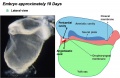
Development of the heart and vascular system begins very early in mesoderm both within (embryonic) and outside (extra embryonic, yolk sac and placental) the embryo. Vascular development therefore occurs in many places, the most obvious though is the early forming heart, which grows rapidly creating an externally obvious cardiac "bulge" on the early embryo. The cardiovascular system is extensively remodelled throughout development, this current page discusses systemic arterial development. Note that placental vessels are discussed in placental notes.
See also the related pages Arterial Development, Venous Development, Placental Villi Blood Vessels and Coronary Circulation Development.
Some Recent Findings
|
| More recent papers |
|---|
 This table allows an automated computer search of the external PubMed database using the listed "Search term" text link.
More? References | Discussion Page | Journal Searches | 2019 References | 2020 References Search term: Arterial Embryology <pubmed limit=5>Arterial Embryology</pubmed> <pubmed limit=5>Artery Development</pubmed> |
Textbooks

- Human Embryology (2nd ed.) Larson Ch7 p151-188 Heart, Ch8 p189-228 Vasculature
- The Developing Human: Clinically Oriented Embryology (6th ed.) Moore and Persaud Ch14: p304-349
- Before we Are Born (5th ed.) Moore and Persaud Ch12; p241-254
- Essentials of Human Embryology Larson Ch7 p97-122 Heart, Ch8 p123-146 Vasculature
- Human Embryology Fitzgerald and Fitzgerald Ch13-17: p77-111
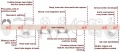
Pharyngeal Arch Arteries

In the head region of the embryo, each pharyngeal arch initially has paired arch arteries. These are extensively remodelled through development and give rise to a range of different arterial structures, as shown in the list below.
- Arch 1 - mainly lost, form part of maxillary artery.
- Arch 2 - stapedial arteries.
- Arch 3 - common carotid arteries, internal carotid arteries.
- Arch 4 - left forms part of aortic arch, right forms part right subclavian artery.
- Arch 6 - left forms part of left pulmonary artery , right forms part of right pulmonary artery.
- Links: Head Development
Renal Venous Development
The renal arterial and venous systems are also reorganised extensively throughout development with changing kidney position.

|

|
| Embryo renal venous | Adult renal venous |
- Links: Renal Development
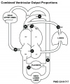
Fetal Blood Flow

Mean Late Fetal Blood Flows[2]
(8 subjects) in the major vessels of the human fetal circulation by phase contrast MRI. (median gestational age 37 weeks, age range of 30–39 weeks)
| (left) Mean flows in ml/kg/min | (right) Proportions of the combined ventricular output in the major vessels of the human fetal circulation by phase contrast MRI. |
|
|
- Cardiovascular Links: Fetal Blood Flow values | Mean Fetal Blood Flow | Proportions Ventricular Output | Ventricular Output (colour) | heart | blood | cardiovascular
References
- ↑ <pubmed>25167202</pubmed>
- ↑ <pubmed>23181717</pubmed>| J Cardiovasc Magn Reson.
Reviews
<pubmed></pubmed> <pubmed></pubmed> <pubmed></pubmed> <pubmed>22449840</pubmed> <pubmed>21593862</pubmed> <pubmed>18607112</pubmed> <pubmed>16565980</pubmed> <pubmed>16236564</pubmed> <pubmed>15614842</pubmed>
Articles
<pubmed>21808168</pubmed> <pubmed>21732277</pubmed> <pubmed>21541028</pubmed> <pubmed>21540552</pubmed> <pubmed>21364285</pubmed> <pubmed>18057862</pubmed>
Search Pubmed
Search May 2010
- Cardiovascular System Development All (63457) Review (10735) Free Full Text (15717)
Search Pubmed: Cardiovascular System Development
Additional Images
See also Category:Heart ILP and Category:Heart


Historic image
Heart Development Timeline
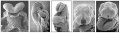
Human heart SEM
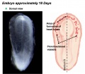
Early Heart Tube (Dorsal)
Early Heart Tube (Lateral)
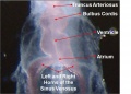
Heart Tube Segments
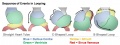
Heart Looping Sequence
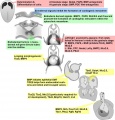
Molecular & Genetic Cardiac Development Factors
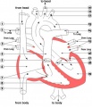
Adult heart blood flow cartoon
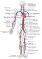
Adult human cardiovascular system cartoon
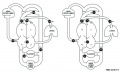
Fetal Blood Flow
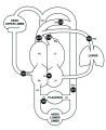
Fetal Blood Flow
Fetal Blood Flow


.jpg)
.jpg)


External Links
External Links Notice - The dynamic nature of the internet may mean that some of these listed links may no longer function. If the link no longer works search the web with the link text or name. Links to any external commercial sites are provided for information purposes only and should never be considered an endorsement. UNSW Embryology is provided as an educational resource with no clinical information or commercial affiliation.
Glossary Links
- Glossary: A | B | C | D | E | F | G | H | I | J | K | L | M | N | O | P | Q | R | S | T | U | V | W | X | Y | Z | Numbers | Symbols | Term Link
Cite this page: Hill, M.A. (2024, June 27) Embryology Cardiovascular - Arterial Development. Retrieved from https://embryology.med.unsw.edu.au/embryology/index.php/Cardiovascular_-_Arterial_Development
- © Dr Mark Hill 2024, UNSW Embryology ISBN: 978 0 7334 2609 4 - UNSW CRICOS Provider Code No. 00098G