Scanning Electron Microscopy: Difference between revisions
No edit summary |
mNo edit summary |
||
| (50 intermediate revisions by 2 users not shown) | |||
| Line 1: | Line 1: | ||
{{ | {{Header}} | ||
==Introduction== | ==Introduction== | ||
[[File:Microscopy_LM_and_SEM_cartoon.jpg|thumb|Scanning Electron Microscopy (MyScope)]] | |||
[[File:Stage12 sem1c.jpg|thumb|'''Human Embryo''' <br>(stage 12, week 4) SEM]] | [[File:Stage12 sem1c.jpg|thumb|'''Human Embryo''' <br>(stage 12, week 4) SEM]] | ||
The Scanning Electron Microscope (SEM) was a development of the electron microscope. Unlike a light microscope, using light, the electron microscope uses a focussed beam of electrons to image materials. The first version of this technology was the transmission electron microscope (TEM). | The Scanning Electron Microscope (SEM) was a development of the electron microscope. Unlike a light microscope, using light, the electron microscope uses a focussed beam of electrons to image materials. The first version of this technology was the transmission electron microscope (TEM). Some example SEM images are shown on this current page. | ||
: ''On this site the acronym "SEM" is used to denote a '''S'''canning '''E'''lectron '''M'''icrograph, the image produced by this form of microscopy.'' | |||
A new technique called Helium Ion Microscopy (HIM) gives similar high resolution 3D images, using a scanning beam of He<sup>+</sup> ions, from fixed tissue without the coating required for generating SEM images.{{#pmid:23505418|PMID23505418}} | |||
:'''Links:''' [[:Category:Scanning EM|Category:Scanning EM]] | |||
== Microscopy Timeline == | == Microscopy Timeline == | ||
* '''1665''' - Robert Hooke publishes Micrographia, a collection of biological micrographs. | * '''1665''' - Robert Hooke publishes Micrographia, a collection of biological micrographs. | ||
| Line 19: | Line 18: | ||
* '''1898''' - Golgi first saw and described the Golgi apparatus by staining cells with silver nitrate. | * '''1898''' - Golgi first saw and described the Golgi apparatus by staining cells with silver nitrate. | ||
* '''1931''' - Ernst Ruska first transmission electron microscope, (TEM). | * '''1931''' - Ernst Ruska first transmission electron microscope, (TEM). | ||
* ''' | * '''1938''' - Von Ardenne first scanning electron microscope (SEM) | ||
* '''1965''' - first commercial scanning electron microscopes. | |||
* ''' | |||
* '''1986''' - Ernst Ruska, Gerd Binnig and Heinrich Rohrer receive the Nobel Prize in Physics for invention of the electron microscope (ER) and scanning tunneling microscope (GB and HR). | * '''1986''' - Ernst Ruska, Gerd Binnig and Heinrich Rohrer receive the Nobel Prize in Physics for invention of the electron microscope (ER) and scanning tunneling microscope (GB and HR). | ||
==Human Embryo SEM Images== | |||
There are a series of beautiful SEM images made available by Prof Kathy Sulik of the early developing human embryo between week 3 to 5 (Carnegie stage 7 to 14) available: [[Carnegie_stage_7|7]], [[Carnegie_stage_8|8]], [[Carnegie_stage_9|9]], [[Carnegie_stage_10|10]], [[Carnegie_stage_11|11]], [[Carnegie_stage_12|12]], [[Carnegie_stage_13|13]] and [[Carnegie_stage_14|14]]. | |||
<gallery> | |||
File:Stage7-sem2.jpg|[[Carnegie_stage_7|Stage 7]] | |||
File:Stage8_SEM1.jpg|[[Carnegie_stage_8|Stage 8]] | |||
File:Stage9_sem4c.jpg|[[Carnegie_stage_9|Stage 9]] | |||
File:Stage10_sem6.jpg|[[Carnegie_stage_10|Stage 10]] | |||
File:Stage11_sem5.jpg|[[Template:Stage11SEM|Stage 11 SEM images]] | |||
File:Stage12_sem1.jpg|[[Template:Stage 12 SEM images|Stage 12 SEM images]] | |||
File:Stage13_sem1.jpg|[[Template:Stage13SEM|Stage 13 SEM images]] | |||
File:Stage14_sem5.jpg|[[Template:Stage 14 SEM images|Stage 14 SEM images]] | |||
</gallery> | |||
:'''Links:''' [[Template:Stage11SEM|Stage 11 SEM images]] | [[Template:Stage 12 SEM images|Stage 12 SEM images]] | [[Template:Stage13SEM|Stage 13 SEM images]] | [[Template:Stage 14 SEM images|Stage 14 SEM images]] | [[:Category:Scanning EM|Category:Scanning EM]] | |||
{{Carnegie stages}} | {{Carnegie stages}} | ||
==Other Species SEM Images== | |||
{| | |||
| [[File:Sea urchin SEM01.jpg|400px]] | |||
| [[File:Sea urchin SEM02.jpg|400px]] | |||
|- | |||
| [[Sea Urchin Development|Sea Urchin]] (2 cell stage) | |||
| [[Sea Urchin Development|Sea Urchin]] (16 cell stage) | |||
|- | |||
| [[File:Chicken- PGC grown in vitro 01.jpg|400px]] | |||
| [[File:Hamster_oocyte_and_spermatozoa.jpg|400px]] | |||
|- | |||
| [[Chicken Development|Chicken]] primordial germ cell (PGC) grown in vitro{{#pmid:20886037|PMID20886037}} | |||
| [[Hamster Development|Hamster]] oocyte and spermatozoa | |||
|- | |||
| [[File:Fly_wild-type_head.jpg|400px]] | |||
| [[File:Fly_antennapedia_head.jpg|400px]] | |||
|- | |||
| [[Fly Development|Fly]] wild-type head | |||
| [[Fly Development|Fly]] antennapedia head | |||
|} | |||
==Helium Ion Microscopy== | |||
[[File:Rat kidney HIM 01.jpg|thumb|300px|Adult rat kidney glomerulus]] | |||
A new technique called Helium Ion Microscopy (HIM) gives similar high resolution 3D images, using a scanning beam of He<sup>+</sup> ions, from fixed tissue without the coating required for generating SEM images. | |||
Some research images examples from the rat kidney.{{#pmid:23505418|PMID23505418}} | |||
<gallery> | |||
Rat_kidney_HIM_01.jpg|Kidney Glomerulus | |||
Rat_kidney_HIM_04.jpg|Collecting Tubule - Principal and Intercalated Cell | |||
Rat_kidney_HIM_02.jpg|Principal Cell - cilium and microvilli | |||
Rat_kidney_HIM_03.jpg|Principal Cell - cilium and microvilli | |||
</gallery> | |||
== References == | |||
<references/> | |||
===Reviews=== | |||
{{#pmid:4571700}} | |||
{{#pmid:14667297}} | |||
{{#pmid:4571700}} | |||
===Articles=== | |||
{{#pmid:19493101}} | |||
{{#pmid:16089300}} | |||
{{#pmid:6798043}} | |||
===Search PubMed=== | |||
'''Search Pubmed:''' [http://www.ncbi.nlm.nih.gov/sites/entrez?db=pubmed&cmd=search&term=Scanning%20Electron%20Microscopy Scanning Electron Microscopy] | |||
==External Links== | ==External Links== | ||
{{External Links}} | {{External Links}} | ||
* '''Nobel Prize''' [http://nobelprize.org/nobel_prizes/physics/laureates/1986/ The Nobel Prize in Physics 1986] Ernst Ruska, Gerd Binnig, Heinrich Rohrer [http://nobelprize.org/nobel_prizes/physics/laureates/1986/presentation-speech.html Nobel Speech] | |||
* [http://www.ammrf.org.au AMMFR] [http://www.ammrf.org.au/myscope MyScope] [http://www.ammrf.org.au/myscope/sem/background/whatissem What is an SEM] | |||
* '''Dartmouth Electron Microscope Facility''' [http://remf.dartmouth.edu/imagesindex.html A variety of scanning and transmission electron microscope images] (These images are in the public domain) | |||
* '''University of Minnesota''' [http://www.charfac.umn.edu/instruments/sem_primer.pdf Bob Hafner - Scanning Electron Microscopy Primer (PDF)] | |||
* '''Molecular Expressions''' [http://micro.magnet.fsu.edu/primer/java/electronmicroscopy/magnify1/index.html Virtual Scanning Electron Microscopy] | |||
{{Glossary}} | {{Glossary}} | ||
{{Footer}} | {{Footer}} | ||
[[Category:Scanning EM]] [[Category:Method]] | [[Category:Scanning EM]] [[Category:Method]] | ||
Latest revision as of 10:19, 12 April 2019
| Embryology - 15 Jun 2024 |
|---|
| Google Translate - select your language from the list shown below (this will open a new external page) |
|
العربية | català | 中文 | 中國傳統的 | français | Deutsche | עִברִית | हिंदी | bahasa Indonesia | italiano | 日本語 | 한국어 | မြန်မာ | Pilipino | Polskie | português | ਪੰਜਾਬੀ ਦੇ | Română | русский | Español | Swahili | Svensk | ไทย | Türkçe | اردو | ייִדיש | Tiếng Việt These external translations are automated and may not be accurate. (More? About Translations) |
Introduction


The Scanning Electron Microscope (SEM) was a development of the electron microscope. Unlike a light microscope, using light, the electron microscope uses a focussed beam of electrons to image materials. The first version of this technology was the transmission electron microscope (TEM). Some example SEM images are shown on this current page.
- On this site the acronym "SEM" is used to denote a Scanning Electron Micrograph, the image produced by this form of microscopy.
A new technique called Helium Ion Microscopy (HIM) gives similar high resolution 3D images, using a scanning beam of He+ ions, from fixed tissue without the coating required for generating SEM images.[1]
- Links: Category:Scanning EM
Microscopy Timeline
- 1665 - Robert Hooke publishes Micrographia, a collection of biological micrographs.
- 1674 - Anton van Leeuwenhoek improved simple microscope for biological specimens.
- 1833 - Brown published a microscopic observation of orchids, describing the cell nucleus.
- 1898 - Golgi first saw and described the Golgi apparatus by staining cells with silver nitrate.
- 1931 - Ernst Ruska first transmission electron microscope, (TEM).
- 1938 - Von Ardenne first scanning electron microscope (SEM)
- 1965 - first commercial scanning electron microscopes.
- 1986 - Ernst Ruska, Gerd Binnig and Heinrich Rohrer receive the Nobel Prize in Physics for invention of the electron microscope (ER) and scanning tunneling microscope (GB and HR).
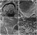
Human Embryo SEM Images
There are a series of beautiful SEM images made available by Prof Kathy Sulik of the early developing human embryo between week 3 to 5 (Carnegie stage 7 to 14) available: 7, 8, 9, 10, 11, 12, 13 and 14.


- Links: Stage 11 SEM images | Stage 12 SEM images | Stage 13 SEM images | Stage 14 SEM images | Category:Scanning EM
- Carnegie Stages: 1 | 2 | 3 | 4 | 5 | 6 | 7 | 8 | 9 | 10 | 11 | 12 | 13 | 14 | 15 | 16 | 17 | 18 | 19 | 20 | 21 | 22 | 23 | About Stages | Timeline
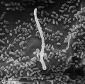
Other Species SEM Images

|

|
| Sea Urchin (2 cell stage) | Sea Urchin (16 cell stage) |

|

|
| Chicken primordial germ cell (PGC) grown in vitro[2] | Hamster oocyte and spermatozoa |

|

|
| Fly wild-type head | Fly antennapedia head |
Helium Ion Microscopy

A new technique called Helium Ion Microscopy (HIM) gives similar high resolution 3D images, using a scanning beam of He+ ions, from fixed tissue without the coating required for generating SEM images.
Some research images examples from the rat kidney.[1]
Kidney Glomerulus

Collecting Tubule - Principal and Intercalated Cell
Principal Cell - cilium and microvilli
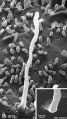
Principal Cell - cilium and microvilli


References
- ↑ 1.0 1.1 Rice WL, Van Hoek AN, Păunescu TG, Huynh C, Goetze B, Singh B, Scipioni L, Stern LA & Brown D. (2013). High resolution helium ion scanning microscopy of the rat kidney. PLoS ONE , 8, e57051. PMID: 23505418 DOI.
- ↑ Choi JW, Kim S, Kim TM, Kim YM, Seo HW, Park TS, Jeong JW, Song G & Han JY. (2010). Basic fibroblast growth factor activates MEK/ERK cell signaling pathway and stimulates the proliferation of chicken primordial germ cells. PLoS ONE , 5, e12968. PMID: 20886037 DOI.
Reviews
Hollenberg MJ & Erickson AM. (1973). The scanning electron microscope: potential usefulness to biologists. A review. J. Histochem. Cytochem. , 21, 109-30. PMID: 4571700 DOI.
Stokes DJ. (2003). Recent advances in electron imaging, image interpretation and applications: environmental scanning electron microscopy. Philos Trans A Math Phys Eng Sci , 361, 2771-87. PMID: 14667297 DOI.
Hollenberg MJ & Erickson AM. (1973). The scanning electron microscope: potential usefulness to biologists. A review. J. Histochem. Cytochem. , 21, 109-30. PMID: 4571700 DOI.
Articles
Sim KS, Ting HY, Lai MA & Tso CP. (2009). Improvement to the scanning electron microscope image colorization by adaptive tuning. J Microsc , 234, 243-50. PMID: 19493101 DOI.
Oho E, Sugawara T & Suzuki K. (2005). An improved scanning method based on characteristics of the human visual system for scanning electron microscopy. Scanning , 27, 170-5. PMID: 16089300
Pease DC & Porter KR. (1981). Electron microscopy and ultramicrotomy. J. Cell Biol. , 91, 287s-292s. PMID: 6798043
Search PubMed
Search Pubmed: Scanning Electron Microscopy
External Links
External Links Notice - The dynamic nature of the internet may mean that some of these listed links may no longer function. If the link no longer works search the web with the link text or name. Links to any external commercial sites are provided for information purposes only and should never be considered an endorsement. UNSW Embryology is provided as an educational resource with no clinical information or commercial affiliation.
- Nobel Prize The Nobel Prize in Physics 1986 Ernst Ruska, Gerd Binnig, Heinrich Rohrer Nobel Speech
- AMMFR MyScope What is an SEM
- Dartmouth Electron Microscope Facility A variety of scanning and transmission electron microscope images (These images are in the public domain)
- University of Minnesota Bob Hafner - Scanning Electron Microscopy Primer (PDF)
- Molecular Expressions Virtual Scanning Electron Microscopy
Glossary Links
- Glossary: A | B | C | D | E | F | G | H | I | J | K | L | M | N | O | P | Q | R | S | T | U | V | W | X | Y | Z | Numbers | Symbols | Term Link
Cite this page: Hill, M.A. (2024, June 15) Embryology Scanning Electron Microscopy. Retrieved from https://embryology.med.unsw.edu.au/embryology/index.php/Scanning_Electron_Microscopy
- © Dr Mark Hill 2024, UNSW Embryology ISBN: 978 0 7334 2609 4 - UNSW CRICOS Provider Code No. 00098G