Carnegie stage 7: Difference between revisions
mNo edit summary |
|||
| (68 intermediate revisions by 3 users not shown) | |||
| Line 1: | Line 1: | ||
{{Header}} | |||
== Introduction == | == Introduction == | ||
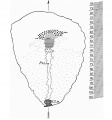
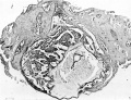
[[File:Stage7-bf1.jpg|300px | {| | ||
| valign=top width=310px|[[File:Stage7-bf1.jpg|300px]] | |||
| | |||
===Facts=== | ===Facts=== | ||
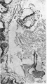
Human embryonic stage 7 occurs during week 3 between 15 to 17 days. | Human embryonic stage 7 occurs during week 3 between 15 to 17 days. | ||
| Line 9: | Line 11: | ||
The initial images are displayed unlabeled to allow you to explore the embryo for yourself, linked labeled versions are also available for some images. | The initial images are displayed unlabeled to allow you to explore the embryo for yourself, linked labeled versions are also available for some images. | ||
=== | ===Summary=== | ||
Gastrulation is continuing as cells migrate from the epiblast, continuing to form mesoderm. | Gastrulation is continuing as cells migrate from the epiblast, continuing to form mesoderm. | ||
| Line 17: | Line 19: | ||
From the primitive node a tube extends under the ectoderm in the opposite direction to the primitive streak. This tube forms first the axial process then notochordal process, then finally the notochord. | From the primitive node a tube extends under the ectoderm in the opposite direction to the primitive streak. This tube forms first the axial process then notochordal process, then finally the notochord. | ||
The notochord is a key to embryonic folding and regulation of ectoderm and mesoderm differentiation. It lies in the rostrocordal axis and the embryonic disc will fold either side ventrally, pinching off a portion of the yolk sac to form the lining of the gastrointestinal tract. | The notochord is a key to embryonic folding and regulation of {{ectoderm}} and {{mesoderm}} differentiation. It lies in the rostrocordal axis and the embryonic disc will fold either side ventrally, pinching off a portion of the yolk sac to form the lining of the gastrointestinal tract. | ||
{{ | See also [[#Events|Events]] | ||
|} | |||
<br> | |||
{{Carnegie stage 7 links}} | |||
{{Carnegie_stage_table_1}} | |||
{{Carnegie_stages}} | |||
== Bright Field Lateral== | == Bright Field Lateral== | ||
| Line 27: | Line 37: | ||
[[File:Stage7-bf4.jpg|300px]] | [[File:Stage7-bf4.jpg|300px]] | ||
==Scanning EM== | ==Scanning EM== | ||
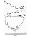
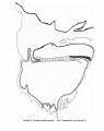
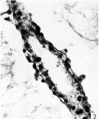
[[File:Stage7-sem1.jpg|300px]] | {| | ||
[[File:Stage7-sem4.jpg|300px]] | | [[File:Stage7-sem1.jpg|300px]] | ||
| [[File:Stage7-sem4.jpg|300px]] | |||
|- | |||
|''' Human embryonic disc''' | |||
* epiblast layer, dorsal view | |||
* primitive streak (left) | |||
* connecting stalk (far left) | |||
* amniotic sac removed | |||
|''' Human embryonic disc''' | |||
* epiblast layer, dorsolateral view | |||
* primitive streak (left) | |||
* connecting stalk (far left) | |||
* amniotic sac removed | |||
|- | |||
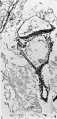
| [[File:Stage7-sem2.jpg|300px]] | |||
| [[File:Stage7-sem5.jpg|300px]] | |||
'''Primitive streak''' detail (left) | |||
|- | |||
|''' Human embryonic disc''' | |||
* epiblast layer, dorsal view | |||
* primitive streak (bottom) | |||
* connecting stalk (bottom) | |||
* amniotic sac removed | |||
| | |||
|} | |||
See also human ectopic embryos (stage 7) images.{{#pmid:21665543|PMID21665543z}} | |||
==Kyoto Collection== | ==Kyoto Collection== | ||
{| | {| | ||
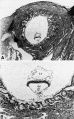
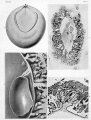
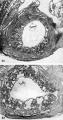
| [[Image:Stage7 features.jpg| | | [[Image:Stage7 features.jpg]] | ||
| '''View:''' embryonic disc, showing the epiblast viewed from the amniotic (dorsal) side. Head to tail orientation is Cranial (image top) and Caudal (image bottom). | | [[File:Stage7.jpg]] | ||
|} | |||
'''View:''' embryonic disc, showing the epiblast viewed from the amniotic (dorsal) side. Head to tail orientation is Cranial (image top) and Caudal (image bottom). | |||
'''Features:''' embryonic disc, primitive node, primative streak, primative groove, connecting stalk | '''Features:''' embryonic disc, primitive node, primative streak, primative groove, connecting stalk | ||
Alternate View: embryonic disc, probably from from the ventral side. Showing the connecting stalk to the left. | Alternate View: embryonic disc, probably from from the ventral side. Showing the connecting stalk to the left. | ||
{{Kyoto collection}} | |||
==Carnegie Collection== | |||
{| | |||
| [[File:Stage7_bf7.jpg]] | |||
Surface view of implantation site | |||
| [[File:Stage7_bf8.jpg]] | |||
Surface lateral view of implantation site | |||
|} | |} | ||
[[File:Stage7_bf6.jpg|300px]][[File:Stage7_bf9.jpg|500px]] | |||
{{Carnegie Collection stage 7 table}} | |||
{{Carnegie Embryo iBook table}} | |||
==Manchester Collection== | |||
'''Manchester No. 1285''' | |||
Described by Florian and Hill (1935)<ref name=Florian1935>{{Ref-FlorianHill1935}}</ref>. | |||
* Specimen is housed in Department of Anatomy, University of Manchester. | |||
* Hysterectomy. | |||
* Chorionic cavity, 4.28 x 3.28 mm. | |||
* Embryonic disc (narrow type), 0.87 x 0.625 mm. | |||
* Primitive streak, 0.39 mm, and node, 0.05 mm. | |||
* Notochordal process, 0.125 mm. Prechordal plate, 0.03 mm. | |||
* Connecting stalk attached to chorion at decidua capsularis (suggesting polar variety of velamentous insertion of umbilical cord). | |||
Dorsal and median projections published (Hill and Florian, 1931b, figs. 5 and 13; Florian and Hill, 1935, figs. 1-3; Mazanec, 1959, fig. 52). | |||
<gallery caption="Text Figures"> | |||
File:Florian1935 textfig01.jpg|1. Graphic reconstruction of the dorsal view of the embryonal shield | |||
File:Florian1935 textfig02.jpg|2. Graphic reconstruction of the median section | |||
File:Florian1935 textfig03.jpg|3. Idealised Median Section | |||
</gallery> | |||
<gallery caption="Plate 1"> | |||
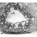
File:Florian1935 fig01.jpg|Fig. 1. Section 49 | |||
File:Florian1935 fig02.jpg|Fig. 2. Section 43 | |||
File:Florian1935 fig03.jpg|Fig. 3. Section 40 | |||
File:Florian1935 fig04.jpg|Fig. 4. Section 37 | |||
</gallery> | |||
<gallery caption="Plate 2"> | |||
File:Florian1935 fig05.jpg|Fig. 5. Section 32 | |||
File:Florian1935 fig06.jpg|Fig. 6. Section 79 | |||
File:Florian1935 fig07.jpg|Fig. 7. Section 97 | |||
File:Florian1935 fig08.jpg|Fig. 8. Section 75 | |||
</gallery> | |||
<gallery caption="Plate 3"> | |||
File:Florian1935 fig09.jpg|
Fig.9. Section 122 | |||
File:Florian1935 fig10.jpg|
Fig. 10. Section 23 | |||
File:Florian1935 fig11.jpg|
Fig.11. Section 99 | |||
</gallery> | |||
==Photograph== | ==Photograph== | ||
{| | |||
| [[File:Stage7 bf5.jpg|400px]] | |||
| [[File:Stage7 bf51.jpg|400px]] | |||
|- | |||
| [[File:Stage7 bf52.jpg|400px]] | |||
| [[File:Stage7 bf53.jpg|400px]] | |||
|} | |||
[[ | {{Ed Uthman}} section of [[Abnormal_Development_-_Ectopic_Implantation|ectopic (tubal) pregnancy]]. | ||
:'''Image Links:''' [[:File:Stage7 bf5.jpg|Original full image]] | [[:File:Stage7 bf51.jpg|Trilaminar embryo excerpt]] | [[:File:Stage7 bf52.jpg|Villi excerpt 1]] | [[:File:Stage7 bf53.jpg|Villi excerpt 2]] | |||
'''Image | :'''Full Image versions:''' [[:File:Stage7 bf5.jpg|ExtraLarge 1712x1206px]] | [[:File:Stage7 bf5a.jpg|Large 1024x721px]] | [[:File:Stage7 bf5b.jpg|Medium 500x352px]] | ||
===Embryo Virtual Slide=== | |||
{| | |||
| valign=bottom|{{SlideStage7bf5}} | |||
| valign=bottom|{{SlideStage7bf51}} | |||
|} | |||
== Stage 7 Images == | == Stage 7 Images == | ||
| Line 56: | Line 156: | ||
'''Scanning Electron Images:''' [[:File:Stage7-sem1.jpg|Embryonic Disc 1]] | [[:File:Stage7-sem2.jpg|Embryonic Disc 2]] | [[:File:Stage7-sem3.jpg|Embryonic Disc 3]] | [[:File:Stage7-sem4.jpg|Primitive Streak]] | '''Scanning Electron Images:''' [[:File:Stage7-sem1.jpg|Embryonic Disc 1]] | [[:File:Stage7-sem2.jpg|Embryonic Disc 2]] | [[:File:Stage7-sem3.jpg|Embryonic Disc 3]] | [[:File:Stage7-sem4.jpg|Primitive Streak]] | ||
== | ==Events== | ||
* {{Gastrulation}} | |||
* {{Notochord}} | |||
==Historic Stage 7 Embryos== | |||
Historic stage 7 embryo data modified from O'Rahilly and Müller (1987).<ref name=O'RahillyMüller1987>{{Ref-O'RahillyMüller1987}}</ref> | |||
===By Length of Notochordal Process=== | |||
'''HEB-37''' - Summarized by Mazanec (1959)<ref name=Mazanec1959>{{Ref-Mazanec1959}}</ref>. Chorionic cavity, 2.25 x 1.29 x 0.4 mm. Embryonic disc, 0.4 mm. Primitive streak, 0.104 mm, and node, 0.04 mm. Notochordal process, 0.032 mm. Stalk of umbilical vesicle (ibid., fig. 77). Median projection published (ibid., fig. 45). | |||
[[Paper - An Early Human Embryo, with 0.55 mm long Embryonic Shield|'''H.R. 1.''']] (Hesketh Roberts) - Described by Johnston (1940)<ref name=Johnston1940>{{Ref-Johnston1940}}</ref>, who believed that a notochordal process (0.04 mm) and a prechordal plate (0.075 mm) were present. Florian in an appendix to the article disagreed, and his interpretation is followed here (see [[Carnegie stage 6|stage 6]]). | |||
[[Paper - Two Early Human Embryos|'''Biggart''']] (Prof. JH. Biggart) - Described by Morton (1949).<ref name=Morton1949>{{Ref-Morton1949}}</ref> Curettage. Embryonic disc (narrow type), 0.27 x 0.16 mm. Primitive streak and node, 0.059 mm. A notochordal process is not referred to in the text but is mapped on a dorsal projection of the embryo (ibid., fig. 2) and is approximately 0.04 mm in length. The specimen is said to resemble the Yale embryo. | |||
<gallery caption="Biggart Embryo"> | |||
File:Morton1949 fig01.jpg|embryo in situ | |||
File:Morton1949 fig02.jpg|duct part of yolk-sac | |||
File:Morton1949 fig03.jpg|distal expansion of yolk-sac | |||
File:Morton1949 fig04.jpg|embryonic rudiment | |||
File:Morton1949 fig05.jpg|opening of duct part into distal expansion of yolk-sac | |||
</gallery> | |||
'''Guá''' (Guálberto) - Described by Lordy (1931). Hysterectomy. Chorionic cavity, 8 x 7.5 mm. Embryonic disc, 0.776 x 0.0465 mm. Primitive streak, 0.09 mm. Notochordal process, 0.045 mm. Possible notochordal canal. Said to resemble Hugo embryo. Probably belongs either to stage 7 or to stage 8. | |||
'''Carnegie No.''' {{CE7802}} - (figs. 7-4 to 7-12). An important specimen described and illustrated by Heuser, Rock, and Hertig (1945)<ref name=HeuserRockHertig1945>{{Ref-HeuserRockHertig1945}}</ref>. Hysterectomy. Chorion, 3.75 x 2.35 x 2.2 mm. Chorionic cavity, 2.3 x 1.4 x 1.1 mm. Embryonic disc (broad type), 0.42 x 0.35 x 0.05 mm. Primitive streak, 0.11 mm, and node, 0.03 mm. Notochordal process, 0.048 mm. Presumed age, 16 days. Median projection published (ibid., plate 6; Mazanec, 1959<ref name=Mazanec1959>{{Ref-Mazanec1959}}</ref>, fig. 46) and dorsal projection has been prepared by the present writers. | |||
<gallery caption="{{CE7802}} Embryo"> | |||
Hertig1946b fig05.jpg|Hertig1946 Fig. 5 | |||
Hertig1946b fig05a.jpg|Hertig1946 Fig. 5a | |||
Hertig1946b fig05b.jpg|Hertig1946 Fig. 5b | |||
Stage7 bf6.jpg | |||
Stage7 bf7.jpg | |||
Stage7 bf8.jpg | |||
Stage7 bf9.jpg | |||
</gallery> | |||
'''P.M.''' - Described by Meyer (1924). Curettage. Measurements have been criticized by Stieve (1926) but defended by Mazanec (1959)<ref name=Mazanec1959>{{Ref-Mazanec1959}}</ref>. Chorion, 3.9 x 3.77 x 2.5 mm. Chorionic cavity, 2.7 x 2.6 x 2.1 mm. Embryonic disc (circular), 0.41 x 0.41 mm. Primitive streak, 0.12 mm, and node, 0.02 mm. Notochordal process, 0.06 mm, acknowledged by Mazanec (1959) although denied by Fahrenholz (1927). No notochordal canal. Median projection published (Mazanec, 1959, fig. 47). | |||
'''Hugo''' - Described by Stieve (1926), who reproduced a photomicrograph of every second section. Hysterectomy. Surrounded by 942 chorionic villi ranging in length from 0.3 to 1 mm. Chorion, 6.4 x 5.9 x 5.6 mm. Chorionic cavity, 4.7 x 4.4 x 3.8 mm. Embryonic disc (broad type), 0.635 (Florian, 1931)<ref name=Florian1931>{{Ref-Florian1931}}</ref> x 0.63 mm. Primitive streak, 0.245 mm, and node, 0.05 mm. Notochordal process, 0.07 (0.11?) mm. (Florian, 1934c). No notochordal canal. Prechordal plate probably not yet developed (Hill and Florian, 1931a). Dorsal and median projections published (Florian, 1934c, fig. 1; Hill and Florian, 1931b, figs. 44 and 11; Mazanec, 1959<ref name=Mazanec1959>{{Ref-Mazanec1959}}</ref>, fig. 49). | |||
Robertson, O’Neill, and Chappell (1948) described a hysterectomy specimen that possessed a chorion of 3.816 x 3.639 x 2.687 mm. Chorionic cavity, 2.718 x 2.239 x 1.679 mm. Embryonic disc (broad type), 0.462 x 0.485 mm. Primitive streak, 0.138 mm, and node (situated halfway), 0.03 mm. Notochordal process, 0.072 mm. Suggestion of notochordal canal in one or two sections. Assigned to horizon VIII by authors but probably belongs to stage 7. Median projection published (ibid., fig. 12). | |||
D’Arrigo (1961) described an embryonic disc of 0.47 mm, which showed a notochordal process of 0.075 mm. Canalization of the process is “doubtful,” but the presence of a prechordal plate is “probable.” The specimen “could be recorded in Streeter’s horizon VII.” | |||
'''Goodwin''' - Described by Kindred (1933). Tubal. Chorion, 5.8 x 2.72 x 2.25 mm. Chorionic cavity, 2.44 x 2.25 x 0.75 mm. Embryonic disc, 0.588 mm in width. Primitive streak, 0.215 mm, and node, 0.078 mm. Notochordal process, 0.078 mm. No notochordal canal and no prechordal plate. | |||
'''Pha I''' - Described by Mazanec (1949).<ref name=Mazanec1949>{{Ref-Mazanec1949}}</ref> Chorionic cavity, 7.872 x 5.475 x 2.032 mm. Embryonic disc, 0.66 x 0.52 mm. Primitive streak, 0.145 mm, and node, 0.06 mm. Notochordal process, 0.09 mm. No prechordal plate. Median projection published (ibid., fig. 51). | |||
'''H. Schm. 10''' - (H. Schmid). Described briefly by Grosser (1931c). Embryonic disc (almost circular), 0.51 x 0.58 mm. Primitive streak, 0.14 mm, and node, 0.1 mm. Notochordal process, 0.1 mm. Probably belongs to stage 7, although a cavity in one section was thought to represent “Lieberkühn’s canal.” | |||
'''Bi 24''' (Bittmann) - Described by Hill and Florian (1931b). Chorionic cavity, 3.05 x 3.036 x 3.029 mm. Embryonic disc (narrow type), 0.62 x 0.39 mm. Primitive streak, 0.28 mm, and node (Mazanec, 1959<ref name=Mazanec1959>{{Ref-Mazanec1959}}</ref>, fig. 105), 0.05 mm. Notochordal process, 0.105 mm, consists of median chord and lateral mesoblastic wings. Prechordal plate, 0.03 mm. Possible primordial germ cells in endoderm of region of cloacal membrane and in endoderm of umbilical vesicle caudally (Florian, 1931). Politzer (1933) counted 41 germ cells in the region of the allanto-enteric diverticulum in this embryo, and 19 such cells in another presomite specimen (Bi 25). Dorsal and median projections published (Hill and Florian, 1931b, figs. 4 and 12; Florian, 1945, plate 5, fig. 43; Mazanec, 1959<ref name=Mazanec1959>{{Ref-Mazanec1959}}</ref>, fig. 50). | |||
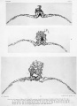
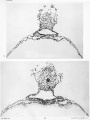
'''Manchester No. 1285''' (fig. 7-1). Described by Florian and Hill (m̩)<ref name=Florian1935>{{Ref-FlorianHill1935}}</ref>. Hysterectomy. Chorionic cavity, 4.28 x 3.28 mm. Embryonic disc (narrow type), 0.87 x 0.625 mm. Primitive streak, 0.39 mm, and node, 0.05 mm. Notochordal process, 0.125 mm. Prechordal plate, 0.03 mm. Connecting stalk attached to chorion at decidua capsularis (suggesting polar variety of velamentous insertion of umbilical cord). Dorsal and median projections published (Hill and Florian, 1931b, figs. 5 and 13; Florian and Hill, 1935, figs. 1-3; Mazanec, 1959, fig. 52). Specimen is housed in Department of Anatomy, University of Manchester. | |||
'''Pha II.''' - Summarized by Mazanec (1959)<ref name=Mazanec1959>{{Ref-Mazanec1959}}</ref>. Chorionic cavity, 4.985 x 3.882 x 3.52 mm. Embryonic disc, 0.895 x 0.62 mm. Primitive streak, 0.37 mm, and node, 0.06 mm. Notochordal process, 0.13 mm. No prechordal plate. Median projection published (ibid., fig. 53). | |||
Thompson and Brash (1923) described a specimen that showed a notochordal process of 0.3 mm. It is described in the present work under stage 8. | |||
===By Year of Publication=== | |||
No measurements of notochordal process available for the following historic embryos. | |||
Debeyre (1912) described in detail a specimen that possessed a chorionic cavity of 5.6 x 2.1 mm. Embryonic disc, 0.9 x 0.6 x 0.95 mm. Primitive streak stated to be 0.54 mm in length. Chorionic villi (0.4-1.6 mm) showed some branching. Unsuitable plane of section makes it impossible to assess the specimen precisely. | |||
'''Carnegie No.''' {{CE1399}} '''Mateer''' - Described by Streeter (1920).<ref name=Ref-Streeter1920a>{{Ref-Streeter1920a}}</ref> Hysterectomy. Angiogenesis in villi described by Hertig (1935). Chorion, 9 x 8 x 3.5 mm. Chorionic cavity, 6.1 x 5.6 x 2.5 mm. Embryonic disc, 1 x 0.75 mm. Primitive streak and groove present. Although the notochordal process was originally thought probably to be absent, Hill and Florian (1931b) have no doubt that it is present. More advanced than Hugo (Florian and Völker, 1929). A very small twin embryo was originally described but that “interpretation has become open to doubt” (Corner, 1955). Drawing of every section reproduced by Turner (1920). A median drawing (Davis, 1927, fig. 5A) and a projection have been published (Mazanec, 1959, fig. 56). | |||
<gallery caption="{{CE1399}} Embryo"> | |||
File:Streeter1920 Plate 1.jpg|Streeter Plate 1 | |||
File:Streeter1920 Plate 2.jpg|Streeter Plate 2 | |||
File:Streeter1920 Plate 3.jpg|Streeter Plate 3 | |||
</gallery> | |||
'''Ho''' (Hodiesne) - Described by Fahrenholz (1927). Abortion. Chorionic cavity, 6.5 x 6 x 3 mm. Embryonic disc (deformed), 0.6 mm (0.725 mm by flexible scale). Primitive streak, 0.22 mm (0.345 mm by flexible scale). Notochordal process just beginning (“undoubtedly present,” Hill and Florian, 1931b). Possible prechordal plate claimed (disputed by Waldeyer, 1929a, but supported by Hill and Florian, 1963). “Lieberkühn’s canal” (0.065 mm) is an artificial folding of the embryonic disc (Hill and Florian, 1931b). Dorsal and median projections published (Fahrenholz, 1927, figs. 32, 6 and 7; Mazanec, 1959, fig. 54). | |||
Debeyre (1933) described a specimen (0.9 mm) that possessed a primitive streak and probably belonged to stage 6 or stage 7. Large cells near the opening of the allantois were identified as primordial germ cells. | |||
'''Falkiner''' - Described by Martin and Falkiner (1938).<ref name=MartinFalkiner1938>{{Ref-MartinFalkiner1938}}</ref> Curettage. Embryo damaged and not in good condition. Measurements seem too small (see Mazanec, 1959)<ref name=Mazanec1959>{{Ref-Mazanec1959}}</ref>. Chorionic cavity, 1.5 x 1.4 mm. Embryonic disc, 0.15 x 0.29 mm. Primitive streak, 0.07 mm. Notochordal process contains “no definite lumen.” Cells rostral to notochordal process are “probably” the prechordal plate. Development “agrees most closely” with that of Bi I. Median projection published (Martin and Falkiner, 1938, fig. 8; Mazanec, 1959, fig. 39). | |||
<gallery caption="Falkiner Embryo"> | |||
File:MartinFalkiner1938 fig01.jpg|Fig. 1 | |||
File:MartinFalkiner1938 fig02.jpg|Fig. 2 | |||
File:MartinFalkiner1938 fig03.jpg|Fig. 3 | |||
File:MartinFalkiner1938 fig04.jpg|Fig. 4 | |||
File:MartinFalkiner1938 fig05.jpg|Fig. 5 | |||
File:MartinFalkiner1938 fig06.jpg|Fig. 6 | |||
File:MartinFalkiner1938 fig07.jpg|Fig. 7 | |||
File:MartinFalkiner1938 fig08.jpg|Fig. 8 Reconstruction | |||
</gallery> | |||
'''Gar''' (Green-Armytage) - Described by West (1952).<ref name=West1952>{{Ref-West1952}}</ref> Hysterectomy. Chorionic cavity, 3 x 2.6 x 2 mm. Embryonic disc (broad type), 0.56 x 0.69 mm. Primitive streak, node, and groove present. Short notochordal process. No notochordal plate. Said to resemble Hugo embryo. Trophoblast described by Hamilton and Boyd (1960). | |||
'''Mal''' (Maliphant) - Described by West (1952).<ref name=West1952>{{Ref-West1952}}</ref> Hysterectomy. Chorionic cavity, 3 x 1.8 mm. Embryonic disc (broad type), 0.45 x 0.6 mm. Primitive streak, node, and groove present. Notochordal process present (Mazanec, 1959); small cavity in node (West, 1952) or in notochordal process (Mazanec, 1959) “hardly sufficient to warrant the name chorda canal.” No prechordal plate. Belongs either to stage 7 or to stage 8 (said to resemble Jones-Brewer I, which is in stage 8). | |||
'''Carnegie No.''' {{CE8602}} - Photomicrograph reproduced by Hertig, Rock, and Adams (1956)<ref name=Hertig1956>{{Ref-Hertig1956}}</ref> ([[:Hertig1956 plate10.jpg|plate 10]], [[:Hertig1956 fig53.jpg|fig. 53]]). Chorion, 2.73 x 2.43 mm. Chorionic cavity, 1.83 x 1.33 mm. Embryonic disc, 0.3 x 0.06 mm. Presumed age, 16-17 days. | |||
<gallery caption="{{CE8602}} Embryo"> | |||
Hertig1956 fig53.jpg | |||
Hertig1956 plate10.jpg | |||
</gallery> | |||
'''Missen''' - Trophoblast described by Hamilton and Boyd (1960). Curettage. Chorion, 1.66 x 1.43 mm. Embryonic disc, 0.28 x 0.214 mm. Primitive streak and node. Notochordal process. Said to resemble No. 7801 and Edwards Jones-Brewer (stage 6). Presumed age, about 14 days. | |||
Other embryos probably belonging to stage 7 include Fitzgerald, Fitzgerald-Brewer II, and Jones-Brewer II (Brewer and Fitzgerald, 1937).<ref name=BrewerFitzgerald1937>{{Ref-BrewerFitzgerald1937}}</ref> | |||
===References=== | |||
<references/> | |||
==Additional Images== | |||
{{Carnegie_stages}} | |||
{{Glossary}} | |||
{{ | {{Footer}} | ||
[[Category:Human Embryo]] [[Category:Carnegie Stage]] [[Category:Carnegie Stage 7]] [[Category:Week 3]] | [[Category:Human Embryo]] [[Category:Carnegie Stage]] [[Category:Carnegie Stage 7]] [[Category:Week 3]] | ||
[[Category:Carnegie Embryo 7802]] | |||
Latest revision as of 11:08, 3 April 2020
| Embryology - 15 Jun 2024 |
|---|
| Google Translate - select your language from the list shown below (this will open a new external page) |
|
العربية | català | 中文 | 中國傳統的 | français | Deutsche | עִברִית | हिंदी | bahasa Indonesia | italiano | 日本語 | 한국어 | မြန်မာ | Pilipino | Polskie | português | ਪੰਜਾਬੀ ਦੇ | Română | русский | Español | Swahili | Svensk | ไทย | Türkçe | اردو | ייִדיש | Tiếng Việt These external translations are automated and may not be accurate. (More? About Translations) |
Introduction

|
FactsHuman embryonic stage 7 occurs during week 3 between 15 to 17 days. The embryo is now 0.4 mm diameter in size. The initial images are displayed unlabeled to allow you to explore the embryo for yourself, linked labeled versions are also available for some images. SummaryGastrulation is continuing as cells migrate from the epiblast, continuing to form mesoderm. Mesoderm lies between the ectoderm and endoderm as a continuous sheet except at the buccopharyngeal and cloacal membranes. These membranes have ectoderm and endoderm only and will lie at the rostral (head) and caudal (tail) of the gastrointestinal tract. From the primitive node a tube extends under the ectoderm in the opposite direction to the primitive streak. This tube forms first the axial process then notochordal process, then finally the notochord. The notochord is a key to embryonic folding and regulation of ectoderm and mesoderm differentiation. It lies in the rostrocordal axis and the embryonic disc will fold either side ventrally, pinching off a portion of the yolk sac to form the lining of the gastrointestinal tract. See also Events |
| Stage 7 Links: Week 3 | Gastrulation | Lecture | Practical | Carnegie Embryos | Category:Carnegie Stage 7 | Next Stage 8 |
| Historic Papers: 1923 head-process | 1933 tubal | 1940 | 1949 |
| Week: | 1 | 2 | 3 | 4 | 5 | 6 | 7 | 8 |
| Carnegie stage: | 1 2 3 4 | 5 6 | 7 8 9 | 10 11 12 13 | 14 15 | 16 17 | 18 19 | 20 21 22 23 |
- Carnegie Stages: 1 | 2 | 3 | 4 | 5 | 6 | 7 | 8 | 9 | 10 | 11 | 12 | 13 | 14 | 15 | 16 | 17 | 18 | 19 | 20 | 21 | 22 | 23 | About Stages | Timeline
Bright Field Lateral


Scanning EM

|

|
Human embryonic disc
|
Human embryonic disc
|

|

Primitive streak detail (left) |
Human embryonic disc
|
See also human ectopic embryos (stage 7) images.[1]
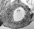
Kyoto Collection

|

|
View: embryonic disc, showing the epiblast viewed from the amniotic (dorsal) side. Head to tail orientation is Cranial (image top) and Caudal (image bottom).
Features: embryonic disc, primitive node, primative streak, primative groove, connecting stalk
Alternate View: embryonic disc, probably from from the ventral side. Showing the connecting stalk to the left.
Image source: The Kyoto Collection images are reproduced with the permission of Prof. Kohei Shiota and Prof. Shigehito Yamada, Anatomy and Developmental Biology, Kyoto University Graduate School of Medicine, Kyoto, Japan for educational purposes only and cannot be reproduced electronically or in writing without permission.
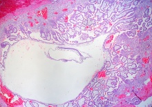
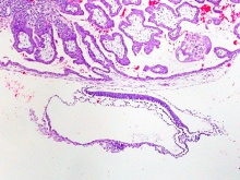
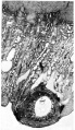
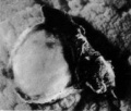
Carnegie Collection

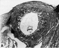
Surface view of implantation site |

Surface lateral view of implantation site |


| Carnegie Collection - Stage 7 | ||||||||||
|---|---|---|---|---|---|---|---|---|---|---|
| Serial No. | Grade | Fixative | Embedding Medium | Thinness (µm) | Stain | Year | Notes | |||
| 7802 | Exc. | Alc. & Bouin | C-P | 6 | (Stain - Haematoxylin Eosin) | 1940 | Heuser et al. (1945) | |||
| 8206 | Good | p | C-P | 6 | (Stain - Haematoxylin Eosin) | 1943 | ||||
| 8361 | Good | Bouin | C-P | 10 | p | 1946 | Abnormal | |||
| 8602 | Exc. | Alc. | C-P | 8 | (Stain - Haematoxylin Eosin) | 1948 | ||||
| 8752 | Exc. | ? | C-P | 10 | (Stain - Haematoxylin Eosin) | 1950 | ||||
| 8755 | Exc. | Formol | C-P | 10 | (Stain - Haematoxylin Eosin) | 1950 | ||||
| 9217 | Exc. | p | P | 10 | (Stain - Haematoxylin Eosin) | 1954 | ||||
Abbreviations
| ||||||||||
| iBook - Carnegie Embryos | |
|---|---|

|
|
Manchester Collection
Manchester No. 1285
Described by Florian and Hill (1935)[2].
- Specimen is housed in Department of Anatomy, University of Manchester.
- Hysterectomy.
- Chorionic cavity, 4.28 x 3.28 mm.
- Embryonic disc (narrow type), 0.87 x 0.625 mm.
- Primitive streak, 0.39 mm, and node, 0.05 mm.
- Notochordal process, 0.125 mm. Prechordal plate, 0.03 mm.
- Connecting stalk attached to chorion at decidua capsularis (suggesting polar variety of velamentous insertion of umbilical cord).
Dorsal and median projections published (Hill and Florian, 1931b, figs. 5 and 13; Florian and Hill, 1935, figs. 1-3; Mazanec, 1959, fig. 52).
- Text Figures
1. Graphic reconstruction of the dorsal view of the embryonal shield
2. Graphic reconstruction of the median section
3. Idealised Median Section


- Plate 1
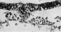
Fig. 1. Section 49
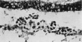
Fig. 2. Section 43
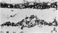
Fig. 3. Section 40
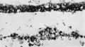
Fig. 4. Section 37


- Plate 2
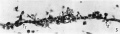
Fig. 5. Section 32
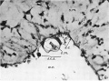
Fig. 6. Section 79
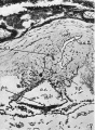
Fig. 7. Section 97
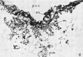
Fig. 8. Section 75


- Plate 3
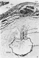
Fig.9. Section 122
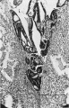
Fig. 10. Section 23
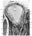
Fig.11. Section 99


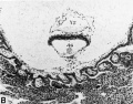
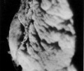
Photograph

|

|

|

|
Image: Dr Ed Uthman (Houston, Texas) - other pathology images section of ectopic (tubal) pregnancy.
- Image Links: Original full image | Trilaminar embryo excerpt | Villi excerpt 1 | Villi excerpt 2
- Full Image versions: ExtraLarge 1712x1206px | Large 1024x721px | Medium 500x352px
Embryo Virtual Slide
|
|
Stage 7 Images
Bright Field Images: Stage7-bf1.jpg | Stage7-bf2.jpg | Stage7-bf3.jpg | Stage7-bf4.jpg
Scanning Electron Images: Embryonic Disc 1 | Embryonic Disc 2 | Embryonic Disc 3 | Primitive Streak
Events
Historic Stage 7 Embryos
Historic stage 7 embryo data modified from O'Rahilly and Müller (1987).[3]
By Length of Notochordal Process
HEB-37 - Summarized by Mazanec (1959)[4]. Chorionic cavity, 2.25 x 1.29 x 0.4 mm. Embryonic disc, 0.4 mm. Primitive streak, 0.104 mm, and node, 0.04 mm. Notochordal process, 0.032 mm. Stalk of umbilical vesicle (ibid., fig. 77). Median projection published (ibid., fig. 45).
H.R. 1. (Hesketh Roberts) - Described by Johnston (1940)[5], who believed that a notochordal process (0.04 mm) and a prechordal plate (0.075 mm) were present. Florian in an appendix to the article disagreed, and his interpretation is followed here (see stage 6).
Biggart (Prof. JH. Biggart) - Described by Morton (1949).[6] Curettage. Embryonic disc (narrow type), 0.27 x 0.16 mm. Primitive streak and node, 0.059 mm. A notochordal process is not referred to in the text but is mapped on a dorsal projection of the embryo (ibid., fig. 2) and is approximately 0.04 mm in length. The specimen is said to resemble the Yale embryo.
- Biggart Embryo
embryo in situ
duct part of yolk-sac
distal expansion of yolk-sac
embryonic rudiment
opening of duct part into distal expansion of yolk-sac


Guá (Guálberto) - Described by Lordy (1931). Hysterectomy. Chorionic cavity, 8 x 7.5 mm. Embryonic disc, 0.776 x 0.0465 mm. Primitive streak, 0.09 mm. Notochordal process, 0.045 mm. Possible notochordal canal. Said to resemble Hugo embryo. Probably belongs either to stage 7 or to stage 8.
Carnegie No. 7802 - (figs. 7-4 to 7-12). An important specimen described and illustrated by Heuser, Rock, and Hertig (1945)[7]. Hysterectomy. Chorion, 3.75 x 2.35 x 2.2 mm. Chorionic cavity, 2.3 x 1.4 x 1.1 mm. Embryonic disc (broad type), 0.42 x 0.35 x 0.05 mm. Primitive streak, 0.11 mm, and node, 0.03 mm. Notochordal process, 0.048 mm. Presumed age, 16 days. Median projection published (ibid., plate 6; Mazanec, 1959[4], fig. 46) and dorsal projection has been prepared by the present writers.
- 7802 Embryo
Hertig1946 Fig. 5
Hertig1946 Fig. 5a
Hertig1946 Fig. 5b


P.M. - Described by Meyer (1924). Curettage. Measurements have been criticized by Stieve (1926) but defended by Mazanec (1959)[4]. Chorion, 3.9 x 3.77 x 2.5 mm. Chorionic cavity, 2.7 x 2.6 x 2.1 mm. Embryonic disc (circular), 0.41 x 0.41 mm. Primitive streak, 0.12 mm, and node, 0.02 mm. Notochordal process, 0.06 mm, acknowledged by Mazanec (1959) although denied by Fahrenholz (1927). No notochordal canal. Median projection published (Mazanec, 1959, fig. 47).
Hugo - Described by Stieve (1926), who reproduced a photomicrograph of every second section. Hysterectomy. Surrounded by 942 chorionic villi ranging in length from 0.3 to 1 mm. Chorion, 6.4 x 5.9 x 5.6 mm. Chorionic cavity, 4.7 x 4.4 x 3.8 mm. Embryonic disc (broad type), 0.635 (Florian, 1931)[8] x 0.63 mm. Primitive streak, 0.245 mm, and node, 0.05 mm. Notochordal process, 0.07 (0.11?) mm. (Florian, 1934c). No notochordal canal. Prechordal plate probably not yet developed (Hill and Florian, 1931a). Dorsal and median projections published (Florian, 1934c, fig. 1; Hill and Florian, 1931b, figs. 44 and 11; Mazanec, 1959[4], fig. 49).
Robertson, O’Neill, and Chappell (1948) described a hysterectomy specimen that possessed a chorion of 3.816 x 3.639 x 2.687 mm. Chorionic cavity, 2.718 x 2.239 x 1.679 mm. Embryonic disc (broad type), 0.462 x 0.485 mm. Primitive streak, 0.138 mm, and node (situated halfway), 0.03 mm. Notochordal process, 0.072 mm. Suggestion of notochordal canal in one or two sections. Assigned to horizon VIII by authors but probably belongs to stage 7. Median projection published (ibid., fig. 12).
D’Arrigo (1961) described an embryonic disc of 0.47 mm, which showed a notochordal process of 0.075 mm. Canalization of the process is “doubtful,” but the presence of a prechordal plate is “probable.” The specimen “could be recorded in Streeter’s horizon VII.”
Goodwin - Described by Kindred (1933). Tubal. Chorion, 5.8 x 2.72 x 2.25 mm. Chorionic cavity, 2.44 x 2.25 x 0.75 mm. Embryonic disc, 0.588 mm in width. Primitive streak, 0.215 mm, and node, 0.078 mm. Notochordal process, 0.078 mm. No notochordal canal and no prechordal plate.
Pha I - Described by Mazanec (1949).[9] Chorionic cavity, 7.872 x 5.475 x 2.032 mm. Embryonic disc, 0.66 x 0.52 mm. Primitive streak, 0.145 mm, and node, 0.06 mm. Notochordal process, 0.09 mm. No prechordal plate. Median projection published (ibid., fig. 51).
H. Schm. 10 - (H. Schmid). Described briefly by Grosser (1931c). Embryonic disc (almost circular), 0.51 x 0.58 mm. Primitive streak, 0.14 mm, and node, 0.1 mm. Notochordal process, 0.1 mm. Probably belongs to stage 7, although a cavity in one section was thought to represent “Lieberkühn’s canal.”
Bi 24 (Bittmann) - Described by Hill and Florian (1931b). Chorionic cavity, 3.05 x 3.036 x 3.029 mm. Embryonic disc (narrow type), 0.62 x 0.39 mm. Primitive streak, 0.28 mm, and node (Mazanec, 1959[4], fig. 105), 0.05 mm. Notochordal process, 0.105 mm, consists of median chord and lateral mesoblastic wings. Prechordal plate, 0.03 mm. Possible primordial germ cells in endoderm of region of cloacal membrane and in endoderm of umbilical vesicle caudally (Florian, 1931). Politzer (1933) counted 41 germ cells in the region of the allanto-enteric diverticulum in this embryo, and 19 such cells in another presomite specimen (Bi 25). Dorsal and median projections published (Hill and Florian, 1931b, figs. 4 and 12; Florian, 1945, plate 5, fig. 43; Mazanec, 1959[4], fig. 50).
Manchester No. 1285 (fig. 7-1). Described by Florian and Hill (m̩)[2]. Hysterectomy. Chorionic cavity, 4.28 x 3.28 mm. Embryonic disc (narrow type), 0.87 x 0.625 mm. Primitive streak, 0.39 mm, and node, 0.05 mm. Notochordal process, 0.125 mm. Prechordal plate, 0.03 mm. Connecting stalk attached to chorion at decidua capsularis (suggesting polar variety of velamentous insertion of umbilical cord). Dorsal and median projections published (Hill and Florian, 1931b, figs. 5 and 13; Florian and Hill, 1935, figs. 1-3; Mazanec, 1959, fig. 52). Specimen is housed in Department of Anatomy, University of Manchester.
Pha II. - Summarized by Mazanec (1959)[4]. Chorionic cavity, 4.985 x 3.882 x 3.52 mm. Embryonic disc, 0.895 x 0.62 mm. Primitive streak, 0.37 mm, and node, 0.06 mm. Notochordal process, 0.13 mm. No prechordal plate. Median projection published (ibid., fig. 53).
Thompson and Brash (1923) described a specimen that showed a notochordal process of 0.3 mm. It is described in the present work under stage 8.
By Year of Publication
No measurements of notochordal process available for the following historic embryos.
Debeyre (1912) described in detail a specimen that possessed a chorionic cavity of 5.6 x 2.1 mm. Embryonic disc, 0.9 x 0.6 x 0.95 mm. Primitive streak stated to be 0.54 mm in length. Chorionic villi (0.4-1.6 mm) showed some branching. Unsuitable plane of section makes it impossible to assess the specimen precisely.
Carnegie No. 1399 Mateer - Described by Streeter (1920).[10] Hysterectomy. Angiogenesis in villi described by Hertig (1935). Chorion, 9 x 8 x 3.5 mm. Chorionic cavity, 6.1 x 5.6 x 2.5 mm. Embryonic disc, 1 x 0.75 mm. Primitive streak and groove present. Although the notochordal process was originally thought probably to be absent, Hill and Florian (1931b) have no doubt that it is present. More advanced than Hugo (Florian and Völker, 1929). A very small twin embryo was originally described but that “interpretation has become open to doubt” (Corner, 1955). Drawing of every section reproduced by Turner (1920). A median drawing (Davis, 1927, fig. 5A) and a projection have been published (Mazanec, 1959, fig. 56).
- 1399 Embryo
Streeter Plate 1
Streeter Plate 2
Streeter Plate 3


Ho (Hodiesne) - Described by Fahrenholz (1927). Abortion. Chorionic cavity, 6.5 x 6 x 3 mm. Embryonic disc (deformed), 0.6 mm (0.725 mm by flexible scale). Primitive streak, 0.22 mm (0.345 mm by flexible scale). Notochordal process just beginning (“undoubtedly present,” Hill and Florian, 1931b). Possible prechordal plate claimed (disputed by Waldeyer, 1929a, but supported by Hill and Florian, 1963). “Lieberkühn’s canal” (0.065 mm) is an artificial folding of the embryonic disc (Hill and Florian, 1931b). Dorsal and median projections published (Fahrenholz, 1927, figs. 32, 6 and 7; Mazanec, 1959, fig. 54).
Debeyre (1933) described a specimen (0.9 mm) that possessed a primitive streak and probably belonged to stage 6 or stage 7. Large cells near the opening of the allantois were identified as primordial germ cells.
Falkiner - Described by Martin and Falkiner (1938).[11] Curettage. Embryo damaged and not in good condition. Measurements seem too small (see Mazanec, 1959)[4]. Chorionic cavity, 1.5 x 1.4 mm. Embryonic disc, 0.15 x 0.29 mm. Primitive streak, 0.07 mm. Notochordal process contains “no definite lumen.” Cells rostral to notochordal process are “probably” the prechordal plate. Development “agrees most closely” with that of Bi I. Median projection published (Martin and Falkiner, 1938, fig. 8; Mazanec, 1959, fig. 39).
- Falkiner Embryo
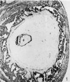
Fig. 1
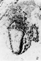
Fig. 2
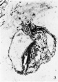
Fig. 3
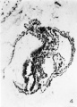
Fig. 4
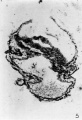
Fig. 5
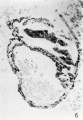
Fig. 6
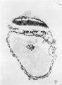
Fig. 7
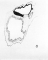
Fig. 8 Reconstruction


Gar (Green-Armytage) - Described by West (1952).[12] Hysterectomy. Chorionic cavity, 3 x 2.6 x 2 mm. Embryonic disc (broad type), 0.56 x 0.69 mm. Primitive streak, node, and groove present. Short notochordal process. No notochordal plate. Said to resemble Hugo embryo. Trophoblast described by Hamilton and Boyd (1960).
Mal (Maliphant) - Described by West (1952).[12] Hysterectomy. Chorionic cavity, 3 x 1.8 mm. Embryonic disc (broad type), 0.45 x 0.6 mm. Primitive streak, node, and groove present. Notochordal process present (Mazanec, 1959); small cavity in node (West, 1952) or in notochordal process (Mazanec, 1959) “hardly sufficient to warrant the name chorda canal.” No prechordal plate. Belongs either to stage 7 or to stage 8 (said to resemble Jones-Brewer I, which is in stage 8).
Carnegie No. 8602 - Photomicrograph reproduced by Hertig, Rock, and Adams (1956)[13] (plate 10, fig. 53). Chorion, 2.73 x 2.43 mm. Chorionic cavity, 1.83 x 1.33 mm. Embryonic disc, 0.3 x 0.06 mm. Presumed age, 16-17 days.
- 8602 Embryo


Missen - Trophoblast described by Hamilton and Boyd (1960). Curettage. Chorion, 1.66 x 1.43 mm. Embryonic disc, 0.28 x 0.214 mm. Primitive streak and node. Notochordal process. Said to resemble No. 7801 and Edwards Jones-Brewer (stage 6). Presumed age, about 14 days.
Other embryos probably belonging to stage 7 include Fitzgerald, Fitzgerald-Brewer II, and Jones-Brewer II (Brewer and Fitzgerald, 1937).[14]
References
- ↑ Sathananthan H, Selvaraj K & Clark J. (2011). The fine structure of human germ layers in vivo: clues to the early differentiation of embryonic stem cells in vitro. Reprod. Biomed. Online , 23, 227-33. PMID: 21665543 DOI.
- ↑ 2.0 2.1 Florian J. and Hill JP. An early human embryo (no. 1285, Manchester Collection), with capsular attachment of the connecting stalk. (1935) J Anat. 69(4): 399-411. PMID 17104547
- ↑ O'Rahilly R. and Müller F. Developmental Stages in Human Embryos. Contrib. Embryol., Carnegie Inst. Wash. 637 (1987).
- ↑ 4.0 4.1 4.2 4.3 4.4 4.5 4.6 4.7 Mazanec K. Blastogenese des Menschen. (1959) Fischer, Jena.
- ↑ Johnston TB. An early human embryo, with 0.55 mm. long embryonic shield. (1940) J. Anat., 75:1-49.
- ↑ Morton WRM. Two early human embryos. (1949) J. Anat., 83: 308-314.
- ↑ Heuser CH. Rock J. and Hertig AT. Two human embryos showing early stages of the definitive yolk sac. (1945) Contrib. Embryol., Carnegie Inst. Wash. Publ. 557, 31: 85-99.
- ↑ Template:Ref-Florian1931
- ↑ Mazanec K. A young human embryo "Pha I" with the head process 0.090 mm long. (1949) Bull, internat. Acad. tchique Sci, 50: 1-18.
- ↑ Streeter GL. A human embryo (Mateer) of the pre-somite period. (1920) Contrib. Embryol., Carnegie Inst. Wash. Publ. 272, 9: 389-424.
- ↑ Martin CP. and Falkiner N. Mcl. The Falkiner ovum. (1938) Amer. J Anat., 63: 251-271.
- ↑ 12.0 12.1 West CM. Two presomite human embryos. (1952) J. of Gynaecol. Geat Brit. Emp., 59: 336-351.
- ↑ Hertig AT. Rock J. and Adams EC. A description of 34 human ova within the first 17 days of development. (1956) Amer. J Anat., 98:435-493.
- ↑ Brewer JI. and Fitzgerald JE. Six normal and complete presomite human ova. (1937) Amer. J. Obstet. Gynecol, 34: 210-224.
Additional Images
- Carnegie Stages: 1 | 2 | 3 | 4 | 5 | 6 | 7 | 8 | 9 | 10 | 11 | 12 | 13 | 14 | 15 | 16 | 17 | 18 | 19 | 20 | 21 | 22 | 23 | About Stages | Timeline
Glossary Links
- Glossary: A | B | C | D | E | F | G | H | I | J | K | L | M | N | O | P | Q | R | S | T | U | V | W | X | Y | Z | Numbers | Symbols | Term Link
Cite this page: Hill, M.A. (2024, June 15) Embryology Carnegie stage 7. Retrieved from https://embryology.med.unsw.edu.au/embryology/index.php/Carnegie_stage_7
- © Dr Mark Hill 2024, UNSW Embryology ISBN: 978 0 7334 2609 4 - UNSW CRICOS Provider Code No. 00098G