BGDB Face and Ear - Early Embryo: Difference between revisions
mNo edit summary |
mNo edit summary |
||
| (24 intermediate revisions by the same user not shown) | |||
| Line 5: | Line 5: | ||
During week 4 a number of features appear visible on the embryo surface: | During week 4 a number of features appear visible on the embryo surface: | ||
* | * Body - heart, liver, somite bulges and limb buds appear. | ||
* | * Head - buccopharyngeal membrane breaks down, sensory placodes and pharyngeal arches appear. | ||
<br> | |||
{{Buccopharyngeal Membrane timeline EM gallery}} | |||
==Carnegie Stage 12 to 14== | ==Carnegie Stage 12 to 14== | ||
| Line 28: | Line 31: | ||
| [[File:Stage11 sem8.jpg|250px|right]] | | [[File:Stage11 sem8.jpg|250px|right]] | ||
| [[File:Stage11 sem81.jpg|500px]] | | [[File:Stage11 sem81.jpg|500px]] | ||
| | |} | ||
{| | |||
| [[File:Stage11 bf9.jpg|400px]] | |||
| [[File:Stage11 sem3b.jpg]] | | [[File:Stage11 sem3b.jpg]] | ||
| [[File:Stage11 sem3b.gif]] | | [[File:Stage11 sem3b.gif]] | ||
| Line 54: | Line 59: | ||
[[File:Stage13_sem1c.jpg]] [[File:Stage13_bf1c.jpg]] [[File:Stage13_sem2c.jpg]] | [[File:Stage13_sem1c.jpg]] [[File:Stage13_bf1c.jpg]] [[File:Stage13_sem2c.jpg]] | ||
{| | |||
! Surface view | |||
! Central Nervous System | |||
|- | |||
| <html5media height="620" width="480">File:Stage 13 MRI 3D04.mp4</html5media> | |||
| <html5media height="600" width="400">File:Stage 13 MRI 3D02.mp4</html5media> | |||
|} | |||
==Sensory Placodes== | ==Sensory Placodes== | ||
| Line 65: | Line 76: | ||
===Stage 13=== | ===Stage 13=== | ||
Identify the structure and position of the otic vesicle (otocyst) relative to other head structures. | |||
[[File:Stage13_otocyst.jpg|thumb|400px|Otic placode forms the otocyst]] | [[File:Stage13_otocyst.jpg|thumb|400px|Otic placode forms the otocyst]] | ||
<gallery caption="Otic Placode - Otocyst"> | |||
File:Stage 13 image 051.jpg| | |||
File:Stage 13 image 052.jpg| | |||
File:Stage 13 image 053.jpg| | |||
File:Stage 13 image 054.jpg| | |||
</gallery> | |||
<br> | |||
<gallery caption="Optic Placode"> | |||
File:Stage 13 image 059.jpg| | |||
File:Stage 13 image 060.jpg| | |||
File:Stage 13 image 061.jpg| | |||
</gallery> | |||
<br> | |||
<gallery caption="Nasal Placode"> | |||
File:Stage 13 image 061.jpg| | |||
File:Stage 13 image 062.jpg| | |||
File:Stage 13 image 063.jpg| | |||
File:Stage 13 image 064.jpg| | |||
</gallery> | |||
<br> | |||
<gallery caption="Pituitary Placode"> | |||
File:Stage 13 image 058.jpg| | |||
File:Stage 13 image 059.jpg| | |||
File:Stage 13 image 060.jpg| | |||
</gallery> | |||
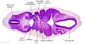
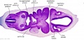
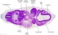
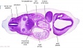
===Stage 15=== | |||
[[Carnegie_stage_15|Stage 15]] | |||
[[File: | [[File:Stage15 sagittal section upper half 01.jpg|400px]] | ||
Later week 5 development showing a sagittal section upper half of human embryo. | |||
==Pharyngeal Arches== | ==Pharyngeal Arches== | ||
[[File:Head arches cartoon.jpg]] | [[File:Head arches cartoon.jpg]] | ||
| Line 99: | Line 130: | ||
==Structures derived from Arches== | ==Structures derived from Arches== | ||
{{Pharyngeal Arch table}} | |||
<br> | |||
{{BGDB Practical 6 - Early Embryo Interactive}} | |||
{{BGDB Face}} | |||
{ | |||
==Additional Information== | ==Additional Information== | ||
| Line 205: | Line 141: | ||
===Other Sensory Systems=== | ===Other Sensory Systems=== | ||
The links below are to '''additional information''' providing background about each of the sensory systems. Only hearing is covered in today's class. | The links below are to '''additional information''' providing background about each of the sensory systems. Only hearing is covered in today's class. | ||
{ | {| | ||
| [[File:Hearing cartoon.jpg|160px|link=Sensory_-_Hearing_and_Balance_Development]] | |||
| [[File:Stage_22_image_153.jpg|160px|link=Sensory_-_Vision_Development]] | |||
| [[File:Stage_22_image_209.jpg|160px|link=Sensory_-_Smell_Development]] | |||
| [[File:Tongue_-_taste_cartoon.jpg|160px|link=Sensory - Taste Development]] | |||
|- | |||
| <center>[[Sensory_-_Hearing_and_Balance_Development|Hearing Development]]</center> | |||
| <center>[[Sensory_-_Vision_Development|Vision Development]]</center> | |||
| <center>[[Sensory_-_Smell_Development|Smell Development]]</center> | |||
| <center>[[Sensory - Taste Development|Taste Development]]</center> | |||
|} | |||
{{Hearing Links}} | |||
{{Vision Links}} | |||
{{Smell Links}} | |||
===Neural Crest=== | ===Neural Crest=== | ||
During this period neural crest cells migrate into the pharyngeal arches and other head locations, and have an important contribution to many different head structures. Neural crest cells at other levels contribute to body many structures. There are also many developmental abnormalities associated with abnormal neural crest development and/or migration. This topic is beyond the scope of the current class. | During this period neural crest cells migrate into the pharyngeal arches and other head locations, and have an important contribution to many different head structures. Neural crest cells at other levels contribute to body many structures. There are also many developmental abnormalities associated with abnormal neural crest development and/or migration. This topic is beyond the scope of the current class. | ||
:'''Links:''' [[Neural Crest Development]] | :'''Links:''' [[Neural Crest Development]] | ||
| Line 234: | Line 176: | ||
Some bones, including the squamosal (SQ), alisphenoid (AS), and pterygoid (PT), are shown with mixed contribution from different NCC populations. Note that in mammals the frontal (FR) and parietal (PA) bones have been reported to be of neural crest and mesodermal origin, respectively. | Some bones, including the squamosal (SQ), alisphenoid (AS), and pterygoid (PT), are shown with mixed contribution from different NCC populations. Note that in mammals the frontal (FR) and parietal (PA) bones have been reported to be of neural crest and mesodermal origin, respectively. | ||
There has been controversy about the neural crest embryonic contribution to the parietal region. A recent transcriptional analysis of second trimester human cranial compartments{{#pmid:26188427|PMID26188427}} suggests that "a gene expression signature of neural crest origin still exists in frontal and metopic compartments while gene expression of parietal and sagittal compartments is more similar to mesoderm."{{#pmid:14523380|PMID14523380}} | |||
* frontal bone - {{neural crest}} | |||
* parietal bone - {{mesoderm}} | |||
|} | |} | ||
| Line 255: | Line 199: | ||
|} | |} | ||
<references/> | |||
===Terms=== | |||
{{Head terms}} | |||
| Line 261: | Line 210: | ||
{{ | {{BGDBFooter}} | ||
Latest revision as of 20:56, 12 May 2019
Week 4
During week 4 a number of features appear visible on the embryo surface:
- Body - heart, liver, somite bulges and limb buds appear.
- Head - buccopharyngeal membrane breaks down, sensory placodes and pharyngeal arches appear.
- Human Buccopharyngeal Membrane Timeline Gallery (Week 4)
Stage 10

Stage 11
Stage 12
Stage 13


Carnegie Stage 12 to 14
Week 4


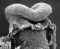
This is a scanning EM of the embryo superior dorsal view showing the paired otic placodes sinking into the surface at the level of the hindbrain between day 24 and day 25
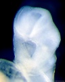
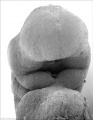
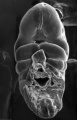
Light microscope ventrolateral view
EM ventrolateral view of the Buccopharyngeal Membrane

EM ventral view of the Buccopharyngeal Membrane

EM close up view of the degenerating Buccopharyngeal Membrane


| Human Embryo (Stage 11) | |
|---|---|
 |

|

|

|

|


Week 5


| Surface view | Central Nervous System |
|---|---|
| <html5media height="620" width="480">File:Stage 13 MRI 3D04.mp4</html5media> | <html5media height="600" width="400">File:Stage 13 MRI 3D02.mp4</html5media> |
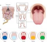
Sensory Placodes
Sensory placodes develop as small patches of ectodermal thickenings.
The placodes are laterally paired and contribute key components to sensory structures of the ear, eye and nose.
Named by the sensory system and components they will form: otic placode, optic (lens) placode and nasal placode.
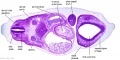
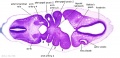
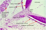
Stage 13
Identify the structure and position of the otic vesicle (otocyst) relative to other head structures.

- Otic Placode - Otocyst


- Optic Placode


- Nasal Placode


- Pituitary Placode

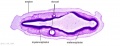
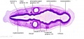
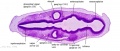
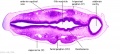
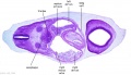
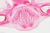
Stage 15

Later week 5 development showing a sagittal section upper half of human embryo.
Pharyngeal Arches


Stage 13 Pharyngeal Arches

Section Images: A6L A7L B1L B2L B3L B4L B5L B6L
Look through the above cross-sections of the stage 13 embryo observing and identifying structures of the face and ear visible at this stage.

Structures derived from Arches
| Pharyngeal Arch | Nerve | Artery | Neural Crest (Skeletal Structures) |
Muscles | Ligaments |
|---|---|---|---|---|---|
| 1 (maxillary/mandibular) |
trigeminal (CN V) | maxillary artery (terminal branches) | mandible, maxilla, malleus, incus | muscles of mastication, mylohyoid, tensor tympanic, ant. belly digastric | ant lig of malleus, sphenomandibular ligament |
| 2 (hyoid) |
facial (CN VII) | stapedial (embryonic) corticotympanic (adult) |
stapes, styloid process, lesser cornu of hyoid, upper part of body of hyoid bone | muscles of facial expression, stapedius, stylohyoid, post. belly digastric | stylohyoid ligament |
| 3 | glossopharyngeal (CN IX) | common carotid, internal carotid arteries | greater cornu of hyoid, lower part of body of hyoid bone | stylopharyngeus | |
| 4 | vagus (CN X) superior laryngeal branch | part of aortic arch (left), part right subclavian artery (right) | thyroid, cricoid, arytenoid, corniculate and cuneform cartilages | crycothyroid, soft palate levator veli palatini (not tensor veli palatini) | |
| 6 | vagus (CN X) recurrent laryngeal branch | part of left pulmonary artery (left), part of right pulmonary artery (right) | thyroid, cricoid, arytenoid, corniculate and cuneform cartilages | larynx intrinsic muscles (not cricothyroid muscle) |
Early Embryo Interactive Component
| Attempt the Quiz - Early Embryo | |
|---|---|

Here are a few simple Quiz questions that relate to Early Embryo from the lecture and practical.
|
Additional Information
| Additional Information - Content shown under this heading is not part of the material covered in this class. It is provided for those students who would like to know about some concepts or current research in topics related to the current class page. |
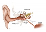
Other Sensory Systems
The links below are to additional information providing background about each of the sensory systems. Only hearing is covered in today's class.
|
|
|
|
| Vision Links: vision | lens | retina | placode | extraocular muscle | cornea | eyelid | lacrima gland | vision abnormalities | Student project 1 | Student project 2 | Category:Vision | sensory | ||
|
| Smell Links: Introduction | placode | Rhinencephalon | head | respiratory | Student project | taste | sensory | Category:Smell | ||
|
Neural Crest
During this period neural crest cells migrate into the pharyngeal arches and other head locations, and have an important contribution to many different head structures. Neural crest cells at other levels contribute to body many structures. There are also many developmental abnormalities associated with abnormal neural crest development and/or migration. This topic is beyond the scope of the current class.
- Links: Neural Crest Development

|
Cranial neural crest contribution to skeletal structures
There has been controversy about the neural crest embryonic contribution to the parietal region. A recent transcriptional analysis of second trimester human cranial compartments[1] suggests that "a gene expression signature of neural crest origin still exists in frontal and metopic compartments while gene expression of parietal and sagittal compartments is more similar to mesoderm."[2]
|
|
|
- ↑ Homayounfar N, Park SS, Afsharinejad Z, Bammler TK, MacDonald JW, Farin FM, Mecham BH & Cunningham ML. (2015). Transcriptional analysis of human cranial compartments with different embryonic origins. Arch. Oral Biol. , 60, 1450-60. PMID: 26188427 DOI.
- ↑ Santagati F & Rijli FM. (2003). Cranial neural crest and the building of the vertebrate head. Nat. Rev. Neurosci. , 4, 806-18. PMID: 14523380 DOI.
Terms
| Head Terms (expand to view) |
|---|
|
| Other Terms Lists |
|---|
| Terms Lists: ART | Birth | Bone | Cardiovascular | Cell Division | Endocrine | Gastrointestinal | Genital | Genetic | Head | Hearing | Heart | Immune | Integumentary | Neonatal | Neural | Oocyte | Palate | Placenta | Radiation | Renal | Respiratory | Spermatozoa | Statistics | Tooth | Ultrasound | Vision | Historic | Drugs | Glossary |
BGDB: Lecture - Gastrointestinal System | Practical - Gastrointestinal System | Lecture - Face and Ear | Practical - Face and Ear | Lecture - Endocrine | Lecture - Sexual Differentiation | Practical - Sexual Differentiation | Tutorial
Glossary Links
- Glossary: A | B | C | D | E | F | G | H | I | J | K | L | M | N | O | P | Q | R | S | T | U | V | W | X | Y | Z | Numbers | Symbols | Term Link
Cite this page: Hill, M.A. (2024, June 17) Embryology BGDB Face and Ear - Early Embryo. Retrieved from https://embryology.med.unsw.edu.au/embryology/index.php/BGDB_Face_and_Ear_-_Early_Embryo
- © Dr Mark Hill 2024, UNSW Embryology ISBN: 978 0 7334 2609 4 - UNSW CRICOS Provider Code No. 00098G