Gastrointestinal Tract Development: Difference between revisions
mNo edit summary |
mNo edit summary |
||
| (17 intermediate revisions by the same user not shown) | |||
| Line 3: | Line 3: | ||
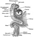
[[File:Gray0982a.jpg|thumb|300px|The early gastrointestinal tract.]] | [[File:Gray0982a.jpg|thumb|300px|The early gastrointestinal tract.]] | ||
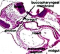
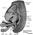
[[File:Stage11_sem4.jpg|thumb|300px|Human head ([[Week 4]], [[Carnegie stage 11|Stage 11]]) showing buccopharyngeal membrane breakdown.]] | [[File:Stage11_sem4.jpg|thumb|300px|Human head ([[Week 4]], [[Carnegie stage 11|Stage 11]]) showing buccopharyngeal membrane breakdown.]] | ||
The {{gastrointestinal tract}} (GIT) arises initially during the process of gastrulation from the | The {{gastrointestinal tract}} (GIT) arises initially during the process of gastrulation from the {{endoderm}} of the trilaminar embryo (week 3) and extends from the {{buccopharyngeal membrane}} to the {{cloacal membrane}}. The tract and associated organs later have contributions from all the germ cell layers. | ||
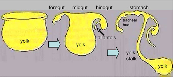
During the 4th week three distinct regions (fore-, mid- and hind-gut) extend the length of the embryo and will contribute different components of the GIT. The large mid-gut is generated by lateral embryonic folding which "pinches off" a pocket of the yolk sac, the 2 compartments continue to communicate through the vitelline duct. | During the 4th week three distinct regions (fore-, mid- and hind-gut) extend the length of the embryo and will contribute different components of the GIT. The large mid-gut is generated by lateral embryonic folding which "pinches off" a pocket of the {{yolk sac}}, the 2 compartments continue to communicate through the vitelline duct. | ||
The oral cavity (mouth) is formed following breakdown of the buccopharyngeal membrane (oropharyngeal or oral membrane) and contributed to mainly by the pharynx lying within the pharyngeal | The oral cavity (mouth) is formed following breakdown of the {{buccopharyngeal membrane}} (oropharyngeal or oral membrane) and contributed to mainly by the pharynx lying within the {{pharyngeal arch}}es (More? [[Head Development]]). Loss of buccopharyngeal membrane opens the tract to amniotic fluid through the remainder of development, and during the fetal period is actively swallowed. | ||
From the oral cavity the next portion of the foregut is initially the pharynx, a single gastrointestinal (oesophagus) and respiratory (trachea) common tube, that lies behind the heart. Note that the respiratory tract will form from a ventral bud arising at this level (More? {{respiratory}}). | From the oral cavity the next portion of the foregut is initially the pharynx, a single gastrointestinal ({{oesophagus}}{{) and respiratory (trachea) common tube, that lies behind the heart. Note that the respiratory tract will form from a ventral bud arising at this level (More? {{respiratory}}). | ||
This current page provides an introductory overview, use the links below for descriptions of specific components and regions as well as developmental abnormalities. | This current page provides an introductory overview, use the links below for descriptions of specific components and regions as well as developmental abnormalities. | ||
| Line 19: | Line 19: | ||
{{Gastrointestinal Tract Links}} | {{Gastrointestinal Tract Links}} | ||
Note that in historic texts the term ''entoderm'' is used to describe '''endoderm''' and other terminology may also differ from current descriptions. | Note that in historic texts the term ''entoderm'' is used to describe '''{{endoderm}}''' and other terminology may also differ from current descriptions. | ||
[[File:Endoderm_cartoon.jpg|600px|link=One Minute Embryology#Endoderm Development]] | [[File:Endoderm_cartoon.jpg|600px|link=One Minute Embryology#Endoderm Development]] | ||
| Line 26: | Line 26: | ||
|-bgcolor="F5FAFF" | |-bgcolor="F5FAFF" | ||
| | | | ||
* ''' | * '''Mapping of extrinsic innervation of the gastrointestinal tract in the mouse embryo'''{{#pmid:32690615|PMID32690615}} "Precise extrinsic afferent (visceral sensory) and efferent (sympathetic and parasympathetic) innervation of the gut is fundamental for gut-brain crosstalk. Owing to the limitation of intrinsic markers to distinctively visualize the three classes of extrinsic axons, which intimately associate within the gut mesentery, detailed information on the development of extrinsic gut-innervating axons remains relatively sparse. Here, we mapped extrinsic innervation of the gut and explored the relationships among various types of extrinsic axons during embryonic development in mice. Visualization with characterized intrinsic markers revealed that visceral sensory, sympathetic, and parasympathetic axons arise from different anatomical locations, project in close association via the gut mesentery, and form distinctive innervation patterns within the gut from embryonic day {{ME10.5}} to {{ME16.5}}. Genetic ablation of visceral sensory trajectories results in the erratic extension of both sympathetic and parasympathetic axons, implicating that afferent axons provide an axonal scaffold to route efferent axons. Co-culture assay further confirmed the attractive effect of sensory axons on sympathetic axons. Taken together, our study provides key information regarding the development of extrinsic gut-innervating axons occurring through heterotypic axonal interactions and provides an anatomical basis to uncover neural circuit assembly in the gut-brain axis." | ||
* '''The Digestive Tract in Human Embryos Between Carnegie Stages | * '''{{Stomach}} curvature is generated by left-right asymmetric gut morphogenesis'''{{#pmid:28242610|PMID28242610}} "Left-right (LR) asymmetry is a fundamental feature of internal anatomy, yet the emergence of morphological asymmetry remains one of the least understood phases of organogenesis. Asymmetric rotation of the intestine is directed by forces outside the gut, but the morphogenetic events that generate anatomical asymmetry in other regions of the digestive tract remain unknown. Here, we show in mouse and Xenopus that the mechanisms that drive the curvature of the stomach are intrinsic to the gut tube itself. The left wall of the primitive stomach expands more than the right wall, as the left epithelium becomes more polarized and undergoes radial rearrangement. These asymmetries exist across several species, and are dependent on LR patterning genes, including Foxj1, Nodal and Pitx2 Our findings have implications for how LR patterning manifests distinct types of morphological asymmetries in different contexts." | ||
* '''The Digestive Tract in Human Embryos Between Carnegie Stages {{CS11}} and {{CS13}}'''{{#pmid:26995337|PMID26995337}} "The digestive tract was initially formed by a narrowing of the yolk sac, and then several derived primordia such as the pharynx, lung, stomach, liver, and dorsal pancreas primordia differentiated during {{CS12}} (21-29 somites) and CS13 (≥ 30 somites). The differentiation of four pairs of pharyngeal pouches was complete in all {{CS13}} embryos. The respiratory primordium was recognized in ≥ 26-somite embryos and it flattened and then branched at {{CS13}}. The trachea formed and then elongated in ≥ 35-somite embryos. The stomach adopted a spindle shape in all ≥ 34-somite embryos, and the liver bud was recognized in ≥ 27-somite embryos. The dorsal pancreas appeared as definitive buddings in all but three CS13 embryos, and around these buddings, the small intestine bent in ≥ 33-somite embryos. In ≥ 35-{{somite}} embryos, the small intestine rotated around the cranial-caudal axis and had begun to form a primitive intestinal loop, which led to umbilical herniation." | |||
|} | |} | ||
| Line 37: | Line 39: | ||
| [[File:Mark_Hill.jpg|90px|left]] {{Most_Recent_Refs}} | | [[File:Mark_Hill.jpg|90px|left]] {{Most_Recent_Refs}} | ||
Search term: [http://www.ncbi.nlm.nih.gov/pubmed/?term=Gastrointestinal+Tract+Embryology ''Gastrointestinal Tract Embryology''] | Search term: [http://www.ncbi.nlm.nih.gov/pubmed/?term=Gastrointestinal+Tract+Embryology ''Gastrointestinal Tract Embryology''] | [http://www.ncbi.nlm.nih.gov/pubmed/?term=Gastrointestinal+Tract+Development''Gastrointestinal Tract Development''] | ||
|} | |} | ||
| Line 45: | Line 46: | ||
! Older papers | ! Older papers | ||
|- | |- | ||
| | | {{Older papers}} | ||
*''' Three-dimensional reconstructions of intrahepatic bile duct tubulogenesis in human liver'''{{#pmid:21943389|PMID21943389}} In the developing human liver, three-dimensional reconstructions using multiple marker proteins confirmed that the human intrahepatic biliary tree forms through several developmental stages involving an initial transition of primitive hepatocytes into cholangiocytes shaping the ductal plate followed by a process of maturation and remodeling where the intrahepatic biliary tree develops through an asymmetrical form of cholangiocyte tubulogenesis. | *''' Three-dimensional reconstructions of intrahepatic bile duct tubulogenesis in human liver'''{{#pmid:21943389|PMID21943389}} In the developing human liver, three-dimensional reconstructions using multiple marker proteins confirmed that the human intrahepatic biliary tree forms through several developmental stages involving an initial transition of primitive hepatocytes into cholangiocytes shaping the ductal plate followed by a process of maturation and remodeling where the intrahepatic biliary tree develops through an asymmetrical form of cholangiocyte tubulogenesis. {{Liver}} | ||
* '''Endocrine Pancreas'''{{#pmid:20377917|PMID20377917}} "The transcription factor Pax6 functions in the specification and maintenance of the differentiated cell lineages in the endocrine pancreas. It has two DNA binding domains, the paired domain and the homeodomain, in addition to a C-terminal transactivation domain. The phenotype of Pax6-/- knockout mice suggests non-redundant functions of the transcription factor in the development of glucagon-expressing alpha-cells as this cell type is absent in the mutants." {{endocrine pancreas}} | * '''Endocrine Pancreas'''{{#pmid:20377917|PMID20377917}} "The transcription factor Pax6 functions in the specification and maintenance of the differentiated cell lineages in the endocrine pancreas. It has two DNA binding domains, the paired domain and the homeodomain, in addition to a C-terminal transactivation domain. The phenotype of Pax6-/- knockout mice suggests non-redundant functions of the transcription factor in the development of glucagon-expressing alpha-cells as this cell type is absent in the mutants." {{endocrine pancreas}} | ||
| Line 103: | Line 104: | ||
{{GIT cartoons}} | {{GIT cartoons}} | ||
== | ==Gastrointestinal Tract Divisions== | ||
[[File:GIT_blood_supply.jpg|left|300px]] During the 4th week the 3 distinct | [[File:GIT_blood_supply.jpg|left|300px]] During the 4th week the 3 distinct divisions ({{foregut}}, {{midgut}} and {{hindgut}}) extend the length of the embryo from oral membrane to cloacal membrane and will contribute different components of the GIT. These 3 divisions are also later anatomically defined by the vascular (artery) supply to each of theses divisions. | ||
The large mid-gut is generated by lateral embryonic folding which "pinches off" a pocket of the yolk sac, the 2 compartments continue to communicate through the vitelline duct. | The large mid-gut is generated by lateral embryonic folding which "pinches off" a pocket of the yolk sac, the 2 compartments continue to communicate through the vitelline duct. | ||
| Line 111: | Line 112: | ||
===Foregut=== | ===Foregut=== | ||
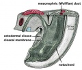
[[File:Gitbpm.jpg|thumb|Stage | [[File:Gitbpm.jpg|thumb|Stage {{CS11}} foregut]] | ||
First embryonic division of gastrointestinal tract, extending from the oral (buccopharyngeal) membrane and contributing oesophagus, {{stomach}}, duodenum (to bile duct opening), {{liver}}, biliary apparatus (hepatic ducts, {{gallbladder}}, and bile duct), and {{pancreas}}. The forgut blood supply is the celiac artery (trunk) excluding the pharynx, lower respiratory tract, and most of the oesophagus. | |||
From the oral cavity the next portion of the foregut is initially a single gastrointestinal (oesophagus) and respiratory (trachea) common tube, the pharynx which lies behind the heart. Note that the respiratory tract will form from a ventral bud arising at this level. | |||
* Oral cavity | * Oral cavity | ||
| Line 120: | Line 125: | ||
===Midgut=== | ===Midgut=== | ||
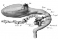
[[File:Gray0986.jpg|thumb|midgut herniation]] | [[File:Gray0986.jpg|thumb|midgut herniation]] | ||
The middle embryonic division of gastrointestinal tract contributing the small intestine (including duodenum beneath distal bile duct opening), cecum, appendix, ascending colon, and part of the transverse colon (right half to two thirds). The midgut blood supply is the superior mesenteric artery. | |||
Much of the '''midgut is herniated''' at the umbilicus external to the abdomen through development. A key step in development is the rotation of this midgut that must occur to place the GIT in the correct abdominal position with its associated mesentry. The GIT itself differentiates to form significantly different structures along its length: oesophagus, stomach, duodenum, jejunum, ilium (small intestine), colon (large intestine). | |||
The '''mesenteries''' of the GIT are generated from the common dorsal mesentery, with the ventral mesentry contributing to the '''lesser omentum''' and '''falciform ligament'''. | |||
Note the duodenum is commonly divided into 4 anatomical sequential parts (superior, descending, horizontal, ascending). | |||
:'''Links:''' {{Intestine}} | {{mesentery}} | |||
===Hindgut=== | ===Hindgut=== | ||
The | |||
The final embryonic division of gastrointestinal tract consisting initially of the {{cloaca}} snd extending to the cloacal membrane. The hindgut contributes part of the transverse colon (left half to one third), descending colon, sigmoid colon, rectum, part of anal canal (superior), urinary epithelium (bladder and most urethra). | |||
The initial {{cloaca}} space will later become partitioned by a septum into a dorsal gastrointestinal component (rectum) and ventral urogenital sinus (renal/genital component). | |||
:'''Links:''' {{cloaca}} | {{Intestine}} | {{bladder}} | {{genital}} | |||
== Development Overview == | == Development Overview == | ||
GIT shown in green anchored by dosal and ventral mesogastrium. The space ouside this will be the peritoneal cavity. | GIT shown in green anchored by dosal and ventral mesogastrium. The space ouside this will be the peritoneal cavity. | ||
Red ring-neural tube with neural | Red ring - neural tube with neural crest Blue ring - notocord Orange - somites | ||
Differentiation of associated organs at the level of the forming stomach occurs both dorsally (spleen) and ventrally (liver). | Differentiation of associated organs at the level of the forming stomach occurs both dorsally (spleen) and ventrally (liver). | ||
| Line 142: | Line 156: | ||
| | ||
Large blue ring- dorsal | Large blue ring - dorsal aorta Dark green ring - {{Liver}} | ||
Continued growth of the GIT and the organs leads to organ movements and bending of tract. | Continued growth of the GIT and the organs leads to organ movements and bending of tract. | ||
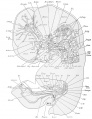
== Carnegie stage 13 Embryo Overview == | == Carnegie stage 13 Embryo Overview == | ||
Below is an overview of the sections starting at the level of pharynx compressed dorsoventrally, following the GIT through to the rectum. The most obvious feature is that of a continuous tube initially, attached by dorsoventral mesentry. Outside this tube and mesentry (at the levels below the lung buds) is the intraembryonic coelom that will form the peritoneal cavity. The hepatic diverticulum (liver bud) lies under the septum transversum is the earliest associated GIT organ that has differentiated, and now occupies a substantial region of the abdomen. Clicking on sections below will open the original images. | Below is an overview of the sections starting at the level of pharynx compressed dorsoventrally, following the GIT through to the rectum. The most obvious feature is that of a continuous tube initially, attached by dorsoventral mesentry. Outside this tube and mesentry (at the levels below the lung buds) is the intraembryonic coelom that will form the peritoneal cavity. The hepatic diverticulum (liver bud) lies under the septum transversum is the earliest associated GIT organ that has differentiated, and now occupies a substantial region of the abdomen. Clicking on sections below will open the original images. | ||
| Line 164: | Line 179: | ||
The gastrointestinal tract has both intrinsic and extrinsic innervation. (see the recent review{{#pmid:27112528|PMID27112528}}) | The gastrointestinal tract has both intrinsic and extrinsic innervation. (see the recent review{{#pmid:27112528|PMID27112528}}) | ||
* The intrinsic innervation, the enteric plexus, is derived from neural crest cells migrating into and along the wall of the gastrointestinal tract. | * The intrinsic innervation, the enteric plexus, is derived from {{neural crest}} cells migrating into and along the wall of the gastrointestinal tract. | ||
** mainly vagal region neural crest - generating both neurons and glia. | ** mainly vagal region neural crest - generating both neurons and glia. | ||
** some sacral neural crest - in chicken.{{#pmid:10677253|PMID10677253}} | ** some sacral neural crest - in chicken.{{#pmid:10677253|PMID10677253}} | ||
| Line 301: | Line 316: | ||
===Search PubMed=== | ===Search PubMed=== | ||
{| | |||
! Year | |||
! Total articles | |||
! Reviews | |||
! Free fun text | |||
|- | |||
| Mar 2007 | |||
| 29,361 | |||
| 3,494 | |||
| | |||
|- | |||
| Apr 2010 | |||
| 35980 | |||
| 4707 | |||
| 8086 | |||
|- | |||
| Dec 2018 | |||
| 58103 | |||
| 9188 | |||
| 44728 | |||
|} | |||
'''Search Pubmed:''' [http://www.ncbi.nlm.nih.gov/sites/entrez?db=pubmed&cmd=search&term=gastrointestinal%20tract%20development Gastrointestinal Tract Development] | '''Search Pubmed:''' [http://www.ncbi.nlm.nih.gov/sites/entrez?db=pubmed&cmd=search&term=gastrointestinal%20tract%20development Gastrointestinal Tract Development] | ||
| Line 309: | Line 343: | ||
==Additional Images== | ==Additional Images== | ||
<gallery> | <gallery> | ||
File:Gitbpm.jpg|buccopharyngeal membrane | |||
File:Stage14-git.jpg|serial sections stage 13 | |||
</gallery> | |||
===Historic=== | |||
{{Historic Disclaimer}} | |||
<gallery> | |||
File:Gitbpm.jpg | File:Gitbpm.jpg | ||
File:Gray0982a.jpg | File:Gray0982a.jpg | ||
Latest revision as of 10:36, 27 August 2020
| Embryology - 10 Jun 2024 |
|---|
| Google Translate - select your language from the list shown below (this will open a new external page) |
|
العربية | català | 中文 | 中國傳統的 | français | Deutsche | עִברִית | हिंदी | bahasa Indonesia | italiano | 日本語 | 한국어 | မြန်မာ | Pilipino | Polskie | português | ਪੰਜਾਬੀ ਦੇ | Română | русский | Español | Swahili | Svensk | ไทย | Türkçe | اردو | ייִדיש | Tiếng Việt These external translations are automated and may not be accurate. (More? About Translations) |
Introduction


The gastrointestinal tract (GIT) arises initially during the process of gastrulation from the endoderm of the trilaminar embryo (week 3) and extends from the buccopharyngeal membrane to the cloacal membrane. The tract and associated organs later have contributions from all the germ cell layers.
During the 4th week three distinct regions (fore-, mid- and hind-gut) extend the length of the embryo and will contribute different components of the GIT. The large mid-gut is generated by lateral embryonic folding which "pinches off" a pocket of the yolk sac, the 2 compartments continue to communicate through the vitelline duct.
The oral cavity (mouth) is formed following breakdown of the buccopharyngeal membrane (oropharyngeal or oral membrane) and contributed to mainly by the pharynx lying within the pharyngeal arches (More? Head Development). Loss of buccopharyngeal membrane opens the tract to amniotic fluid through the remainder of development, and during the fetal period is actively swallowed.
From the oral cavity the next portion of the foregut is initially the pharynx, a single gastrointestinal (oesophagus{{) and respiratory (trachea) common tube, that lies behind the heart. Note that the respiratory tract will form from a ventral bud arising at this level (More? respiratory).
This current page provides an introductory overview, use the links below for descriptions of specific components and regions as well as developmental abnormalities.
Note that in historic texts the term entoderm is used to describe endoderm and other terminology may also differ from current descriptions.
Some Recent Findings
|
| More recent papers |
|---|
 This table allows an automated computer search of the external PubMed database using the listed "Search term" text link.
More? References | Discussion Page | Journal Searches | 2019 References | 2020 References Search term: Gastrointestinal Tract Embryology | Gastrointestinal Tract Development |
| Older papers |
|---|
| These papers originally appeared in the Some Recent Findings table, but as that list grew in length have now been shuffled down to this collapsible table.
See also the Discussion Page for other references listed by year and References on this current page.
|
Textbooks
- Human Embryology Larson Chapter 9 p229-260
- The Developing Human: Clinically Oriented Embryology (6th ed.) Moore and Persaud Chapter 12 p271-302
- Before We Are Born (5th ed.) Moore and Persaud Chapter 13 p255-287
- Essentials of Human Embryology Larson Chapter 9 p123-146
- Human Embryology Fitzgerald and Fitzgerald Chapter 19,20 p119-123
More? References | Online Textbooks | Historic Textbooks
| UNSW Students | |||||||
|---|---|---|---|---|---|---|---|
|
|
You have access the following online Embryology resources and textbooks through the UNSW Library. | ||||||

|
Hill, M.A. (2020). UNSW Embryology (20th ed.) Retrieved June 10, 2024, from https://embryology.med.unsw.edu.au
| ||||||

|
Moore, K.L., Persaud, T.V.N. & Torchia, M.G. (2015). The developing human: clinically oriented embryology (10th ed.). Philadelphia: Saunders. (links only function with UNSW connection)
Chapter 11 Alimentary System
| ||||||

|
Schoenwolf, G.C., Bleyl, S.B., Brauer, P.R., Francis-West, P.H. & Philippa H. (2015). Larsen's human embryology (5th ed.). New York; Edinburgh: Churchill Livingstone.(links only function with UNSW connection)
Chapter 14 Development of the Gastrointestinal Tract
| ||||||


Objectives
- Understanding of germ layer contributions to the early gastrointestinal tract (GIT)
- Understanding of the folding of the GIT
- Understanding of three main GIT embryonic divisions
- Understanding of associated organ development (liver, pancreas, spleen)
- Brief understanding of mechanical changes (rotations) during GIT development
- Brief understanding of gastrointestinal abnormalities
Germ Layer Contributions
- Endoderm - epithelium and associated glands
- Mesoderm (splanchnic) - mesentry, connective tissues, smooth muscle, blood vessels
- Ectoderm (neural crest) - enteric nervous system (neural tube) - extrinsic innervation
Both endoderm and mesoderm will contribute to associated organs.
Gastrointestinal Tract Movies
| Gastrointestinal Tract Movies | |||||||||||||||||||
|---|---|---|---|---|---|---|---|---|---|---|---|---|---|---|---|---|---|---|---|
|
|
|
|
| |||||||||||||||
|
|
|
|
| |||||||||||||||
|
|
|
|
| |||||||||||||||
| Stage 13 (week 5) | Stage 22 (week 8) | Stage 23 (week 8) | GIT Abnormalities Ultrasound | ||||||||||||||||

Gastrointestinal Tract Divisions

During the 4th week the 3 distinct divisions (foregut, Midgut and hindgut) extend the length of the embryo from oral membrane to cloacal membrane and will contribute different components of the GIT. These 3 divisions are also later anatomically defined by the vascular (artery) supply to each of theses divisions.
The large mid-gut is generated by lateral embryonic folding which "pinches off" a pocket of the yolk sac, the 2 compartments continue to communicate through the vitelline duct.
The oral cavity (mouth) is formed following breakdown of the buccopharyngeal membrane (oropharyngeal, oral membrane) and contributed to mainly by the pharynx lying within the pharyngeal arches. The opening of the GIT means that it contains amniotic fluid, which is also swallowed later in development.
Foregut

First embryonic division of gastrointestinal tract, extending from the oral (buccopharyngeal) membrane and contributing oesophagus, stomach, duodenum (to bile duct opening), liver, biliary apparatus (hepatic ducts, gallbladder, and bile duct), and pancreas. The forgut blood supply is the celiac artery (trunk) excluding the pharynx, lower respiratory tract, and most of the oesophagus.
From the oral cavity the next portion of the foregut is initially a single gastrointestinal (oesophagus) and respiratory (trachea) common tube, the pharynx which lies behind the heart. Note that the respiratory tract will form from a ventral bud arising at this level.
- Oral cavity
- Pharynx (esophagus, trachea)
- Respiratory tract
- Stomach
Midgut

The middle embryonic division of gastrointestinal tract contributing the small intestine (including duodenum beneath distal bile duct opening), cecum, appendix, ascending colon, and part of the transverse colon (right half to two thirds). The midgut blood supply is the superior mesenteric artery.
Much of the midgut is herniated at the umbilicus external to the abdomen through development. A key step in development is the rotation of this midgut that must occur to place the GIT in the correct abdominal position with its associated mesentry. The GIT itself differentiates to form significantly different structures along its length: oesophagus, stomach, duodenum, jejunum, ilium (small intestine), colon (large intestine).
The mesenteries of the GIT are generated from the common dorsal mesentery, with the ventral mesentry contributing to the lesser omentum and falciform ligament.
Note the duodenum is commonly divided into 4 anatomical sequential parts (superior, descending, horizontal, ascending).
Hindgut
The final embryonic division of gastrointestinal tract consisting initially of the cloaca snd extending to the cloacal membrane. The hindgut contributes part of the transverse colon (left half to one third), descending colon, sigmoid colon, rectum, part of anal canal (superior), urinary epithelium (bladder and most urethra).
The initial cloaca space will later become partitioned by a septum into a dorsal gastrointestinal component (rectum) and ventral urogenital sinus (renal/genital component).
Development Overview
GIT shown in green anchored by dosal and ventral mesogastrium. The space ouside this will be the peritoneal cavity.
Red ring - neural tube with neural crest Blue ring - notocord Orange - somites
Differentiation of associated organs at the level of the forming stomach occurs both dorsally (spleen) and ventrally (liver).
Large blue ring - dorsal aorta Dark green ring - liver
Continued growth of the GIT and the organs leads to organ movements and bending of tract.
Carnegie stage 13 Embryo Overview
Below is an overview of the sections starting at the level of pharynx compressed dorsoventrally, following the GIT through to the rectum. The most obvious feature is that of a continuous tube initially, attached by dorsoventral mesentry. Outside this tube and mesentry (at the levels below the lung buds) is the intraembryonic coelom that will form the peritoneal cavity. The hepatic diverticulum (liver bud) lies under the septum transversum is the earliest associated GIT organ that has differentiated, and now occupies a substantial region of the abdomen. Clicking on sections below will open the original images.

| |||
| Bifurcation of the pharynx into anterior respiratory and posterior oesophagous. | The stomach forming beneath the lung buds and adjacent to the developing liver. | Below the stomach the GIT has a large dorsal mesogastrium and finer ventral mesogastrium. Associated with the tract is the large portal blood vessel derived from the vitelline circulation. | At the bottom curvature of the embryo the mesentry association with the GIT shows extensive vitelline vessels running out through the umbilicus. The hindgut can then be seen, ending at the common urogenital sinus, the cloaca. |
Innervation

The gastrointestinal tract has both intrinsic and extrinsic innervation. (see the recent review[6])
- The intrinsic innervation, the enteric plexus, is derived from neural crest cells migrating into and along the wall of the gastrointestinal tract.
- mainly vagal region neural crest - generating both neurons and glia.
- some sacral neural crest - in chicken.[7]
- The extrinsic innervation occurs by efferent and afferent nerves, from the vagus and sympathetic chain and pelvic nerves.
- Vagus - sensory and motor fibers project from oesophagus to small intestine.
- Sympathetic and parasympathetic - lower oesophagus to large intestine.
- Pelvic nerves - large intestine, rectum.
| Myenteric plexus | Submucosal plexus |
|---|---|
| Auerbach's plexus | Meissner's plexus |
| Leopold Auerbach (1828–1897) a German anatomist and neuropathologist. | Georg Meissner (1829–1905) a German anatomist and physiologist. |
|
|
| Links: enteric nervous system | intestine | neural crest | PMID 25428846 |
Neural History
- 1857 Meissner was the first to describe a nerve plexus in the submucosa of the bowel wall.
- 1864 Auerbach described the myenteric plexus between the longitudinal and circular muscle layers.
- 1981 LeDouarin describes neural crest contribution to both plexuses.
Myenteric Plexus
- Peristalsis
- Coordinated waves of descending inhibition followed by waves of descending excitation
+ Extrinsic parasympathetic cholinergic nerves (vagal and sacral) excite peristalsis and stimulate
- Sympathetic noradrenergic nerves inhibit the transit of gut contents
Submucosal Plexus
- epithelial movements
- secretion and absorption
- Links: neural crest
Associated Organs

The early tract develops as a simple tube, then a number of endodermal outgrowths from this tube at different levels and contribute to a range of additional organs and tissues. The gastrointestinal associated organs liver, gall bladder and pancreas. Development of these organs is described on separate pages.
There are also a number of additional non-gastrointestinal structures including the respiratory tract and development within the mesentery such as the spleen.
Gastrointestinal Tract Abnormalities
Only a brief description is given on this current page, for more details see Gastrointestinal Tract - Abnormalities.
Lumen Abnormalities
There are several types of abnormalities that impact upon the continuity of the gastrointestinal tract lumen.
- Atresia - interuption of the lumen (esophageal atresia, duodenal atresia, extrahepatic biliary atresia, anorectal atresia)
- Stenosis - narrowing of the lumen (duodenal stenosis, pyloric stenosis).
- Duplication - incomplete recanalization resulting in parallel lumens, this is really a specialized form of stenosis.
Meckel's Diverticulum
This GIT abnormality is a very common and results from improper closure and absorption of the omphalomesenteric duct (vitelline duct) in development. This transient developmental duct connects the yolk to the primitive GIT.
Intestinal Malrotation

- Links: Intestinal Malrotation
Intestinal Aganglionosis
(intestinal aganglionosis, Hirschsprung's disease, aganglionic colon, megacolon, congenital aganglionic megacolon, congenital megacolon) A condition caused by the lack of enteric nervous system (neural ganglia) in the intestinal tract responsible for gastric motility (peristalsis).
Gastroschisis
Gastroschisis (omphalocele, paraomphalocele, laparoschisis, abdominoschisis, abdominal hernia) is a congenital abdominal wall defect which results in herniation of fetal abdominal viscera (intestines and/or organs) into the amniotic cavity. Incidence of gastroschisis has been reported at 1.66/10,000, occuring more frequently in young mothers (less than 20 years old).
By definition, it is a body wall musculoskeletal defect, not a gastrointestinal tract defect, which in turn impacts upon GIT development.
Molecular
The endoderm of the developing gastrointestinal tract is a source for patterning signals for both within the tract and also for the surrounding organs and tissues.
- Sox2 - expressed in the anterior part of the primitive gut[8]
- Cdx2 - expressed in the posterior part of the primitive gut[8]
- GDNF - regulate migration of enteric neural crest cells[9]
- endothelin - regulate migration of enteric neural crest cells[9]
References
- ↑ Niu X, Liu L, Wang T, Chuan X, Yu Q, Du M, Gu Y & Wang L. (2020). Mapping of extrinsic innervation of the gastrointestinal tract in the mouse embryo. J. Neurosci. , , . PMID: 32690615 DOI.
- ↑ Davis A, Amin NM, Johnson C, Bagley K, Ghashghaei HT & Nascone-Yoder N. (2017). Stomach curvature is generated by left-right asymmetric gut morphogenesis. Development , 144, 1477-1483. PMID: 28242610 DOI.
- ↑ Ueno S, Yamada S, Uwabe C, Männer J, Shiraki N & Takakuwa T. (2016). The Digestive Tract and Derived Primordia Differentiate by Following a Precise Timeline in Human Embryos Between Carnegie Stages 11 and 13. Anat Rec (Hoboken) , 299, 439-49. PMID: 26995337 DOI.
- ↑ Vestentoft PS, Jelnes P, Hopkinson BM, Vainer B, Møllgård K, Quistorff B & Bisgaard HC. (2011). Three-dimensional reconstructions of intrahepatic bile duct tubulogenesis in human liver. BMC Dev. Biol. , 11, 56. PMID: 21943389 DOI.
- ↑ Dames P, Puff R, Weise M, Parhofer KG, Göke B, Götz M, Graw J, Favor J & Lechner A. (2010). Relative roles of the different Pax6 domains for pancreatic alpha cell development. BMC Dev. Biol. , 10, 39. PMID: 20377917 DOI.
- ↑ Uesaka T, Young HM, Pachnis V & Enomoto H. (2016). Development of the intrinsic and extrinsic innervation of the gut. Dev. Biol. , 417, 158-67. PMID: 27112528 DOI.
- ↑ Burns AJ, Champeval D & Le Douarin NM. (2000). Sacral neural crest cells colonise aganglionic hindgut in vivo but fail to compensate for lack of enteric ganglia. Dev. Biol. , 219, 30-43. PMID: 10677253 DOI.
- ↑ 8.0 8.1 Raghoebir L, Bakker ER, Mills JC, Swagemakers S, Kempen MB, Munck AB, Driegen S, Meijer D, Grosveld F, Tibboel D, Smits R & Rottier RJ. (2012). SOX2 redirects the developmental fate of the intestinal epithelium toward a premature gastric phenotype. J Mol Cell Biol , 4, 377-85. PMID: 22679103 DOI.
- ↑ 9.0 9.1 Goto A, Sumiyama K, Kamioka Y, Nakasyo E, Ito K, Iwasaki M, Enomoto H & Matsuda M. (2013). GDNF and endothelin 3 regulate migration of enteric neural crest-derived cells via protein kinase A and Rac1. J. Neurosci. , 33, 4901-12. PMID: 23486961 DOI.
Online Textbooks
- Developmental Biology (6th ed) Gilbert, Scott F. Sunderland (MA): Sinauer Associates, Inc.; c2000. The Digestive Tube and Its Derivatives | Endodermal development of a human embryo
- The Gastrointestinal Circulation Peter R. Kvietys. San Rafael (CA): Morgan & Claypool Publishers; 2010. Table of Contents
- Motor Function of the Pharynx, Esophagus, and its Sphincters. Mittal RK. San Rafael (CA): Morgan & Claypool Life Sciences; 2011. Table of Contents
- Search NLM Online Textbooks "gastrointestinal tract" : Developmental Biology | Endocrinology | Molecular Biology of the Cell | The Cell- A molecular Approach
Historic Textbooks
- The Elements of Embryology by Foster, M., Balfour, F. M., Sedgwick, A., & Heape, W. (1883) The Alimentary Canal and its Appendages
- Text-Book of the Embryology of Man and Mammals by Dr Oscar Hertwig (1892) The Organs of the Inner Germ-Layer The Alimentary Tube with its Appended Organs
- Atlas of the Development of Man Volume 2 by Julius Kollmann (1907) Gastrointestinal
- Text-Book of Embryology by Bailey, F.R. and Miller, A.M. (1921) Alimentary tube and organs
| Historic Disclaimer - information about historic embryology pages |
|---|
|
Reviews
Le Guen L, Marchal S, Faure S & de Santa Barbara P. (2015). Mesenchymal-epithelial interactions during digestive tract development and epithelial stem cell regeneration. Cell. Mol. Life Sci. , 72, 3883-96. PMID: 26126787 DOI.
Browning KN & Travagli RA. (2014). Central nervous system control of gastrointestinal motility and secretion and modulation of gastrointestinal functions. Compr Physiol , 4, 1339-68. PMID: 25428846 DOI.
Hao MM, Bornstein JC, Vanden Berghe P, Lomax AE, Young HM & Foong JP. (2013). The emergence of neural activity and its role in the development of the enteric nervous system. Dev. Biol. , 382, 365-74. PMID: 23261929 DOI.
Burns AJ, Roberts RR, Bornstein JC & Young HM. (2009). Development of the enteric nervous system and its role in intestinal motility during fetal and early postnatal stages. Semin. Pediatr. Surg. , 18, 196-205. PMID: 19782301 DOI.
Burn SF & Hill RE. (2009). Left-right asymmetry in gut development: what happens next?. Bioessays , 31, 1026-37. PMID: 19708022 DOI.
McLin VA, Henning SJ & Jamrich M. (2009). The role of the visceral mesoderm in the development of the gastrointestinal tract. Gastroenterology , 136, 2074-91. PMID: 19303014 DOI.
Young HM. (2008). On the outside looking in: longitudinal muscle development in the gut. Neurogastroenterol. Motil. , 20, 431-3. PMID: 18416699 DOI.
Rubin DC. (2007). Intestinal morphogenesis. Curr. Opin. Gastroenterol. , 23, 111-4. PMID: 17268237 DOI.
Neu J. (2007). Gastrointestinal development and meeting the nutritional needs of premature infants. Am. J. Clin. Nutr. , 85, 629S-634S. PMID: 17284768
Anderson RB, Newgreen DF & Young HM. (2006). Neural crest and the development of the enteric nervous system. Adv. Exp. Med. Biol. , 589, 181-96. PMID: 17076282 DOI.
Costa RH, Kalinichenko VV, Holterman AX & Wang X. (2003). Transcription factors in liver development, differentiation, and regeneration. Hepatology , 38, 1331-47. PMID: 14647040 DOI.
de Santa Barbara P, van den Brink GR & Roberts DJ. (2003). Development and differentiation of the intestinal epithelium. Cell. Mol. Life Sci. , 60, 1322-32. PMID: 12943221 DOI.
Johnson LR. (1985). Functional development of the stomach. Annu. Rev. Physiol. , 47, 199-215. PMID: 3922287 DOI.
Articles
Ueno S, Yamada S, Uwabe C, Männer J, Shiraki N & Takakuwa T. (2016). The Digestive Tract and Derived Primordia Differentiate by Following a Precise Timeline in Human Embryos Between Carnegie Stages 11 and 13. Anat Rec (Hoboken) , 299, 439-49. PMID: 26995337 DOI.
Wilm B, Ipenberg A, Hastie ND, Burch JB & Bader DM. (2005). The serosal mesothelium is a major source of smooth muscle cells of the gut vasculature. Development , 132, 5317-28. PMID: 16284122 DOI.
Search PubMed
| Year | Total articles | Reviews | Free fun text |
|---|---|---|---|
| Mar 2007 | 29,361 | 3,494 | |
| Apr 2010 | 35980 | 4707 | 8086 |
| Dec 2018 | 58103 | 9188 | 44728 |
Search Pubmed: Gastrointestinal Tract Development
Additional Images
buccopharyngeal membrane
serial sections stage 13
Historic
| Historic Disclaimer - information about historic embryology pages |
|---|
|


Historic image showing midgut herniation
1914 Thyng
1914 Thyng Plate 2


Terms
| Gastrointestinal Tract Terms | ||
|---|---|---|
| ||
|
| System Links: Introduction | Cardiovascular | Coelomic Cavity | Endocrine | Gastrointestinal Tract | Genital | Head | Immune | Integumentary | Musculoskeletal | Neural | Neural Crest | Placenta | Renal | Respiratory | Sensory | Birth |
Glossary Links
- Glossary: A | B | C | D | E | F | G | H | I | J | K | L | M | N | O | P | Q | R | S | T | U | V | W | X | Y | Z | Numbers | Symbols | Term Link
Cite this page: Hill, M.A. (2024, June 10) Embryology Gastrointestinal Tract Development. Retrieved from https://embryology.med.unsw.edu.au/embryology/index.php/Gastrointestinal_Tract_Development
- © Dr Mark Hill 2024, UNSW Embryology ISBN: 978 0 7334 2609 4 - UNSW CRICOS Provider Code No. 00098G