Cardiovascular System - Coronary Circulation Development: Difference between revisions
No edit summary |
|||
| Line 12: | Line 12: | ||
|-bgcolor="F5FAFF" | |-bgcolor="F5FAFF" | ||
| | | | ||
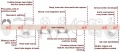
* '''Coronary arteries form by developmental reprogramming of venous cells''' <ref><pubmed>20336138</pubmed></ref> "Here we show, using histological and clonal analysis in mice and cardiac organ culture, that coronary vessels arise from angiogenic sprouts of the sinus venosus-the vein that returns blood to the embryonic heart. Sprouting venous endothelial cells dedifferentiate as they migrate over and invade the myocardium. Invading cells differentiate into arteries and capillaries; cells on the surface redifferentiate into veins. These results show that some differentiated venous cells retain developmental plasticity, and indicate that position-specific cardiac signals trigger their dedifferentiation and conversion into coronary arteries, capillaries and veins." | * '''Coronary arteries form by developmental reprogramming of venous cells''' <ref name="PMID20336138"><pubmed>20336138</pubmed></ref> "Here we show, using histological and clonal analysis in mice and cardiac organ culture, that coronary vessels arise from angiogenic sprouts of the sinus venosus-the vein that returns blood to the embryonic heart. Sprouting venous endothelial cells dedifferentiate as they migrate over and invade the myocardium. Invading cells differentiate into arteries and capillaries; cells on the surface redifferentiate into veins. These results show that some differentiated venous cells retain developmental plasticity, and indicate that position-specific cardiac signals trigger their dedifferentiation and conversion into coronary arteries, capillaries and veins." | ||
|} | |} | ||
==Mouse Coronary Vessel Development== | |||
[[File:Mouse-coronary vessel formation.jpg]] | |||
==References== | ==References== | ||
Revision as of 18:08, 15 October 2010
Introduction

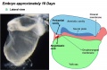
Development of the heart and vascular system begins very early in mesoderm both within (embryonic) and outside (extra embryonic) the embryo. Vascular development therefore occurs in many places, the most obvious though is the early forming heart, which grows rapidly creating an externally obvious cardiac "bulge" on the early embryo.
The heart forms initially in the embryonic disc as a simple paired tube inside the forming pericardial cavity, which when the disc folds, gets carried into the correct anatomical position in the chest cavity.
Some Recent Findings
|
Mouse Coronary Vessel Development

References
- ↑ <pubmed>20336138</pubmed>
Reviews
Articles
Search Pubmed
Search May 2010
- Cardiovascular System Development All (63457) Review (10735) Free Full Text (15717)
Search Pubmed: Coronary Circulation Development
Additional Images
See also Category:Heart ILP and Category:Heart
Historic image
Heart Development Timeline
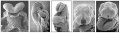
Human heart SEM
Early Heart Tube (Dorsal)
Early Heart Tube (Lateral)
Heart Tube Segments
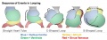
Heart Looping Sequence
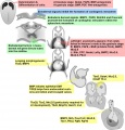
Molecular & Genetic Cardiac Development Factors
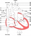
Adult heart blood flow cartoon


.jpg)
.jpg)


External Links
External Links Notice - The dynamic nature of the internet may mean that some of these listed links may no longer function. If the link no longer works search the web with the link text or name. Links to any external commercial sites are provided for information purposes only and should never be considered an endorsement. UNSW Embryology is provided as an educational resource with no clinical information or commercial affiliation.
| System Links: Introduction | Cardiovascular | Coelomic Cavity | Endocrine | Gastrointestinal Tract | Genital | Head | Immune | Integumentary | Musculoskeletal | Neural | Neural Crest | Placenta | Renal | Respiratory | Sensory | Birth |
Glossary Links
- Glossary: A | B | C | D | E | F | G | H | I | J | K | L | M | N | O | P | Q | R | S | T | U | V | W | X | Y | Z | Numbers | Symbols | Term Link
Cite this page: Hill, M.A. (2024, June 20) Embryology Cardiovascular System - Coronary Circulation Development. Retrieved from https://embryology.med.unsw.edu.au/embryology/index.php/Cardiovascular_System_-_Coronary_Circulation_Development
- © Dr Mark Hill 2024, UNSW Embryology ISBN: 978 0 7334 2609 4 - UNSW CRICOS Provider Code No. 00098G