Category:Cardiovascular: Difference between revisions
From Embryology
mNo edit summary |
mNo edit summary |
||
| Line 1: | Line 1: | ||
This | This {{Embryology}} category shows pages, images and media related to cardiovascular system development. | ||
See also the narrower categories [[:Category:Heart]] and [[:Category:Blood]]. | See also the narrower categories [[:Category:Heart]] and [[:Category:Blood]]. | ||
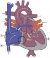
{{Heart Links}} | {{Heart Links}} | ||
Latest revision as of 12:39, 13 February 2017
This Embryology category shows pages, images and media related to cardiovascular system development.
See also the narrower categories Category:Heart and Category:Blood.
Subcategories
This category has the following 13 subcategories, out of 13 total.
Pages in category 'Cardiovascular'
The following 200 pages are in this category, out of 519 total.
(previous page) (next page)H
- HM Practical - Blood Vessel Histology
- HM Practical - Cardiac Histology
- Template:Hofbauer cell
- Template:Hofbauer cells
- Template:Human Embryology 2 18-6 Dorsal segmental artery table1
- Template:Human Embryology 2 18-6 Dorsal segmental artery table2
- Template:Human Embryology 2 18-6 Mesonephric artery table3
- Human System Development
- Template:Hypoplastic left heart
- Hypoplastic Left Heart Syndrome Movie
I
- Template:ICD-10-circulatory system Q20-Q28 table
- Template:Immune
- Intermediate - Atrial Ventricular Septation
- Intermediate - Cardiac Abnormalities
- Intermediate - Heart Tube Looping
- Intermediate - Heart Tube Looping 2
- Intermediate - Heart Tube Looping 3
- Intermediate - Heart Valves
- Intermediate - Outflow Tract
- Intermediate - Primordial Heart Tube
- Intermediate - Vascular Overview
- Intermediate Cardiac Embryology
- Template:Intermediate Cardiac menu
L
M
P
- Paper - A case of early ectopic gestation
- Paper - A congenital anomaly of the heart - truncus arteriosus communis (1927)
- Paper - A contribution to the early development of the heart in mammalia, with special reference to the guinea pig
- Paper - A Contribution to the Embryology of the Liver and Vascular System in Man
- Paper - A rare vascular anomaly-opening of the upper left pulmonary vein into a persistent left superior vena cava (1915)
- Paper - A specimen showing complete remains of the left superior vena cava (1915)
- Paper - A study of wandering mesenchymal cells on the living yolk-sac and their developmental products (1915)
- Paper - Coarctation of the aorta 1942
- Paper - Complete situs inversus of the vena cava superior (1930)
- Paper - Congenital deficiency of the pericardium (1916)
- Paper - Cor biloculare, with a note on the development of the pulmonary veins (1937)
- Paper - Development and vascularization of the testis (1906)
- Paper - Development of postcava and tributaries in the domestic cat (1907)
- Paper - Development of the great anterior veins in man and mammalia
- Paper - Development of the human heart from its earliest appearance to the stage found in embryos of twenty paired somites (1927)
- Paper - Development of the inferior vena cava (1929)
- Paper - development of the postcaval vein in birds (1903)
- Paper - Development of the vascular system in five to twenty-one somite dog embryos
- Paper - Developmental Changes in the Pericardium, the Mesocardia, and the Pleural Sacs in the Human Embryo
- Paper - Developmental defects at the foramen ovale (1938)
- Paper - Equivalence of different hematopoietic anlages 1 (1916)
- Paper - First contractions of the heart without cytological differentiation
- Paper - Four cases of anomalous inferior vena cava with an explanation of their developmental origin (1928)
- Paper - Functional limitations of the foramen ovale in the human foetal heart
- Paper - Further evidence on the origin of the lymphatic endothelium from the endothelium of the blood-vascular system
- Paper - General observations on early superficial lymphatics in living chick embryos (1912)
- Paper - Growth allometry of the myocardium in human embryos from stages 15 to 23
- Paper - Hematopoiesis in young human embryos
- Paper - Histogenesis of the heart muscle of the pig in relation to the appearance and development of the intercalated discs (1919)
- Paper - Histogenesis of the human aorta
- Paper - Initiation and early changes in the character of the heart beat in vertebrate embryos
- Paper - Injection and reconstruction of the jugular lymph sac in the chick (1912)
- Paper - Migration processes during ontogeny with reference to the venous development in the dorsal body wall (1946)
- Paper - Normal haemopoiesis in intra-uterine and neonatal life (1941)
- Paper - Observations on the development of the earliest lymphatics in the region of the posterior lymph heart in living chick embryos (1912)
- Paper - Observations upon the occurrence, structure and function of the giant cells of the marrow (1890)
- Paper - On the cervical veins and lymphatics in four human embryos, with an interpretation of anomalies on the subclavian and jugular veins in the adult (1909)
- Paper - On the development of the aortae cardinal and umbilical veins and the other blood vessels of vertebrate embryos from capillaries (1909)
- Paper - On the development of the atrial septum and the valvular apparatus in the right atrium of the pig embryo (1916)
- Paper - On the development of the blood-vessels of the brain in the human embryo (1905)
- Paper - On the Development of the Human Heart
- Paper - On the earliest blood-vessels in the anterior limb-buds of birds and their relation to the primary subclavian artery
- Paper - On the fate of the posterior cardinal veins and their relation to the development of the vena cava and azygos in the embryo pig (1915)
- Paper - On the origin of the abdominal lymphatics in mammals from the vena cava and the renal veins (1912)
- Paper - On the origin of the lymphatic system from the veins and the development of the lymph hearts and thoracic duct in the pig (1902)
- Paper - On the origin of the pulmonary arteries in mammals
- Paper - On the origin of the pulmonary arteries in mammals 2
- Paper - On the placentation of the macaque (Macaca mulatta), from the time of implantation until the formation of the definitive placenta
- Paper - On the position of the vitelline arteries in human embryos
- Paper - On the time of the post-natal obliteration of the fetal blood-passages (1918)
- Paper - Origin and development of the primitive vessels of the chick and of the pig (1917)
- Paper - Origin of the pulmonary vessels in the chick (1922)
- Paper - Origin, development and degeneration of the blood vessels of the ovary (1899)
- Paper - Persistence of the left posterior cardinal vein (1911)
- Paper - Persistent left superior vena cava, left duct of cuvier and left horn of the sinus venosus
- Paper - Persistent left superior vena cava, left duct of cuvier and left horn of the sinus venosus (1930)
- Paper - Preliminary note on the differentiation of angioblasts in the living chick (1917)
- Paper - Significant superficial anastomoses in the arterial blood supply to the human brain (1959)
- Paper - Six specimens of abnormal heart (1912)
- Paper - Some abnormal developments in the vascular system of the frog (rana temporaria) (1915)
- Paper - Some observations on the cardio-vascular system in the viable foetal lamb (1940)
- Paper - Studies on the area vasculosa of the embryo chick 2 (1937)
- Paper - Teratogenecity in the setting of cardiac development and maldevelopment
- Paper - The aortic arch derivatives in human adult (1951)
- Paper - The circle of Willis - An examination of 700 specimens (1905)
- Paper - The course of the blood flow through the fetal mammalian heart
- Paper - The course of the blood through the heart of the fetal mammal, with a note on the reptilian and amphibian circulations (1909)
- Paper - The development of the aorta and aortic arches in rabbits
- Paper - The development of the arteries of the human lower extremity
- Paper - The Development of the Atrio-Ventricular Valves in Man
- Paper - The development of the cardiac loop in the rabbit with especial reference to the bulboventricular groove and origin of the interventricular septum (1919)
- Paper - The development of the cardiac-coronary circulatory system
- Paper - The development of the cranial arteries in the human embryo
- Paper - The development of the cranial venous system in man, from the viewpoint of comparative anatomy
- Paper - The development of the heart in man
- Paper - The development of the mammalian spleen, with special reference to its hematopoietic activity (1921)
- Paper - The Development of the Pars Membranacea Septi in the Human Heart
- Paper - The development of the principal arterial stems in the forelimb of the pig (1922)
- Paper - The development of the pulmonary vein in the domestic cat (1913)
- Paper - The development of the subcutaneous vascular plexus in the head of the human embryo (1923)
- Paper - The development of the vascular system in the human embryo prior to the establishment of the heart
- Paper - The development of the veins in the limbs of rabbit embryos
- Paper - The development of the vena cava inferior (1902)
- Paper - The development of the vena cava inferior in man (1925)
- Paper - The development of the venous sinuses of the dura mater in the human embryo
- Paper - The developmental alterations in the vascular system of the brain of the human embryo (1921)
- Paper - The ductus arteriosus in the human fetus and newborn infant
- Paper - The ductus venosus in the fetus and in the adult (1923)
- Paper - The Earliest Blood-Vessels in Man
- Paper - The earliest stages of development of the blood-vessels and of the heart in ferret embryos
- Paper - The earliest stages of development of the blood-vessels and of the heart in ferret embryos 2
- Paper - The early development of the sheep heart (1946)
- Paper - The early stages of the development of the pericardium
- Paper - The effect of the heart-beat upon the development of the vascular system in the chick (1918)
- Paper - The equivalence of different homatopoietic anlages by method of stimulation of the different stem cells 1
- Paper - The equivalence of different homatopoietic anlages by method of stimulation of the different stem cells 2
- Paper - The fifth aortic arch of mammalian embryos; the nature of the last pharyngeal evagination
- Paper - The first contractions of the heart in rat embryos
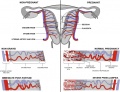
- Paper - The form and the functions of the uterine blood vessels in the rhesus monkey
- Paper - The formation of the cardiac loop in the chick
- Paper - The Formation of the Pars Membranacea Septi (1916)
- Paper - The formation of the venous valves, the foramen secundum and the septum secundum in the human heart
- Paper - The frequency of an opening between the right and left auricles at the seat of the foetal foramen ovale (1900)
- Paper - The fusion of the cardiac anlages and the formation of the cardiac loop in the cat (1916)
- Paper - The genesis, development, and adult anatomy of the nasofrontal region in man
- Paper - The genetic interpretation of the development of the mammalian lymphatic system (1908)
- Paper - The genetic principles of the development of the systemic lymphatic vessels in the mammalian embryo (1910)
- Paper - The human embryonic heart in the ninth week
- Paper - The human embryonic heart in the ninth week (1954)
- Paper - The human embryonic heart in the seventh week (1962)
- Paper - The life-history of the formed elements of the blood, especially the red blood corpuscles (1890)
- Paper - The morphology of human uteroplacental vasculature
- Paper - The nerve supply of the mammalian ductus arteriosus (1941)
- Paper - The origin and development of the carotid body (1924)
- Paper - The origin and early development of the posterior lymph heart in the chick (1915)
- Paper - The origin and occurrence of the single umbilical artery in normal and abnormal human fetuses (1922)
- Paper - The origin of blood and vascular endothelium in embryos without a circulation of the blood and in the normal embryo (1915)
- Paper - The origin of blood cells (1916)
- Paper - The origin of the heart and blood vessels in felis domestica (1924)
- Paper - The origin, development and function of the blood cells in certain marine teleosts 1 (1939)
- Paper - The partitioning of the truncus and conus and the formation of the membranous portion of the interventricular septum in the human heart (1942)
- Paper - The phylogenetic relations of the lymphatic and bloodvascular systems in vertebrates (1910)
- Paper - The physiology of the embryonic mammalian heart before circulation
- Paper - The relation between the size of the artery and the capillary bed in the embryo (1937)
- Paper - The relative role played by the embryonic veins in the development of the mammalian vena cava posterior
- Paper - Three examples of a right aortic arch
- Paper - Time and rate of loss of nuclei by the red blood cells of human embryos
- Paper - Transformation of the aortic-arch system during the development of the human embryo (1922)
- Paper - Transposition of the ventricles and the arterial stems (1931)
- Paper - True congenital diverticulum of the trachea in a subject showing also right aortic arch (1929)
- Paper - Two cases considered from the developmental standpoint in which the right subclavian artery arose from the arch of the aorta (1915)
- Paper - Variations and anomalies of the venous valves of the right atrium of the human heart (1929)
- Paper - Wilhelm His - His relation to the institution of learning
- Paper The development of the subcutaneous vascular plexus in the head of the human embryo (1923)
- Paper- The primary divisions of the myocardium in the human embryo
- Template:Patent ductus arteriosus
- Patent Ductus Venosus Movie
- Template:PDGF
- Template:Persistent right umbilical vein
- Template:Pia mater
- Template:Placenta vascular
- Template:Placenta vascular bed
- Template:Placental cord
- Template:Placental villi
- Template:Pre-eclampsia
Media in category 'Cardiovascular'
The following 95 files are in this category, out of 695 total.
(previous page) (next page) Stage 13 image 093.jpg 1,000 × 454; 92 KB
 Stage 13 image 094.jpg 1,000 × 469; 90 KB
 Stage 13 image 095.jpg 1,000 × 452; 76 KB
 Stage 13 image 096.jpg 1,000 × 469; 75 KB
 Stage 13 image 097.jpg 1,000 × 720; 162 KB
 Stage 13 image 098.jpg 1,000 × 623; 144 KB
 Stage 13 image 101.jpg 1,000 × 649; 65 KB
 Stage 22 image 175.jpg 1,000 × 666; 158 KB
 Stage 22 image 178.jpg 1,000 × 658; 148 KB
 Stage 22 image 179.jpg 1,000 × 658; 131 KB
 Stage 22 image 192.jpg 1,000 × 658; 180 KB
 Stage11 sem9.jpg 1,203 × 1,653; 301 KB
 Stage11 sem9a.jpg 728 × 1,000; 145 KB
 Stage11 sem9b.jpg 582 × 800; 104 KB
 Stage13 bloodflow.jpg 437 × 297; 24 KB
 Streeter-plate01.jpg 1,200 × 950; 138 KB
 Streeter-plate02.jpg 1,200 × 871; 180 KB
 Streeter-plate03.jpg 1,398 × 1,000; 192 KB
 Streeter-plate04.jpg 1,301 × 1,000; 214 KB
 Streeter-plate05.jpg 833 × 1,000; 115 KB
 Streeter1915 fig17.jpg 700 × 513; 76 KB
 Streeter1921 fig01.jpg 981 × 1,000; 103 KB
 Streeter1921 fig02.jpg 1,286 × 1,000; 138 KB
 Streeter1921 fig03.jpg 1,154 × 1,000; 142 KB
 Streeter1921 fig04.jpg 1,030 × 1,000; 101 KB
 Streeter1921 fig05.jpg 600 × 342; 19 KB
 Streeter1921 fig06.jpg 1,007 × 1,000; 106 KB
 Streeter1921 fig07-09.jpg 1,200 × 494; 92 KB
 Streeter1921 fig10.jpg 1,108 × 1,000; 95 KB
 Streeter1921 fig11.jpg 908 × 751; 103 KB
 Streeter1921 fig12.jpg 960 × 745; 117 KB
 Streeter1921 fig13.jpg 800 × 600; 45 KB
 Streeter1921 fig14.jpg 800 × 600; 55 KB
 Streeter1921 fig15.jpg 800 × 600; 54 KB
 Streeter1921 fig16.jpg 800 × 600; 60 KB
 Streeter1921 fig17.jpg 800 × 600; 60 KB
 Streeter1921 fig18.jpg 800 × 600; 48 KB
 Streeter1921 fig19.jpg 800 × 600; 48 KB
 Streeter1921 fig20.jpg 800 × 600; 40 KB
 Streeter1921 fig21.jpg 800 × 600; 40 KB
 Streeter1921 fig22.jpg 917 × 1,000; 157 KB
 Streeter1921 fig23.jpg 917 × 1,000; 152 KB
 Streeter1921 fig24.jpg 921 × 1,000; 130 KB
 Streeter1921 fig25.jpg 921 × 1,000; 118 KB
 Streeter1921 fig26.jpg 1,328 × 1,000; 228 KB
 Streeter1921 fig27.jpg 949 × 1,000; 138 KB
 Streeter1921 table01.jpg 800 × 347; 44 KB
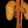
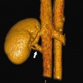 Supernumerary renal vein 01.jpg 800 × 798; 72 KB
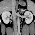
 Supernumerary renal vein 02.jpg 800 × 795; 89 KB
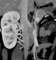
 Supernumerary renal vein 03.jpg 800 × 794; 80 KB
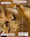
 Supernumerary renal vein 04.jpg 800 × 850; 76 KB
 Tetralogy of Fallot 01.jpg 700 × 873; 113 KB
 Tetralogy of Fallot 02.jpg 800 × 796; 63 KB
 Tetralogy of Fallot.jpg 300 × 350; 17 KB
 Thalidomide - CPS49 vascular effect.jpg 1,000 × 1,002; 197 KB
 Thalidomide - limb signaling.jpg 628 × 540; 88 KB
 Total Anomalous Pulmonary Venous Connection.jpg 295 × 350; 17 KB
 Transposition of the Great Vessels.jpg 299 × 350; 18 KB
 Tricuspid Atresia.jpg 303 × 350; 16 KB
 Trigeminal artery 01.jpg 947 × 800; 102 KB
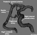 Trigeminal artery 02.jpg 520 × 490; 35 KB
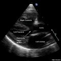
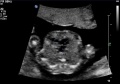
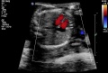
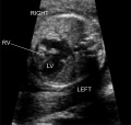
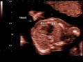 Ultrasound - Hypoplastic left heart syndrome 01.jpg 800 × 600; 53 KB
 Ultrasound - Hypoplastic left heart syndrome 02.jpg 890 × 626; 54 KB
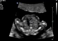 Ultrasound - Hypoplastic left heart syndrome 03.jpg 874 × 612; 51 KB
 Ultrasound - Hypoplastic left heart syndrome 04.jpg 919 × 618; 68 KB
 Uterine and placental vasculature.jpg 614 × 472; 143 KB
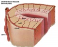 Uterine arterial vessel cartoon.jpg 600 × 483; 44 KB
 Uterine vascular anastomoses.jpg 1,200 × 370; 48 KB
 Vasculature development 01 cartoon.jpg 599 × 900; 106 KB
 Vasculature development 02 cartoon.jpg 601 × 1,002; 93 KB
 Vein valve animation.gif 300 × 200; 54 KB
 Ventricular septal defect 01.jpg 1,024 × 692; 84 KB
 Ventricular Septal Defect.jpg 289 × 350; 16 KB
 Venule microvessel EM.jpg 600 × 626; 91 KB
 Waterston13.jpg 429 × 681; 63 KB
 Waterston14.jpg 438 × 680; 66 KB
 Waterston16.jpg 593 × 675; 73 KB
 Waterston17.jpg 500 × 710; 74 KB
 Waterston18.jpg 500 × 662; 71 KB
 Waterston19.jpg 500 × 642; 66 KB
 Week17 fetal heart rate.mp4 ; 398 KB
 West11.jpg 367 × 963; 27 KB
 Woollard-plate01.jpg 744 × 1,000; 153 KB
 Woollard-plate02.jpg 788 × 1,000; 196 KB
 Woollard001.jpg 739 × 858; 142 KB
 Woollard002.jpg 699 × 873; 125 KB
 Woollard003.jpg 1,107 × 848; 159 KB
 Woollard004.jpg 1,037 × 717; 148 KB
 Woollard005.jpg 1,034 × 655; 144 KB
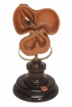 Ziegler model 12.jpg 475 × 700; 39 KB
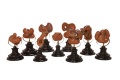 Ziegler model 18.jpg 700 × 465; 42 KB
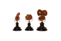 Ziegler model 19.jpg 700 × 465; 23 KB
 ZPulmonary Atresia.jpg 653 × 618; 85 KB
 ZUltrasound Image of Fetal Aorta.jpg 640 × 480; 47 KB


