Neural - Cerebrum Development

From Embryology
Embryology - 19 Mar 2024    Facebook link Pinterest link Twitter link  Expand to Translate  
Google Translate - select your language from the list shown below (this will open a new external page)

العربية | català | 中文 | 中國傳統的 | français | Deutsche | עִברִית | हिंदी | bahasa Indonesia | italiano | 日本語 | 한국어 | မြန်မာ | Pilipino | Polskie | português | ਪੰਜਾਬੀ ਦੇ | Română | русский | Español | Swahili | Svensk | ไทย | Türkçe | اردو | ייִדיש | Tiếng Việt    These external translations are automated and may not be accurate. (More? About Translations)

Introduction

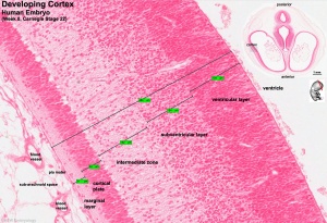
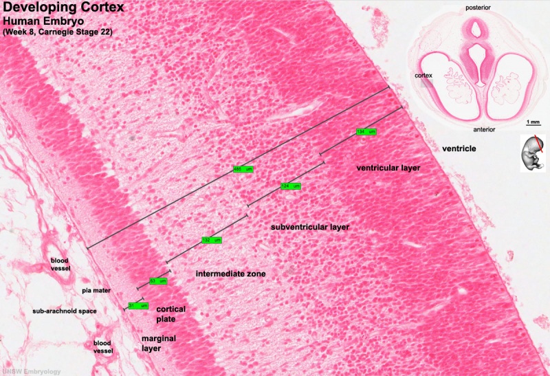
Human embryo developing cortex (week 8, stage 22)
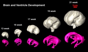
Human cerebrum and underlying ventricular development imaged by MRI[1]
Dev anat 01.jpg
Gray0677.jpg

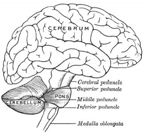
The brain (cerebral cortex, cerebrum, cortex) as it is generally recognised. Though the cerebrum includes the cerebral cortex and the subcortical structures (hippocampus, basal ganglia, and olfactory bulb). The adult cerebral cortex like other neural structures has a laminar organisation, the mammalian neocortex consists of six layers, while the reptilian and avian cortices have only three layers (equivalent to mammalian layers I, V and VI). The adult human cerebrum contains about 16,340,000,000 ± 2,170,000,000 (sixteen billion three hundred forty million) neurons and 60,840,000,000 ± 7,020,000,000 other cell types.[2]

A simplified developmental sequence can be described as cell proliferation, cell migration, and finally cortical organization. In development, lamination occurs in an "inside-out" sequence earlier inside and later born neurons outside. The cortex is divided into areas which serve distinct functions including motor, sensory and cognitive processing. The lamination process requires a range of different signals including; Reelin (Reln, an extracellular protein), Disabled-1 (Dab1, an intracellular signaling molecule), and Cullin-5 (Cul5, an E3 ubiquitin ligase).

The cortex progenitor cell types are either neuron-restricted or bipotent (neuron-glial) progenitors that generate glial-restricted progenitors at mid- and late neurogenesis.

Neural Parts: neural | prosencephalon | telencephalon cerebrum | amygdala | hippocampus | basal ganglia | diencephalon | epithalamus | thalamus | hypothalamus‎ | pituitary | pineal | mesencephalon | tectum | rhombencephalon | metencephalon | pons | cerebellum | myelencephalon | medulla oblongata | spinal cord | neural vascular | ventricular | lateral ventricles | third ventricle | cerebral aqueduct | fourth ventricle | central canal | meninges | Category:Ventricular System | Category:Neural


Neural Links: ectoderm | neural | neural crest | ventricular | sensory | Stage 22 | gliogenesis | neural fetal | Medicine Lecture - Neural | Lecture - Ectoderm | Lecture - Neural Crest | Lab - Early Neural | neural abnormalities | folic acid | iodine deficiency | Fetal Alcohol Syndrome | neural postnatal | neural examination | Histology | Historic Neural | Category:Neural


Historic Cortex Embryology  
1891 Cortex Sensory Areas | 1907 Papilla of Retzius | 1911 human cerebral cortex | 1912 cortex development | 1912 Central Nervous System | 1945 cerebral cortex morphophysiology
Historic Neural Embryology  
1883 Nervous System | 1893 Brain Structure | 1892 Nervous System Development | 1900 fourth ventricle | 1905 Brain Blood-Vessels | 1909 corpus ponto-bulbare | 1912 nuclei pontis - nucleus arcuatus | 1912 Diencephalon | 1921 Neural Development | 1921 Anencephaly | 1921 Brain Weight | 1921 Brain Vascular System | 1921 Cerebellum | 1922 Brain Plan | 1923 Neural Folds | 1904 Brain and Mind | 1904 Brain Structure | 1909 Forebrain Vesicle | 1922 Hippocampal Fissure | 1923 Forebrain | 1927 Anencephaly | 1934 Anencephaly | 1937 Anencephaly | 1945 Spinal Cord | 1945 cerebral cortex | Santiago Ramón y Cajal | Ziegler Neural Models | Historic Embryology Papers | Historic Disclaimer

Some Recent Findings

  • The genetic architecture of the human cerebral cortex[3] "The cerebral cortex underlies our complex cognitive capabilities, yet little is known about the specific genetic loci that influence human cortical structure. To identify genetic variants that affect cortical structure, we conducted a genome-wide association meta-analysis of brain magnetic resonance imaging data from 51,665 individuals. We analyzed the surface area and average thickness of the whole cortex and 34 regions with known functional specializations. We identified 199 significant loci and found significant enrichment for loci influencing total surface area within regulatory elements that are active during prenatal cortical development, supporting the radial unit hypothesis. Loci that affect regional surface area cluster near genes in Wnt signaling pathways, which influence progenitor expansion and areal identity. Variation in cortical structure is genetically correlated with cognitive function, Parkinson's disease, insomnia, depression, neuroticism, and attention deficit hyperactivity disorder."
  • c-Myc controls the fate of neural progenitor cells during cerebral cortex development[4] "The anatomical structure of the mammalian cerebral cortex is the essential foundation for its complex neural activity. This structure is developed by proliferation, differentiation, and migration of neural progenitor cells (NPCs), the fate of which is spatially and temporally regulated by the proper gene. This study was used in utero electroporation and found that the well-known oncogene c-Myc mainly promoted NPCs' proliferation and their transformation into intermediate precursor cells. Furthermore, the obtained results also showed that c-Myc blocked the differentiation of NPCs to postmitotic neurons, and the expression of telomere reverse transcriptase was controlled by c-Myc in the neocortex. These findings indicated c-Myc as a key regulator of the fate of NPCs during the development of the cerebral cortex.}
  • Dopamine as a growth differentiation factor in the mammalian brain[5] "The catecholamine, dopamine, plays an important role in the central nervous system of mammals, including executive functions, motor control, motivation, arousal, reinforcement, and reward. Dysfunctions of the dopaminergic system lead to diseases of the brains, such as Parkinson's disease, Tourette's syndrome, and schizophrenia. In addition to its fundamental role as a neurotransmitter, there is evidence for a role as a growth differentiation factor during development. Recent studies suggest that dopamine regulates the development of γ-aminobutyric acidergic interneurons of the cerebral cortex. Moreover, in adult brains, dopamine increases the production of new neurons in the hippocampus, suggesting the promoting effect of dopamine on proliferation and differentiation of neural stem cells and progenitor cells in the adult brains. In this mini-review, I center my attention on dopaminergic functions in the cortical interneurons during development and further discuss cell therapy against neurodegenerative diseases."
  • The LPA-LPA4 axis is required for establishment of bipolar morphology and radial migration of newborn cortical neurons[6] "Neurons in the developing neocortex undergo radial migration, a process that is coupled with their precise passage from multipolar to bipolar shape. The cell-extrinsic signals that govern this transition are, however, poorly understood. Here, we find that lysophosphatidic acid (LPA) signaling contributes to the establishment of a bipolar shape in mouse migratory neurons through LPA receptor 4 (LPA4)."
More recent papers  
Mark Hill.jpg
PubMed logo.gif

This table allows an automated computer search of the external PubMed database using the listed "Search term" text link.

  • This search now requires a manual link as the original PubMed extension has been disabled.
  • The displayed list of references do not reflect any editorial selection of material based on content or relevance.
  • References also appear on this list based upon the date of the actual page viewing.


References listed on the rest of the content page and the associated discussion page (listed under the publication year sub-headings) do include some editorial selection based upon both relevance and availability.

More? References | Discussion Page | Journal Searches | 2019 References | 2020 References

Search term: Cerebral Cortex Development | Cerebral Cortex Embryology | Cerebrum Development | Cortex Development |

Older papers  
These papers originally appeared in the Some Recent Findings table, but as that list grew in length have now been shuffled down to this collapsible table.

See also the Discussion Page for other references listed by year and References on this current page.

  • Development of the sensorimotor cortex in the human fetus: a morphological description[7] "Twenty-one human fetal brains from 13 to 28 gestational weeks were studied macroscopically to describe the morphological stages of sulcal and gyral development in the sensorimotor cortex....Four chronological stages of sensorimotor cortex development were defined: stage 1: appearance at 18-19 gestational weeks (GWs) of the inferior part of the central cerebral sulcus; stage 2: development of the pericentral lateral regions and the beginning of opercularization at 20-22 GWs; stage 3: development of parietal and temporal cortices and the covering of the postcentral insular region at 24-26 GWs; and finally stage 4: maturation of the central cerebral regions at 27-28 GWs."
  • Local tissue growth patterns underlying normal fetal human brain gyrification quantified in utero[8] "we applied recent advances in fetal MRI motion correction and computational image analysis techniques to 40 normal fetal human brains covering a period of primary sulcal formation (20-28 gestational weeks). Growth patterns were mapped by quantifying tissue locations that were expanding more or less quickly than the overall cerebral growth rate, which reveal increasing structural complexity. We detected increased local relative growth rates in the formation of the precentral and postcentral gyri, right superior temporal gyrus, and opercula, which differentiated between the constant growth rate in underlying cerebral mantle and the accelerating rate in the cortical plate undergoing folding. Analysis focused on the cortical plate revealed greater volume increases in parietal and occipital regions compared to the frontal lobe. Cortical plate growth patterns constrained to narrower age ranges showed that gyrification, reflected by greater growth rates, was more pronounced after 24 gestational weeks. Local hemispheric volume asymmetry was located in the posterior peri-Sylvian area associated with structural lateralization in the mature brain."
  • Development of laminar organization of the fetal cerebrum[9] "Heads of 131 fetal specimens of 14-40 weeks gestational age (GA) were scanned by 3.0T MRI. Eleven fetal specimens of 14-27 weeks GA were scanned by 7.0T MRI. On T(1)-weighted 3.0T MRI, layers could be visualized at 14 weeks GA and appeared clearer after 18 weeks GA. On 7.0T MRI, four zones could be recognized at 14 weeks GA. During 15-22 weeks GA, when laminar organization appeared typical, seven layers including the periventricular zone and external capsule fibers could be differentiated, which corresponded to seven zones in histological stained sections. At 23-28 weeks GA, laminar organization appeared less typical, and borderlines among them appeared obscured. After 30 weeks GA, it disappeared and turned into mature-like structures. The developing lamination appeared the most distinguishable at the parieto-occipital part of brain and peripheral regions of the hippocampus. The migrating thalamocortical afferents were probably delineated as a high signal layer located at the lower, middle, and upper part of the subplate zone at 16-28 weeks GA on T(1)-weighted 3.0T MRI."
  • Correlation of diffusion tensor imaging with histology in the developing human frontal cerebrum[10] "Transient early cerebral laminar organization resulting from normal developmental events has been revealed in human beings through histology and imaging studies. DTI studies have postulated that the fractional anisotropy (FA)-based differentiation of different laminar structures reflects both differing cellular density over the glial fibers and fiber alignment in respective regions. The aim of this study was to correlate FA values in these transient zones with histology. Brain DTI was performed on 50 freshly aborted human fetuses with gestational ages (GA) ranging from 12 to 42 weeks. Regions of interest were placed on the cortical plate, subplate, intermediate and germinal matrix (GMx) zones of the frontal lobe to quantify FA values. Glial fibrillary acidic protein (GFAP), neurofilament (NF) and neuron-specific enolase (NSE) immunohistochemical analyses were performed for the cortical plate, intermediate zone and GMx. In the cortical plate, a significant positive correlation was observed between FA values and percentage area of GFAP expression in fetuses <or=28 weeks of GA (r = 0.56, p = 0.01). FA values showed a significant positive correlation with the percentage area of NF expression in the intermediate zone (r = 0.54, p = 0.05). A significant positive correlation was also observed between FA and the number of NSE-positive cells per mm(2) in the GMx (r = 0.76, p < 0.01) and subplate (r = 0.59, p = 0.03) zones. The results of our study suggest that the FA can be used as noninvasive marker of neurodevelopmental events in the frontal lobe of human fetal brain."

Development Overview

Neuralation begins at the trilaminar embryo with formation of the notochord and somites, both of which underly the ectoderm and do not contribute to the nervous system, but are involved with patterning its initial formation. The central portion of the ectoderm then forms the neural plate that folds to form the neural tube, that will eventually form the entire central nervous system.

Early developmental sequence: Epiblast - Ectoderm - Neural Plate - Neural groove and Neural Crest - Neural Tube and Neural Crest


Neural Tube Development
Neural Tube Primary Vesicles Secondary Vesicles Adult Structures
week 3 week 4 week 5 adult
neural plate
neural groove
neural tube

Brain
prosencephalon (forebrain) telencephalon Rhinencephalon, Amygdala, hippocampus, cerebrum (cortex), hypothalamus‎, pituitary | Basal Ganglia, lateral ventricles
diencephalon epithalamus, thalamus, Subthalamus, pineal, posterior commissure, pretectum, third ventricle
mesencephalon (midbrain) mesencephalon tectum, Cerebral peduncle, cerebral aqueduct, pons
rhombencephalon (hindbrain) metencephalon cerebellum
myelencephalon medulla oblongata, isthmus
spinal cord, pyramidal decussation, central canal

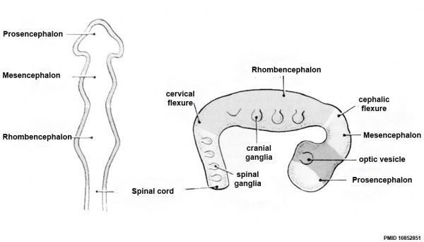
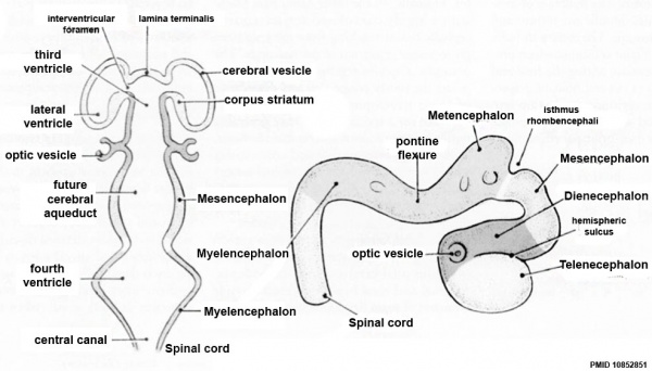
Early Brain Vesicles

Primary Vesicles

CNS primary vesicles.jpg

Secondary Vesicles

CNS secondary vesicles.jpg

Late Embryonic Brain

Stage 22 image 217.jpg

Human embryo developing cortex (Week 8, Carnegie stage 22)

  • small embryo shows approximate level of section.
  • insert top right shows whole head section.
  • small shaded box on whole section shows region of large image.
  • layer thicknesses are shown in microns.

Early Fetal Brain
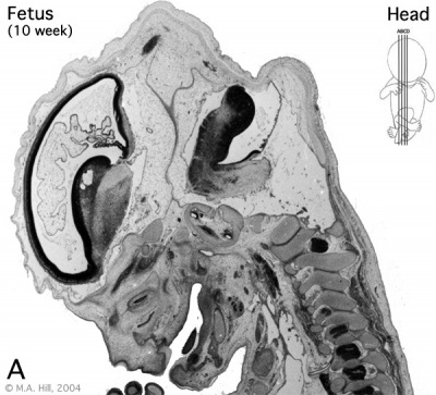
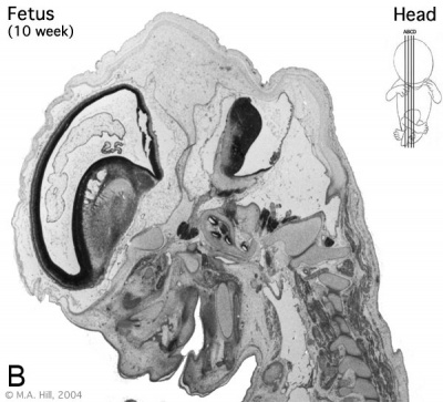
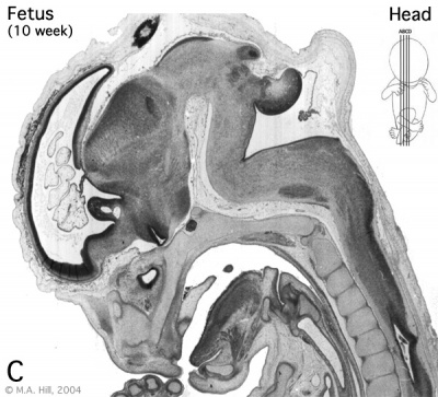
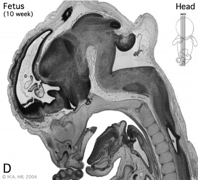
Human- fetal week 10 head A.jpg Human- fetal week 10 head B.jpg
Human- fetal week 10 head C.jpg Human- fetal week 10 head D.jpg

The above images are from a week 10 human fetus.

Fetal Brain
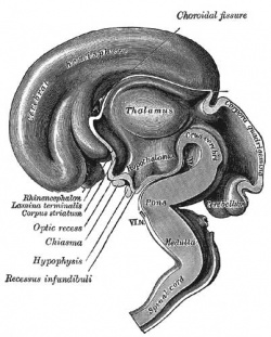
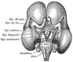
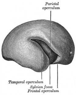
Gray0654.jpg Gray0655.jpg Gray0658.jpg
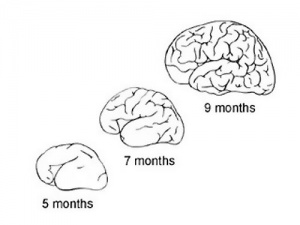
Fetal brain (3 months) Fetal brain (4 months) Fetal brain (5 months)

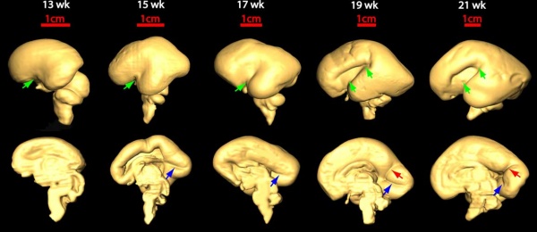
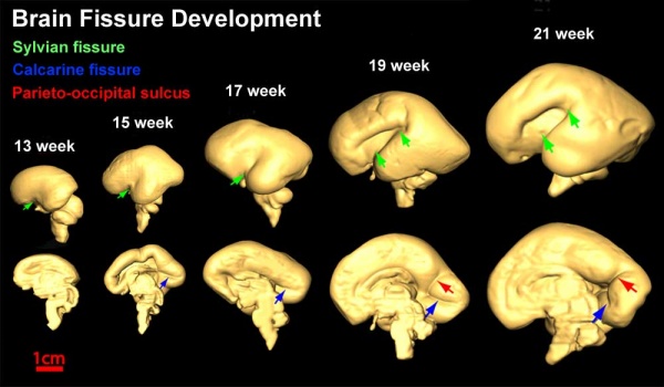
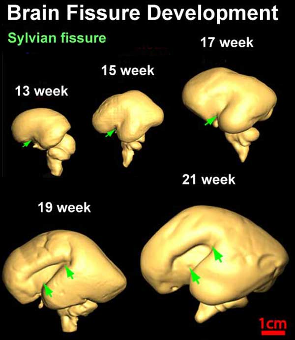
Fissures are the major indentations, sulci (singular sulcus), that divide the brain surface into lobes and appear during fetal development as the brain grows. The images below show MRI analysis of the developing human fetal brain.

Brain fissure development 01.jpg

Brain fissure development 02.jpg

Brain fissure development 03.jpg


Links: Magnetic Resonance Imaging

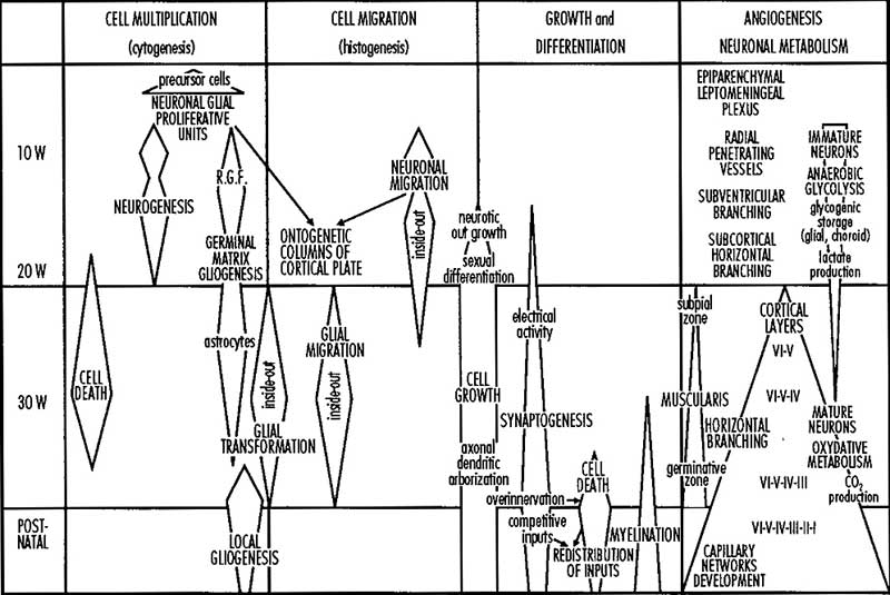
Developmental Overview

Neural-development.jpg

Cortical Neurons

[[File:Telencephalon development signals.jpg|thumb|Telencephalon development signals[11] Neural- cortex Cajal drawing 01.jpg
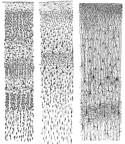
Cortical layers in a historic drawing by Cajal.

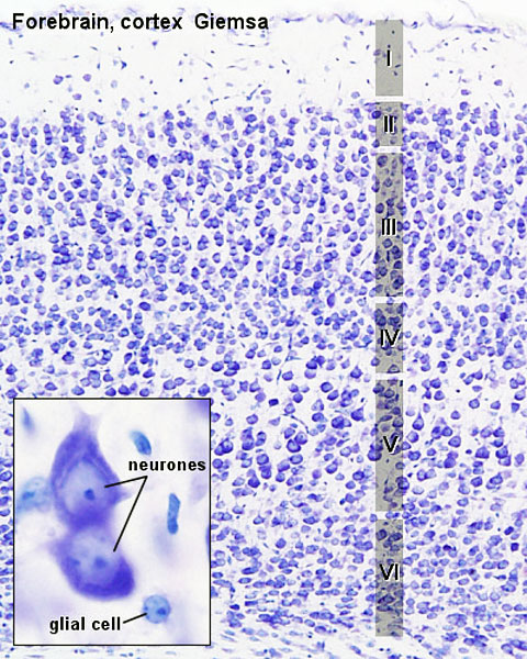
Brain histology 01.jpg

Adult mouse cortex


I molecular layer - few neurons and mainly of extensions of apical dendrites and horizontally-oriented axons.


II external granular layer - small pyramidal neurons and many stellate neurons.


III external pyramidal layer - mainly small and medium-size pyramidal neurons, some non-pyramidal neurons with vertically-oriented intracortical axons.


IV internal granular layer - different types of stellate and pyramidal neurons.


V internal pyramidal layer - large pyramidal neurons.


VI multiform layer - few large pyramidal neurons and many small spindle-like pyramidal and multiform neurons.

Cajal-Retzius Neurons

Cajal-Retzius (CR) cells are some of the earliest generated cortical neurons arising from restricted domains of the pallial ventricular zone, and then migrate from the borders of the developing pallium to cover the cortical primordium. These early forming neurons then control the radial migration of neurons and the formation of cortical layers. In mice, this has been shown by these cells secreting the extracellular glycoprotein Reelin (Reln) and it has been suggested that these cells also fine tune multiple signaling pathways underlying the regulation of cortical regionalization.[11]

Molecular
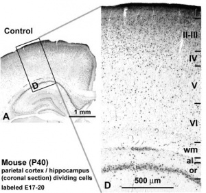
Mouse adult cortex[12]
  • Fgfr1 and Fgfr2 - control excitatory cortical neuron development within the entire cerebral cortex.[13]
  • Fgfr2 - proper formation of the medial prefrontal cortex (mPFC).[13]
  • MARCKS - (myristoylated alanine-rich C-kinase substrate protein) a cellular substrate for PKC modulates radial glial placement and expansion.[14]
  • MicroRNA - noncoding RNAs that regulate mRNA expression, highly expressed during development.[15][16]

Sensorimotor Cortex

The sensorimotor cortex is the region comprising the precentral and postcentral gyri and covers the primary sensory and motor areas of the brain. This region is involved with central integration and processing of the sensory input and motor output of the brain.

A study of this region of the human fetal brain from the end of the first trimester GA 13 weeks (11 weeks) and the to end of the second trimester GA 28 weeks (26 weeks) has identified four stages of development:[7]

Sensorimotor Cortex Timeline
Gestational Age GA weeks Fertilization Age FA weeks Event
18-19 16-17 appearance of the inferior part of the central cerebral sulcus
20-22 18-20 development of the pericentral lateral regions and the beginning of opercularization
24-26 22-24 development of parietal and temporal cortices and the covering of the postcentral insular region
27-28 25-26 maturation of the central cerebral regions
Table data.[7]    Links: sensorimotor cortex | cerebral cortex | second trimester | Sensory System Development


  • central cerebral sulcus - (central fissure, fissure of Rolando, Rolandic fissure) fold in the cerebral cortex.
  • opercularization - during fetal development the insula begins to invaginate from the surface of the immature cerebrum of the brain, until at full term, the opercula completely cover the insula.
  • posterior insula - is composed of the anterior and posterior long insular gyri and the postcentral insular sulcus, which separates them.
Links: Sensory System Development

Corpus Callosum

The corpus callosum is the area of the brain which connects the two cerebral hemispheres. Maximum increase in thickness and width of the corpus callosum occurred between 19 and 21 weeks' gestation.[17]

Human Timeline:

  • 74 days - callosal axons appear.
  • 84 days - subdivisions of the genu and splenium can be identified.
  • 115 days - adult morphology is seen.

Agenesis of Corpus Callosum

Agenesis of the corpus callosum (ACC) is a partial or complete absence of the corpus callosum, a rare cerebral malformation.


Links: NINDS Information

Cerebral Vascular Development

Overview cartoon Early vascular changes
Cerebral brain artery development 01.jpg Cerebral brain artery development 01.jpg

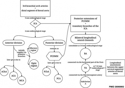
Chronological development of cerebral brain arteries initially with the rise of the internal carotid artery and subsequently with the development of the posterior circulation.[18]

Links: blood vessel | Head Development


Animal Models

Mouse Cortex

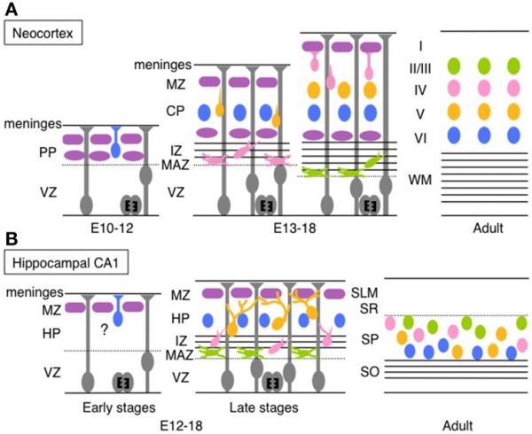
Timeline comparison of the migration and layer arrangement in neocortex and hippocampal CA1 during cortical development[19]
Mouse cortex and hippocampus development (A) Neocortical neurons born between E10 and E12 radially migrate using the somal translocation mode. In contrast, late-born neurons transform their migration mode sequentially to multipolar migration, locomotion mode, and terminal translocation mode during their radial migration. These neurons form neocortical layers in a birthdate-dependent inside-out manner.


(B) Hippocampal CA1 neurons born at late developmental stages change the migration mode to multipolar migration and then to the climbing mode. The migration mode used by early-born CA1 neurons remains unknown (somal translocation mode is a candidate). The layer arrangement in the Ammon's horn is thought to occur roughly in a birth-date dependent inside-out manner.

PP, preplate; VZ, ventricular zone; MZ, marginal zone; CP, cortical plate; IZ, intermediated zone; MAZ, multipolar cell accumulation zone; WM, white matter; HP, hippocampal plate; SLM, stratum lacunosum-moleculare; SR, stratum radiatum; SP, stratum pyramidale; SO, stratum oriens.

(text modified from original figure legend)


Abnormalities

Agenesis of Corpus Callosum

Agenesis of the corpus callosum (ACC) is a partial or complete absence of the corpus callosum, a rare cerebral malformation.


Links: NINDS Information


Hemimegalencephaly

Increased proliferation

Heterotopia

Ectopic migration

Lissencephaly

A malformations derived from abnormal neuronal migration leading to agyria (convolutions of the cerebral cortex are not fully formed) and pachygyria (convolutions of the cerebral cortex unusually thick ).

Pachygyria (Greek, pachy = "thick")

Microlissencephaly

Decreased proliferation

Polymicrogyria

Abnormal organization, numerous small gyri and a thick disorganized cortical plate lacking normal lamination, disruption of microtubule-based processes underlies a large spectrum of neuronal migration.[20]

Schizencephaly

Abnormal organization

References

  1. Habas PA, Kim K, Rousseau F, Glenn OA, Barkovich AJ & Studholme C. (2010). Atlas-based segmentation of developing tissues in the human brain with quantitative validation in young fetuses. Hum Brain Mapp , 31, 1348-58. PMID: 20108226 DOI.
  2. Herculano-Houzel S, Catania K, Manger PR & Kaas JH. (2015). Mammalian Brains Are Made of These: A Dataset of the Numbers and Densities of Neuronal and Nonneuronal Cells in the Brain of Glires, Primates, Scandentia, Eulipotyphlans, Afrotherians and Artiodactyls, and Their Relationship with Body Mass. Brain Behav. Evol. , 86, 145-63. PMID: 26418466 DOI.
  3. Grasby KL, Jahanshad N, Painter JN, Colodro-Conde L, Bralten J, Hibar DP, Lind PA, Pizzagalli F, Ching CRK, McMahon MAB, Shatokhina N, Zsembik LCP, Thomopoulos SI, Zhu AH, Strike LT, Agartz I, Alhusaini S, Almeida MAA, Alnæs D, Amlien IK, Andersson M, Ard T, Armstrong NJ, Ashley-Koch A, Atkins JR, Bernard M, Brouwer RM, Buimer EEL, Bülow R, Bürger C, Cannon DM, Chakravarty M, Chen Q, Cheung JW, Couvy-Duchesne B, Dale AM, Dalvie S, de Araujo TK, de Zubicaray GI, de Zwarte SMC, den Braber A, Doan NT, Dohm K, Ehrlich S, Engelbrecht HR, Erk S, Fan CC, Fedko IO, Foley SF, Ford JM, Fukunaga M, Garrett ME, Ge T, Giddaluru S, Goldman AL, Green MJ, Groenewold NA, Grotegerd D, Gurholt TP, Gutman BA, Hansell NK, Harris MA, Harrison MB, Haswell CC, Hauser M, Herms S, Heslenfeld DJ, Ho NF, Hoehn D, Hoffmann P, Holleran L, Hoogman M, Hottenga JJ, Ikeda M, Janowitz D, Jansen IE, Jia T, Jockwitz C, Kanai R, Karama S, Kasperaviciute D, Kaufmann T, Kelly S, Kikuchi M, Klein M, Knapp M, Knodt AR, Krämer B, Lam M, Lancaster TM, Lee PH, Lett TA, Lewis LB, Lopes-Cendes I, Luciano M, Macciardi F, Marquand AF, Mathias SR, Melzer TR, Milaneschi Y, Mirza-Schreiber N, Moreira JCV, Mühleisen TW, Müller-Myhsok B, Najt P, Nakahara S, Nho K, Olde Loohuis LM, Orfanos DP, Pearson JF, Pitcher TL, Pütz B, Quidé Y, Ragothaman A, Rashid FM, Reay WR, Redlich R, Reinbold CS, Repple J, Richard G, Riedel BC, Risacher SL, Rocha CS, Mota NR, Salminen L, Saremi A, Saykin AJ, Schlag F, Schmaal L, Schofield PR, Secolin R, Shapland CY, Shen L, Shin J, Shumskaya E, Sønderby IE, Sprooten E, Tansey KE, Teumer A, Thalamuthu A, Tordesillas-Gutiérrez D, Turner JA, Uhlmann A, Vallerga CL, van der Meer D, van Donkelaar MMJ, van Eijk L, van Erp TGM, van Haren NEM, van Rooij D, van Tol MJ, Veldink JH, Verhoef E, Walton E, Wang M, Wang Y, Wardlaw JM, Wen W, Westlye LT, Whelan CD, Witt SH, Wittfeld K, Wolf C, Wolfers T, Wu JQ, Yasuda CL, Zaremba D, Zhang Z, Zwiers MP, Artiges E, Assareh AA, Ayesa-Arriola R, Belger A, Brandt CL, Brown GG, Cichon S, Curran JE, Davies GE, Degenhardt F, Dennis MF, Dietsche B, Djurovic S, Doherty CP, Espiritu R, Garijo D, Gil Y, Gowland PA, Green RC, Häusler AN, Heindel W, Ho BC, Hoffmann WU, Holsboer F, Homuth G, Hosten N, Jack CR, Jang M, Jansen A, Kimbrel NA, Kolskår K, Koops S, Krug A, Lim KO, Luykx JJ, Mathalon DH, Mather KA, Mattay VS, Matthews S, Mayoral Van Son J, McEwen SC, Melle I, Morris DW, Mueller BA, Nauck M, Nordvik JE, Nöthen MM, O'Leary DS, Opel N, Martinot MP, Pike GB, Preda A, Quinlan EB, Rasser PE, Ratnakar V, Reppermund S, Steen VM, Tooney PA, Torres FR, Veltman DJ, Voyvodic JT, Whelan R, White T, Yamamori H, Adams HHH, Bis JC, Debette S, Decarli C, Fornage M, Gudnason V, Hofer E, Ikram MA, Launer L, Longstreth WT, Lopez OL, Mazoyer B, Mosley TH, Roshchupkin GV, Satizabal CL, Schmidt R, Seshadri S, Yang Q, Alvim MKM, Ames D, Anderson TJ, Andreassen OA, Arias-Vasquez A, Bastin ME, Baune BT, Beckham JC, Blangero J, Boomsma DI, Brodaty H, Brunner HG, Buckner RL, Buitelaar JK, Bustillo JR, Cahn W, Cairns MJ, Calhoun V, Carr VJ, Caseras X, Caspers S, Cavalleri GL, Cendes F, Corvin A, Crespo-Facorro B, Dalrymple-Alford JC, Dannlowski U, de Geus EJC, Deary IJ, Delanty N, Depondt C, Desrivières S, Donohoe G, Espeseth T, Fernández G, Fisher SE, Flor H, Forstner AJ, Francks C, Franke B, Glahn DC, Gollub RL, Grabe HJ, Gruber O, Håberg AK, Hariri AR, Hartman CA, Hashimoto R, Heinz A, Henskens FA, Hillegers MHJ, Hoekstra PJ, Holmes AJ, Hong LE, Hopkins WD, Hulshoff Pol HE, Jernigan TL, Jönsson EG, Kahn RS, Kennedy MA, Kircher TTJ, Kochunov P, Kwok JBJ, Le Hellard S, Loughland CM, Martin NG, Martinot JL, McDonald C, McMahon KL, Meyer-Lindenberg A, Michie PT, Morey RA, Mowry B, Nyberg L, Oosterlaan J, Ophoff RA, Pantelis C, Paus T, Pausova Z, Penninx BWJH, Polderman TJC, Posthuma D, Rietschel M, Roffman JL, Rowland LM, Sachdev PS, Sämann PG, Schall U, Schumann G, Scott RJ, Sim K, Sisodiya SM, Smoller JW, Sommer IE, St Pourcain B, Stein DJ, Toga AW, Trollor JN, Van der Wee NJA, van 't Ent D, Völzke H, Walter H, Weber B, Weinberger DR, Wright MJ, Zhou J, Stein JL, Thompson PM & Medland SE. (2020). The genetic architecture of the human cerebral cortex. Science , 367, . PMID: 32193296 DOI.
  4. Wang XL, Ma YX, Xu RJ, Ma JJ, Zhang HC, Qi SB, Xu JH, Qin XZ, Zhang HN, Liu CM, Chen JQ, Li B, Yang HL & Saijilafu. (2020). c-Myc controls the fate of neural progenitor cells during cerebral cortex development. J. Cell. Physiol. , 235, 4011-4021. PMID: 31625158 DOI.
  5. Ohira K. (2020). Dopamine as a growth differentiation factor in the mammalian brain. Neural Regen Res , 15, 390-393. PMID: 31571646 DOI.
  6. Kurabayashi N, Tanaka A, Nguyen MD & Sanada K. (2018). The LPA-LPA4 axis is required for establishment of bipolar morphology and radial migration of newborn cortical neurons. Development , 145, . PMID: 30217809 DOI.
  7. 7.0 7.1 7.2 Afif A, Trouillas J & Mertens P. (2015). Development of the sensorimotor cortex in the human fetus: a morphological description. Surg Radiol Anat , 37, 153-60. PMID: 24972575 DOI.
  8. Rajagopalan V, Scott J, Habas PA, Kim K, Corbett-Detig J, Rousseau F, Barkovich AJ, Glenn OA & Studholme C. (2011). Local tissue growth patterns underlying normal fetal human brain gyrification quantified in utero. J. Neurosci. , 31, 2878-87. PMID: 21414909 DOI.
  9. Zhang Z, Liu S, Lin X, Teng G, Yu T, Fang F & Zang F. (2011). Development of laminar organization of the fetal cerebrum at 3.0T and 7.0T: a postmortem MRI study. Neuroradiology , 53, 177-84. PMID: 20981415 DOI.
  10. Trivedi R, Husain N, Rathore RK, Saksena S, Srivastava S, Malik GK, Das V, Pradhan M, Pandey CM & Gupta RK. (2009). Correlation of diffusion tensor imaging with histology in the developing human frontal cerebrum. Dev. Neurosci. , 31, 487-96. PMID: 19622880 DOI.
  11. 11.0 11.1 Griveau A, Borello U, Causeret F, Tissir F, Boggetto N, Karaz S & Pierani A. (2010). A novel role for Dbx1-derived Cajal-Retzius cells in early regionalization of the cerebral cortical neuroepithelium. PLoS Biol. , 8, e1000440. PMID: 20668538 DOI.
  12. Berbel P, Navarro D, Ausó E, Varea E, Rodríguez AE, Ballesta JJ, Salinas M, Flores E, Faura CC & de Escobar GM. (2010). Role of late maternal thyroid hormones in cerebral cortex development: an experimental model for human prematurity. Cereb. Cortex , 20, 1462-75. PMID: 19812240 DOI.
  13. 13.0 13.1 Stevens HE, Smith KM, Maragnoli ME, Fagel D, Borok E, Shanabrough M, Horvath TL & Vaccarino FM. (2010). Fgfr2 is required for the development of the medial prefrontal cortex and its connections with limbic circuits. J. Neurosci. , 30, 5590-602. PMID: 20410112 DOI.
  14. Weimer JM, Yokota Y, Stanco A, Stumpo DJ, Blackshear PJ & Anton ES. (2009). MARCKS modulates radial progenitor placement, proliferation and organization in the developing cerebral cortex. Development , 136, 2965-75. PMID: 19666823 DOI.
  15. Fineberg SK, Kosik KS & Davidson BL. (2009). MicroRNAs potentiate neural development. Neuron , 64, 303-9. PMID: 19914179 DOI.
  16. Kuwahara A, Hirabayashi Y, Knoepfler PS, Taketo MM, Sakai J, Kodama T & Gotoh Y. (2010). Wnt signaling and its downstream target N-myc regulate basal progenitors in the developing neocortex. Development , 137, 1035-44. PMID: 20215343 DOI.
  17. Achiron R & Achiron A. (2001). Development of the human fetal corpus callosum: a high-resolution, cross-sectional sonographic study. Ultrasound Obstet Gynecol , 18, 343-7. PMID: 11778993 DOI.
  18. Menshawi K, Mohr JP & Gutierrez J. (2015). A Functional Perspective on the Embryology and Anatomy of the Cerebral Blood Supply. J Stroke , 17, 144-58. PMID: 26060802 DOI.
  19. Hayashi K, Kubo K, Kitazawa A & Nakajima K. (2015). Cellular dynamics of neuronal migration in the hippocampus. Front Neurosci , 9, 135. PMID: 25964735 DOI.
  20. Jaglin XH, Poirier K, Saillour Y, Buhler E, Tian G, Bahi-Buisson N, Fallet-Bianco C, Phan-Dinh-Tuy F, Kong XP, Bomont P, Castelnau-Ptakhine L, Odent S, Loget P, Kossorotoff M, Snoeck I, Plessis G, Parent P, Beldjord C, Cardoso C, Represa A, Flint J, Keays DA, Cowan NJ & Chelly J. (2009). Mutations in the beta-tubulin gene TUBB2B result in asymmetrical polymicrogyria. Nat. Genet. , 41, 746-52. PMID: 19465910 DOI.

Reviews

Blaauw J & Meiners LC. (2020). The splenium of the corpus callosum: embryology, anatomy, function and imaging with pathophysiological hypothesis. Neuroradiology , , . PMID: 32062761 DOI.

Martínez-Cerdeño V & Noctor SC. (2018). Neural Progenitor Cell Terminology. Front Neuroanat , 12, 104. PMID: 30574073 DOI.

Molnár Z & Clowry G. (2019). Human cerebral cortex development. J. Anat. , 235, 431. PMID: 31435944 DOI.

Fernández V, Llinares-Benadero C & Borrell V. (2016). Cerebral cortex expansion and folding: what have we learned?. EMBO J. , 35, 1021-44. PMID: 27056680 DOI.

Diaz AL & Gleeson JG. (2009). The molecular and genetic mechanisms of neocortex development. Clin Perinatol , 36, 503-12. PMID: 19732610 DOI.

Rakic P. (2009). Evolution of the neocortex: a perspective from developmental biology. Nat. Rev. Neurosci. , 10, 724-35. PMID: 19763105 DOI.

Articles

Zhan J, Dinov ID, Li J, Zhang Z, Hobel S, Shi Y, Lin X, Zamanyan A, Feng L, Teng G, Fang F, Tang Y, Zang F, Toga AW & Liu S. (2013). Spatial-temporal atlas of human fetal brain development during the early second trimester. Neuroimage , 82, 115-26. PMID: 23727529 DOI.

Shoemaker LD, Orozco NM, Geschwind DH, Whitelegge JP, Faull KF & Kornblum HI. (2010). Identification of differentially expressed proteins in murine embryonic and postnatal cortical neural progenitors. PLoS ONE , 5, e9121. PMID: 20161753 DOI.

Zimmer C, Lee J, Griveau A, Arber S, Pierani A, Garel S & Guillemot F. (2010). Role of Fgf8 signalling in the specification of rostral Cajal-Retzius cells. Development , 137, 293-302. PMID: 20040495 DOI.

Simó S, Jossin Y & Cooper JA. (2010). Cullin 5 regulates cortical layering by modulating the speed and duration of Dab1-dependent neuronal migration. J. Neurosci. , 30, 5668-76. PMID: 20410119 DOI.

Stevens HE, Smith KM, Maragnoli ME, Fagel D, Borok E, Shanabrough M, Horvath TL & Vaccarino FM. (2010). Fgfr2 is required for the development of the medial prefrontal cortex and its connections with limbic circuits. J. Neurosci. , 30, 5590-602. PMID: 20410112 DOI.

Kuwahara A, Hirabayashi Y, Knoepfler PS, Taketo MM, Sakai J, Kodama T & Gotoh Y. (2010). Wnt signaling and its downstream target N-myc regulate basal progenitors in the developing neocortex. Development , 137, 1035-44. PMID: 20215343 DOI.

Search PubMed

Search Pubmed: Cerebrum Embryology | Cerebrum Development

External Links

External Links Notice - The dynamic nature of the internet may mean that some of these listed links may no longer function. If the link no longer works search the web with the link text or name. Links to any external commercial sites are provided for information purposes only and should never be considered an endorsement. UNSW Embryology is provided as an educational resource with no clinical information or commercial affiliation.

  • Zagreb Neuroembryological Collection - contains more than 500 prenatal human brains stained with various classical neurohistological, as well as modern histochemical and immunohistochemical methods. The bank is located at the Croatian Institute for Brain Research. http://www.hiim.hr/nova
Neural Terms  
Neural Development
  • 3DMRI - Three-dimensional magnetic resonance imaging. A new technique that allows 3D analysis of embryonic structures. (More? Magnetic Resonance Imaging)
  • 3rd ventricle - a fluid-filled space formed from neural tube lumen, located within the diencephalon (from the primary vesicle prosencephalon, forebrain).
  • 4th ventricle - a fluid-filled space formed from neural tube lumen, located within the rhombencephalon (from the primary vesicle, hindbrain).
  • adenohypophysis - (anterior pituitary) = 3 parts pars distalis, pars intermedia, pars tuberalis.
  • afferent - refers to the direction of conduction from the periphery toward the central nervous system. Efferent is in the opposite direction.
  • alar plate - embryonic dorsolateral region of the neural tube forming at spinal cord level dorsal horns (afferent) and brain level different structures.
  • anlage - (German = primordium) structure or cells that will form a future adult structure.
  • arachnoid mater - (G.) spider web-like used in reference to the middle layer of the brain meninges.
  • astrocytes - cells named by their "star-like" branching appearance, are the most abundant glial cells in the brain, important for the blood-brain barrier.
  • basal ganglia - (basal nuclei) neural structure derived from the secondary vesicle telencephalon (endbrain) structure from the earlier primary vesicle prosencephalon (forebrain).
  • basal plate - embryonic ventrolateral region of the neural tube forming at spinal cord level ventral horns (efferent) and brain level different structures.
  • brachial plexus - mixed spinal nerves innervating the upper limb form a complex meshwork (crossing).
  • brain - general term for the central nervous system formed from 3 primary vesicles.
  • buccopharyngeal membrane - (oral membrane) at cranial (mouth) end of gastrointestinal tract (GIT) where surface ectoderm and GIT endoderm meet. (see also cloacal membrane).
  • cauda equina - (horse's tail) caudal extension of the mature spinal cord.
  • central canal - lumen, cavity of neural tube within the spinal cord. Space is continuous with ventricular system of the brain.
  • central cerebral sulcus - (central fissure, fissure of Rolando, Rolandic fissure) fold in the cerebral cortex associated with the sensorimotor cortex.
  • cerebral aqueduct - ventricular cavity within the mesencephalon.
  • cervical flexure - most caudal brain flexure (of 3) between spinal cord and rhompencephalon.
  • choroid plexus - specialized vascular plexus responsible for secreting ventricular fluid that with further additions becomes cerebrospinal fluid (CSF).
  • cloacal membrane - at caudal (anal) end of gastrointestinal tract (GIT) where surface ectoderm and GIT endoderm meet forms the openings for GIT, urinary, reproductive tracts. (see also buccopharyngeal membrane).
  • connectome - term describing the detailed map of neural connections in the central nervous system.
  • cortex - - CNS structure derived from the secondary vesicle telencephalon (endbrain) from the earlier primary vesicle prosencephalon (forebrain).
  • cortical plate - outer neural tube region which post-mitotic neuroblasts migrate too along radial glia to form adult cortical layers.
  • cranial flexure - (=midbrain flexure) most cranial brain flexure (of 3) between mesencephalon and prosencephalon.
  • diencephalon - the caudal portion of forebrain after it divides into 2 parts in the 5 secondary vesicle brain (week 5). (cavity- 3rd ventricle) Forms the thalmus and other nuclei in the adult brain. (sc-My-Met-Mes-Di-Tel)
  • dorsal root ganglia - (spinal ganglia) sensory ganglia derived from the neural crest lying laterally paired and dorsally to the spinal cord (in the embryo found ventral to the spinal cord). Connects centrally with the dorsal horn of the spinal cord.
  • dura mater- "tough" (Latin, mater = mother) used in reference to the tough outer layer of the brain meninges.
  • efferent - refers to the direction of conduction from the central nervous system toward the periphery. Afferent is in the opposite direction.
  • ependyma - epithelia of remnant cells after neurons and glia have been generated and left the ventricular zone.
  • floorplate - early forming thin region of neural tube closest to the notochord.
  • ganglia - (pl. of ganglion) specialized neural cluster within either the CNS or PNS.
  • glia - supporting, non-neuronal cells of the nervous system. Generated from the same neuroepithelial stem cells that form neurons in ventricular zone of neural tube. Form astrocytes, oligodendrocytes.
  • grey matter - neural regions containing cell bodies (somas) of neurons. In the brain it is the outer layer, in the spinal cord it is inner layer. (see white matter white matter).
  • growth factor - usually a protein or peptide that will bind a cell membrane receptor and then activates an intracellular signaling pathway. The function of the pathway will be to alter the cell directly or indirectly by changing gene expression. (eg SHH).
  • HOX - (homeobox) family of transcription factors that bind DNA and activate gene expression. Expression of different Hox genes along neural tube defines rostral-caudal axis and segmental levels.
  • hydrocephalus - abnormality as the result of an imbalance between the rate at which the CSF is being formed and the rate at which the CSF is passing through the arachnoidal villi back into the blood (hydrocephalus rate is a function of the degree of imbalance in these two). Very small imbalance exhibit subtle, if any, symptoms. Large imbalances will have rapidly evolving symptoms of unmistakable import.
  • isthmus- (G. narrow passage).
  • lamina terminalis - anterior region of brain where cranial neuropore closes.
  • lumbar plexus - mixed spinal nerves innervating the lower limb form a complex meshwork (crossing).
  • mantle layer - layer of cells generated by first neuroblasts migrating from the ventricular zone of the neural tube. Layers are rearranged during development of the brain and spinal cord. (Ven-Man-Mar-CP)
  • marginal zone - layer of processes from neuroblasts in mantle layer. (Ven-Man-Mar-CP)
  • mater - (Latin, mater = mother) used in relation to the 3 layers of the meninges.
  • meninges - mesenchyme surrounding neural tube forms 3 layer (Dura-, pia-, arachnoid- mater) connective tissue sheath of nervous system. (D-P-A-cns)
  • mesencephalon - (midbrain), the middle portion of the 3 primary vesicle brain (week 4). (sc-R-M-P)
  • metencephalon - the cranial portion of hindbrain after it divides into 2 parts in the 5 secondary vesicle brain (week 5). Forms the pons and cerebellum in the adult brain. (sc-My-Met-Mes-Di-Tel)
  • microglia - CNS innate immune cells that have a macrophage function, derive from yolk sac progenitor cells migrating into the CNS. microglia
  • myelencephalon - the caudal portion of hindbrain after it divides into 2 parts in the 5 secondary vesicle brain (week 5). Forms the medulla in the adult brain. (sc-My-Met-Mes-Di-Tel)
  • neural tube - neural plate region of ectoderm pinched off to form hollow ectodermal tube above notochord in mesoderm.
  • neural tube defect - (NTD) any developmental abnormality that affects neural tube development. Commonly failure of neural tube closure.
  • neuroblast - undifferentiated neuron found in ventricular layer of neural tube.
  • neurohypophysis - (posterior pituitary; pas nervosa)
  • neuromere - (prosomere) the model units for segmental brain development regions based upon a series of neural tube transverse subunits.
  • neuron - The cellur "unit" of the nervous system, transmitting signals between neurons and other cells. The post-mitotic cells generated from neuroepithelial stem cells (neuroblasts) in ventricular zone of neural tube.
  • neuropore - opening at either end of neural tube cranial (rostral, anterior) neuropore closes (day 25) about 2 days before caudal (posterior) that closes at somite level 32 to 34. Neural Tube Defects (NTDs) can be due to failure of these two neuropores to close.
  • notochord - rod of cells lying in mesoderm layer ventral to the neural tube, induces neural tube and secretes sonic hedgehog which "ventralizes" the neural tube.
  • olfactory bulb - (cranial nerve I, CN I) bipolar neurons from nasal epithelium project axons through cribiform palate into olfactory bulb of the brain associated with smell.
  • optic nerve - (cranial nerve II, CN II) retinal ganglion neurons project from the retina as a tract into the brain (at the level of the diencephalon) associated with vision.
  • optic vesicle - diencephalon region of neural tube outgrowth that forms the primordia of the retina associated with vision.
  • opercularization - during fetal development of the sensorimotor cortex, the insula (located deep within the lateral sulcus) begins to invaginate from the surface of the immature cerebrum, until at term, the opercula completely cover the insula.
  • otocyst - (otic vesicle) sensory placode that sinks into mesoderm to form spherical vesicle (stage 13/14 embryo) that will form components of the inner ear associated with hearing.
  • pars - (L. part of)
  • pharyngeal arch - (branchial arch, Gk. gill) form the main structures of the head and neck. Humans have 5 arches appearing in week 4 that form 4 external swellings, each arch has a pouch, membrane and cleft.
  • pharynx - uppermost end of GIT, beginning at the buccopharyngeal membrane and at the level of the pharyngeal arches.
  • pia mater - (G.) (L. pius = soft, faithful + mater = mother) delicate vascular membrane which adheres to surface of brain and spinal cord, faithfully following their contours, the inner layer of the brain meninges.
  • placode - specialized regions of ectoderm which form components of the sensory apparatus.
  • pontine flexure - middle brain flexure (of 3) between cervical and cranial flexure in opposite direction, also generates thin roof of rhombencephalon and divides it into myelencephalon and metencephalon. ( sc-^V^ )
  • posterior insula - during sensorimotor cortex development this region is composed of the anterior and posterior long insular gyri and the postcentral insular sulcus, which separates them.
  • prosencephalon - (forebrain), the most cranial portion of the 3 primary vesicle brain (week 4). (sc-R-M-P)
  • prosomere - (neuromere) a model for segmental brain development based upon a series of neural tube transverse subunits. PMID 12948657
  • Rathke's pouch - a portion of the roof of the pharynx pushes upward towards the floor of the brain forming the anterior pituitary (adenohypophysis, pars distalis, pars tuberalis pars intermedia). Where it meets a portion of the brain pushing downward forming the posterior pituitary (neurohypophysis, pars nervosa). Rathke's pouch eventually looses its connection with the pharynx.
  • rhombencephalon - (hindbrain), the most caudal portion of the 3 primary vesicle brain (week 4). (sc-R-M-P)
  • rhombic lip - metencephalon posterior part extending from the roof of the fourth ventricle to dorsal neuroepithelial cells that contributes to the cerebellum.
  • roofplate - early forming thin region of neural tube closest to the overlying ectoderm.
  • spinal cord - caudal end of neural tube that does not contribute to brain. Note: the process of secondary neuralation contributes the caudal end of the spinal cord.
  • spinal ganglia - (dorsal root ganglia, drg) sensory ganglia derived from the neural crest lying laterally paired and dorsally to the spinal cord (in the embryo found ventral to the spinal cord). Connects centrally with the dorsal horn of the spinal cord.
  • spinal nerve - mixed nerve (motor and sensory) arising as latera pairs at each vertebral segmental level.
  • sonic hedgehog - (shh) secreted growth factor that binds patched (ptc) receptor on cell membrane. SHH function is different for different tissues in the embryo. In the nervous system, it is secreted by the notochord, ventralizes the neural tube, inducing the floor plate and motor neurons.
  • sulcus - (L. furrow) groove.
  • sulcus limitans - longitudinal lateral groove in neural tube approx. midway between roofplate and floorplate. Groove divides alar (dorsal) and basal (ventral) plate regions.
  • sympathetic ganglia -
  • telencephalon - the cranial portion of forebrain after it divides into 2 parts in the 5 secondary vesicle brain (week 5). (cavity- lateral ventricles and some of 3rd ventricle) Forms the cerebral hemispheres in the adult brain. (sc-My-Met-Mes-Di-Tel)
  • thalamus - (G. thalamos= bedchamber) cns nucleus, lateral to 3rd ventricle, paired (pl thalami).
  • thyroid hormone - hormone required for brain development. T3 (3,5,3′-triiodothyronine) binding to nuclear receptors then act as a transcription factor in both neurons and glial cells. iodine deficiency
  • transcription factor - a factor (protein or protein with steroid) that binds to DNA to alter gene expression, usually to activate. (eg steroid hormone+receptor, Retinoic acid+Receptor, Hox, Pax, Lim, Nkx-2.2)
  • trigeminal ganglion - (cranial nerve V, CN V) first arch ganglion, very large and has 3 portions.
  • vagal ganglion - (cranial nerve X, CN X) fourth and sixth arch ganglion, innervates the viscera and heart.
  • ventricles - the fluid-filled interconnected cavity system with the brain. Fluid (cerebrospinal fluid, CSF) is generated by the specialized vascular network, the choroid plexus. The ventricles are directly connected to the spinal canal (within the spinal cord).
  • ventricular zone - Neuroepithelial cell layer of neural tube closest to lumen. Neuroepithelial cells generate neurons, glia and ependymal cells. (Ven-Man-Mar-CP)
  • vestibulocochlear nerve - (cranial nerve VIII, CN VIII, also called statoacoustic)
  • white matter - - neural regions containing processes (axons) of neurons. In the brain it is the inner layer, in the spinal cord it is outer layer. (see grey matter).
Other Terms Lists  
Terms Lists: ART | Birth | Bone | Cardiovascular | Cell Division | Endocrine | Gastrointestinal | Genital | Genetic | Head | Hearing | Heart | Immune | Integumentary | Neonatal | Neural | Oocyte | Palate | Placenta | Radiation | Renal | Respiratory | Spermatozoa | Statistics | Tooth | Ultrasound | Vision | Historic | Drugs | Glossary

Glossary Links

Glossary: A | B | C | D | E | F | G | H | I | J | K | L | M | N | O | P | Q | R | S | T | U | V | W | X | Y | Z | Numbers | Symbols | Term Link


Cite this page: Hill, M.A. (2024, March 19) Embryology Neural - Cerebrum Development. Retrieved from https://embryology.med.unsw.edu.au/embryology/index.php/Neural_-_Cerebrum_Development

What Links Here?
© Dr Mark Hill 2024, UNSW Embryology ISBN: 978 0 7334 2609 4 - UNSW CRICOS Provider Code No. 00098G