Lecture - Neural Crest Development
| Embryology - 21 Jul 2026 |
|---|
| Google Translate - select your language from the list shown below (this will open a new external page) |
|
العربية | català | 中文 | 中國傳統的 | français | Deutsche | עִברִית | हिंदी | bahasa Indonesia | italiano | 日本語 | 한국어 | မြန်မာ | Pilipino | Polskie | português | ਪੰਜਾਬੀ ਦੇ | Română | русский | Español | Swahili | Svensk | ไทย | Türkçe | اردو | ייִדיש | Tiếng Việt These external translations are automated and may not be accurate. (More? About Translations) |
Introduction


The neural crest are bilaterally paired strips of cells arising in the ectoderm at the margins of the neural tube. These cells migrate to many different locations and differentiate into many cell types within the embryo. This means that many different systems (neural, skin, teeth, head, face, heart, endocrine, gastrointestinal tract) will also have a contribution fron the neural crest cells.
In the body region, neural crest cells also contribute the peripheral nervous system (both neurons and glia) consisting of sensory ganglia (dorsal root ganglia), sympathetic and parasympathetic ganglia and neural plexuses within specific tissues/organs.
In the head region, neural crest cells migrate into the pharyngeal arches (as shown in movie below) forming ectomesenchyme contributing tissues which in the body region are typically derived from mesoderm (cartilage, bone, and connective tissue). General neural development is also covered in Neural Notes.
The term "neural crest was first used in Marshall AM. The morphology of the vertebrate olfactory organ. (1879) Quarterly Journal of Microscopic Science. 19: 300–340..
Lecture Objectives

- Understand the structures derived from ectoderm.
- Identify the initial location of neural crest cells and pathways of neural crest migration throughout the embryo.
- To know the major tissues to which neural crest cells contribute.
- To know how abnormalities associated with neural crest cell.
Lecture Resources
| Movies | ||||||||||||||||||||||||||||||||
|---|---|---|---|---|---|---|---|---|---|---|---|---|---|---|---|---|---|---|---|---|---|---|---|---|---|---|---|---|---|---|---|---|
|
|
| ||||||||||||||||||||||||||||||
| ||||||||||||||||||||||||||||||||
| References | ||||||
|---|---|---|---|---|---|---|
 |
| |||||

|
Moore, K.L., Persaud, T.V.N. & Torchia, M.G. (2015). The developing human: clinically oriented embryology (10th ed.). Philadelphia: Saunders. (links only function with UNSW connection) | |||||

|
Schoenwolf, G.C., Bleyl, S.B., Brauer, P.R., Francis-West, P.H. & Philippa H. (2015). Larsen's human embryology (5th ed.). New York; Edinburgh: Churchill Livingstone.(links only function with UNSW connection) | |||||
Additional Resources
| ||||||
| 2016 Lecture Video Recording |
|---|
| This 2016 lecture video recording is similar in content to the current 2017 online lecture.
<html5media height="600" width="800">File:2016Lecture-Neural Crest.mp4</html5media> Click to play new window - 2016 Lecture Video (48 MB) |
Neural Crest Migration in the Head
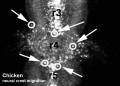
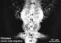
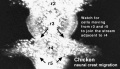
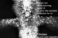
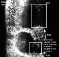
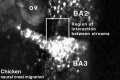
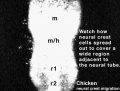
| <html5media height="380" width="410">File:Chicken-neural crest migration 01.mp4</html5media> |
Chicken embryo sequence shows the migration of DiI-labeled neural crest cells towards the branchial arches as the embryo. White rings indicate migration of individual cells. Each image represents 10 confocal sections separated by 10 microns. |
Early Development and Neural Derivatives

- bilaminar embryo- hypoblast
- trilaminar embryo - ectoderm layer
- neural plate - neural groove - neural tube and neural crest
- cranial expansion of neural tube - central nervous system
- caudal remainder of neural tube - spinal cord
Neural Crest - contributes both neural and non-neural cells
- dorsal root ganglia
- parasympathetic / sympathetic ganglia.
- enteric ganglia
Neural Crest Origin
- lateral region of neural plate
- dorsal neural fold->tube
Two main embryo regions
- Head (CNS level) - differentiate slightly earlier, mesencephalic region of neural folds.
- Body (spinal cord level) - lateral edges of fused neural tube.
Neural Crest Generation
- cranial region - Begins when still neural fold
- spinal cord - from day 22 until day 26
- after closure of caudal neuropore
- rostro-caudal gradient of differentiation
Chicken model shows that they are not a segregated population. Interactions between the neural plate and epidermis can generate neural crest cells, since juxtaposition of these tissues at early stages results in the formation of neural crest cells at the interface.
At cranial levels, neuroepithelial cells can regulate to generate neural crest cells when the endogenous neural folds are removed, probably via interaction of the remaining neural tube with the epidermis.
Progenitor cells in the neural folds are multipotent, having the ability to form multiple ectodermal derivatives, including epidermal, neural crest, and neural tube cells the neural crest is an induced population that arises by interactions between the neural plate and the epidermis.
The competence of the neural plate to respond to inductive interactions changes as a function of embryonic age. (Text from: Bronner-Fraser M PNAS 1996 Sep 3;93(18):9352-7)
Neural Crest Derivatives
Note the major regional contributions in the simplified diagram below.

Neural crest contribution[2]
| System | Cell Type |
|---|---|
| Peripheral Nervous System (PNS) | Neurons - sensory ganglia, sympathetic and parasympathetic ganglia, enteric nervous system, and plexuses
Glia (neuroglial cells) - Schwann cells[3], satellite cells, olfactory ensheathing cells[4] |
| endocrine | Adrenal medulla Calcitonin-secreting cells Carotid body type I cells |
| integumentary | Epidermal pigment cells melanocyte |
| Facial cartilage and bone | Facial and anterior ventral skull cartilage and bones |
| Sensory | inner ear, cornea endothelium and stroma |
| Connective tissue | tooth odontoblast
smooth muscle, and adipose tissue of skin in head and neck Connective tissue of meninges, salivary, lachrymal, thymus, thyroid, and pituitary glands Connective tissue and smooth muscle in arteries of aortic arch origin |
| Links: neural crest | Category:Neural Crest | Neural Crest collapsible table | |
Neural Crest - Head
See also Lecture - Head Development


Mesencephalon and caudal Proencephalon
- parasympathetic ganglia CN III
- connective tissue around eye and nerve
- head mesenchyme
- neural connective tissue (meninges)
Mesencephalon and Rhombencephalon
- pharayngeal arches
- look at practical notes on neck and head.
- cartilage rudiments (nose, face, middle ear)
- face and facial skeleton
- dermis, smooth muscle and fat
- odontoblasts of developing teeth
Rhombencephalon
- C cells of thyroid
- cranial nerve ganglia
- neurons and glia
- parasympathetic of VII, IX, X
- sensory ganglia of V, VII, VIII, IX, X
Neural Crest - Peripheral Nervous System
|

Enteric nervous system |
Neural Crest Migration
Head

|

|

|
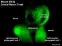
| Hindbrain neural crest migration | Mouse head E9 neural crest GFP | Mouse E9.5 Sox10 |

|

|

|

|

|

|

|
Trunk
Cardiac Outflow Tract


Figure 13.2. Neural crest cell migration in the trunk of the chick embryo
- Neural crest at the level of the body have two general migration pathways, defined by the position of the somite
- medial pathway - between the neural tube and the somite
- lateral pathway - between the somite and the body wall (cardiac NCC)
- Neural crest cells (NCC) in mice guidance show migrate 3 specific pathways.
- SEMA3A and its receptor neuropilin 1 (NRP1) - act as repulsive guidance cues
- migration pathway did not affect specification - differs from the concept of migration pathway specifying the neural crest cell differentiation pathway
Neural crest at the level of the head have a different migration pathway. Figure 13.7. Cranial neural crest cell migration in the mammalian head
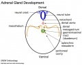
Sympathetic Ganglia and Adrenal Medulla
| <html5media height="500" width="560">File:Adrenal medulla.mp4</html5media> |  The chromatin cells that populate the adrenal medulla are NCC. The chromatin cells that populate the adrenal medulla are NCC.
|
Enteric nervous system
Vagal neural crest cells
|
Myenteric plexus of the gut wall |

Enteric neuron (red) and glia (blue) aggregation.[5] |
Historic Migration Experiments
Key early experiments in understanding the pattern of neural crest migration were carried out by LeDouarin in the 1980's (see Development of the peripheral Nervous system from the neural crest, Ann Rev Cell Biol 4 p375) Quail-Chick Chimeras | Figure 1.11. Neural crest cell migration Chimera experiment
These transplantation studies in chicken/quail chimeras utilised the different nucleoli appearance of cells to differentiate different species. Thus transplanation and subsequent histological processing allowed identification of the migration path and final destination of transplanted neural crest cells.
Similar later experiments have now been carried out using the neural crest cells molecularly tagged with (LacZ).
Abnormalities
Neuroblastoma

Neuroblastoma is the most common childhood cancer diagnosed before the age of 1 year, and accounts for 10 to 15% of all cancer deaths in children arising initially from the adrenal or other tissues.

Childhood cancer survival rates
Neuroblastoma | OMIM - Neuroblastoma
Digeorge Syndrome (DGS)

Digeorge chromosome 22 |
|
Intestinal Aganglionosis


- Intestinal Aganglionosis, Hirschsprung's Disease or Megacolon
- lack of enteric nervous system (neural ganglia) in the intestinal tract responsible for gastric motility (peristalsis).
- severity is dependent upon the amount of the GIT that lacks intrinsic ganglia, due to developmental lack of neural crest migration into those segments.
- first indication in newborns is an absence of the first bowel movement, other symptoms include throwing up and intestinal infections.
- Clinically this is detected by one or more tests (barium enema and x ray, manometry or biopsy) and can currently only be treated by surgery. A temoporary ostomy (Colostomy or Ileostomy) with a stoma is carried out prior to a more permanent pull-through surgery.
Melanoma

- In Australia each year 8,800 people are diagnosed with melanoma, and almost 1000 people die (Data, Cancer Council Australia).
- Two different findings on the reprogramming of melanoma cells, which have a neural crest origin, when transplanted between species into embryos.
Neurofibromatosis Type 1 (NF1)
- Neurofibromatosis Type 1 (von Recklinghausen) occurs in 1 in 3,000 to 4,000 people with characteristic skin blemishes forming in early childhood.
- Multiple café-au-lait spots (flat skin patches darker than the surrounding area) appear in early childhood which increase in both size and number with age.
- tumors can develop along nerves in the skin, brain, and other parts of the body. In the iris of the eye, Lisch nodules (benign growths) also appear
- (French, café-au-lait = coffee with milk)
Atlas of Genetics and Cytogenetics in Oncology- Neurofibroma
Tetralogy of Fallot
Cardiac abnormality possibly stemming from abnormal neural crest migration. Named after Etienne-Louis Arthur Fallot (1888) who described it as "la maladie blue". (More? Cardiovascular System Development | Cardiac Tutorial | Lecture - Heart | Cardiovascular System - Abnormalities)
Treacher Collins syndrome
(TCS) A genetic developmental abnormality results from autosomal dominant mutations of the gene TCOF1 encoding the protein Treacle, identified in 2006. The syndrome is characterized by hypoplasia of the facial bones, cleft palate, and middle and external ear defects. These defects may relate to the effects on neural crest migration. (More? Neural Crest Development | OMIM - TCOF1 | PMID: 8563749)
References
- ↑ 1.0 1.1 Thomas S, Thomas M, Wincker P, Babarit C, Xu P, Speer MC, Munnich A, Lyonnet S, Vekemans M & Etchevers HC. (2008). Human neural crest cells display molecular and phenotypic hallmarks of stem cells. Hum. Mol. Genet. , 17, 3411-25. PMID: 18689800 DOI.
- ↑ Simões-Costa M & Bronner ME. (2013). Insights into neural crest development and evolution from genomic analysis. Genome Res. , 23, 1069-80. PMID: 23817048 DOI.
- ↑ Woodhoo A & Sommer L. (2008). Development of the Schwann cell lineage: from the neural crest to the myelinated nerve. Glia , 56, 1481-90. PMID: 18803317 DOI.
- ↑ Barraud P, Seferiadis AA, Tyson LD, Zwart MF, Szabo-Rogers HL, Ruhrberg C, Liu KJ & Baker CV. (2010). Neural crest origin of olfactory ensheathing glia. Proc. Natl. Acad. Sci. U.S.A. , 107, 21040-5. PMID: 21078992 DOI.
- ↑ Rollo BN, Zhang D, Simkin JE, Menheniott TR & Newgreen DF. (2015). Why are enteric ganglia so small? Role of differential adhesion of enteric neurons and enteric neural crest cells. F1000Res , 4, 113. PMID: 26064478 DOI.
Online Textbooks
- Developmental Biology by Gilbert, Scott F. Sunderland (MA): Sinauer Associates, Inc.; c2000 The Cranial Neural Crest | Figure 13.1. Regions of the neural crest | Figure 13.7. Cranial neural crest cell migration in the mammalian head | Figure 13.2. Neural crest cell migration in the trunk of the chick embryo | Figure 13.10. Separation of the truncus arteriosus into the pulmonary artery and aorta | Figure 22.23. Chick embryo rhombomere neural crest cells and their musculoskeletal packets | Figure 13.4. Segmental restriction of neural crest cells and motor neurons by the ephrin proteins of the sclerotome | Figure 1.3. Pharyngeal arches | Table 13.2. Some derivatives of the pharyngeal arches
- Neural Crest Experiments: Figure 1.11. Neural crest cell migration Chimera experiment | Figure 13.5. Pluripotency of trunk neural crest cells
- Molecular Biology of the Cell Alberts, Bruce; Johnson, Alexander; Lewis, Julian; Raff, Martin; Roberts, Keith; Walter, Peter New York and London: Garland Science; c2002 Figure 21-80. The main pathways of neural crest cell migration Figure 21-91. Diagram of a 2-day chick embryo, showing the origins of the nervous system | Figure 19-23. An example of a more complex mechanism by which cells assemble to form a tissue
- Neuroscience Purves, Dale; Augustine, George J.; Fitzpatrick, David; Katz, Lawrence C.; LaMantia, Anthony-Samuel; McNamara, James O.; Williams, S. Mark. Sunderland (MA): Sinauer Associates, Inc.; c2001Figure 22.1. Neurulation in the mammalian embryo | Figure 22.12. Cell signaling during the migration of neural crest cells
- Madame Curie Bioscience Database Chapters taken from the Madame Curie Bioscience Database (formerly, Eurekah Bioscience Database) Cranial Neural Crest and Development of the Head Skeleton | Neural Crest Cells and the Community of Plan for Craniofacial Development: Historical Debates and Current Perspectives | Figure 1. Diagram of an E10 embryo showing the origins of neural crest cells that colonize the developing gastrointestinal tract
- Basic Neurochemistry: Molecular, Cellular, and Medical Aspects Siegel, George J.; Agranoff, Bernard W.; Albers, R. Wayne; Fisher, Stephen K.; Uhler, Michael D., editors Philadelphia: Lippincott,Williams & Wilkins; c1999Figure 27-10. Neuropoietic model of neural crest cell lineage | Figure 27-11. Growth factor control of neural crest lineage decisions | Figure 27-15. The Schwann cell lineage
Articles
<pubmed></pubmed> <pubmed></pubmed> <pubmed>28780695</pubmed> <pubmed>27923324</pubmed>
Marshall AM. The morphology of the vertebrate olfactory organ. (1879) Quarterly Journal of Microscopic Science. 19: 300–340.
Search
- Bookshelf neural crest
- Pubmed neural crest
External Links
External Links Notice - The dynamic nature of the internet may mean that some of these listed links may no longer function. If the link no longer works search the web with the link text or name. Links to any external commercial sites are provided for information purposes only and should never be considered an endorsement. UNSW Embryology is provided as an educational resource with no clinical information or commercial affiliation.
- The University of Miami Biology Department Lab
- Stowers Institute Kulesa Lab | Trainor Lab
- University College London Mayor Lab
- University of Iowa Cornell Lab
- Washington University in St. Louis, School of Medicine, Department of Pediatrics Heuckeroth Lab