Lecture - 2014 Course Introduction

From Embryology

Course Introduction

Dr Mark Hill

Course coordinator

This first lecture will be a general introduction to the course and the subject of Embryology.
Firstly, an introduction to the course, its content, method of presentation, assessment and an opportunity to ask questions.
Secondly, some historic background to the subject and related current Australian trends. I do not expect you to remember specific historic dates or statistical data, this is provided as an introduction to the topic.


current 2015 Course Introduction

I like my lectures to be interactive, so ask me questions and I will also be asking you questions!

Lecture Objectives

<html5media height="384" width="352">File:Human development 001.mp4</html5media>

Click Here to play on mobile device

  1. Understand the course objectives and assessment.
  2. Brief understanding of the historic background of embryology.
  3. Brief understanding of Australian data.
  4. Broad overview of human development.


ECHO360 icon.gif

Lecture 1 - Rich Media Playback | Vodcast Playback | Podcast Playback


Human development 001 icon.jpg
 ‎‎Human Development
Page | Play

ANAT2341 Course Outline

Human embryonic development week 1 to 8
Human embryonic development (week 1 to 8)

I will spend the first half going through the current course design, online support and assessment criteria. This is an opportunity to ask the coordinator questions about the course.


Course Links: Homepage | Overview | Timetable | Moodle | Lecture 1 PDF

Lecture Archive: 2013 | 2012 | 2011

Email me for any additional information or to make an appointment.

Textbooks

  • Either of the textbooks listed below are recommended for this course and page references to both are given in each lecture.
  • Both textbooks available at campus bookshop and online to UNSW students.
  • There are additional embryology textbooks that can also be used, consult course organizer.
The Developing Human: Clinically Oriented Embryology (10th edn) 
The Developing Human, 10th edn.jpg

UNSW Students have online access to the current 10th edn. through the UNSW Library subscription (with student Zpass log-in).


APA Citation: Moore, K.L., Persaud, T.V.N. & Torchia, M.G. (2015). The developing human: clinically oriented embryology (10th ed.). Philadelphia: Saunders.

Links: PermaLink | UNSW Embryology Textbooks | Embryology Textbooks | UNSW Library
  1. Introduction to the Developing Human
  2. First Week of Human Development
  3. Second Week of Human Development
  4. Third Week of Human Development
  5. Fourth to Eighth Weeks of Human Development
  6. Fetal Period
  7. Placenta and Fetal Membranes
  8. Body Cavities and Diaphragm
  9. Pharyngeal Apparatus, Face, and Neck
  10. Respiratory System
  11. Alimentary System
  12. Urogenital System
  13. Cardiovascular System
  14. Skeletal System
  15. Muscular System
  16. Development of Limbs
  17. Nervous System
  18. Development of Eyes and Ears
  19. Integumentary System
  20. Human Birth Defects
  21. Common Signaling Pathways Used During Development
  22. Appendix : Discussion of Clinically Oriented Problems
Larsen's Human Embryology (5th edn) 
Larsen's human embryology 5th ed.jpg
UNSW students have full access to this textbook edition through UNSW Library subscription (with student Zpass log-in).


APA Citation: Schoenwolf, G.C., Bleyl, S.B., Brauer, P.R., Francis-West, P.H. & Philippa H. (2015). Larsen's human embryology (5th ed.). New York; Edinburgh: Churchill Livingstone.

Links: PermaLink | UNSW Embryology Textbooks | Embryology Textbooks | UNSW Library
  1. Gametogenesis, Fertilization, and First Week
  2. Second Week: Becoming Bilaminar and Fully Implanting
  3. Third Week: Becoming Trilaminar and Establishing Body Axes
  4. Fourth Week: Forming the Embryo
  5. Principles and Mechanisms of Morphogenesis and Dysmorphogenesis
  6. Fetal Development and the Fetus as Patient
  7. Development of the Skin and Its Derivatives
  8. Development of the Musculoskeletal System
  9. Development of the Central Nervous System
  10. Development of the Peripheral Nervous System
  11. Development of the Respiratory System and Body Cavities
  12. Development of the Heart
  13. Development of the Vasculature
  14. Development of the Gastrointestinal Tract
  15. Development of the Urinary System
  16. Development of the Reproductive System
  17. Development of the Pharyngeal Apparatus and Face
  18. Development of the Ears
  19. Development of the Eyes
  20. Development of the Limbs


History

History - Embryologists | Embryology History | Human Embryo Collections

The Position of the Uterus and Fetus at Term (1872)

17-18C Braune - The Position of the Uterus and Fetus at Term (1872)

Human Embryo Collections
Wilhelm His.jpg

Wilhelm His (1831-1904)

His's Normentafel (Normal Table)

Anatomie menschlicher Embryonen (1882)

Keibel Mall 034a.jpg Keibel Mall 034b.jpg
Franz Keibel.jpg

Franz Keibel (1861 - 1929)

Franz Keibel and Curt Elze (1908) Normal Plates of the Development of the Human Embryo

Keibel1908 plate01.jpg Keibel1908 plate02.jpg
Franklin Mall 03.jpg

Franklin Mall (1862-1917)

Carnegie Collection

Human Carnegie stage 10-23.jpg
Begun by Dr. Hideo Nishimura (1912–1995)

Shiota Hill Yamada.jpg Developed by Kohei Shiota and currently curated by Shigehito Yamada.

Kyoto Collection

Human Carnegie stage 1-23.jpg
Animal Models
Frog-icon.png
Frog Links: Frog Development | 2009 Student Project | 1897 Development of the Frog's Egg | Hans Spemann | Wilhelm Roux | 1921 Early Frog Development | 1951 Rana pipiens Development | Rana pipiens Images | Frog Glossary | John Gurdon | Category:Frog | Animal Development
  • The frog was used by many of the early embryology investigators and currently there are many different molecular mechanisms concerning development of the frog.
  • The eggs develop independently, in relative synchrony and are relatively see-through making staging and observation fairly easy.
  • The frog was a key model for the study of the process of gastrulation.
Chick icon.jpg

chicken

  • The chicken embryo develops and hatches in 20-21 days and historically these were one of the first embryos to be studied. Cutting a window in the egg shell allows direct observation of the embryo. The Hamburger & Hamilton chicken development staging allowed researchers to develop this model as a key embryological tool.
  • Key research involved the transplanting of quail cells into chick embryos, to later identify their contribution to different embryonic structures, particularly for somite, neural tube and neural crest development.
Mouse.jpg
mouse
  • The mouse has always been a good embryological model, easy to generate (litters 8-20) and quick (21d).
  • Mouse embryology really expanded when molecular biologists used mice for gene knockouts.
Fly-icon.png
Fly Development - The fruitfly (drosophila) was and is the traditional geneticist's tool. It has been transformed to an magnificent embryologist's tool, with developmental mechanisms being uncovered in this system combined with homolgy gene searches in other species. The fly genome was one of the first to be been completely sequenced. In early development nurse cells sacrifice their cytoplasmic contents to allow egg growth and early pattern formation is through the localization of maternal messenger RNAs (mRNAs).
C elegans.jpg
Worm Development - Early embryological studies of the worm Caenorhabditis elegans (C.Elegans, so called because of its "elegant" curving movement) characterized the fate of each and every cell in the worm through all stages of development. This worm has recently had its entire genome sequenced.
Zebrafish-icon.png
Zebrafish Development - Zebrafish are seen as the latest and greatest "model' for embryological development studies. They can be easily genetically altered and develop as practically "see through" embryos, all internal development can be clearly observed from the outside in the living embryo.
In Vitro Fertilization (1978) Stem Cells (1981) Molecular Development
Intracytoplasmic sperm insemination.jpg Hematopoietic and stromal cell differentiation.jpg Hedgehog signaling pathway.jpg

Australian Data

1 August 2014 at 03:53:30 PM (Canberra time), the resident population of Australia is projected to be: 23,550,233.

Australian-births 2011.jpg

Australian Statistics
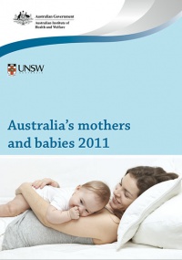
Australia mothers and babies 2011.jpg Assisted reproductive technology in Australia and New Zealand 2010.jpg
Australia’s mothers and babies (2011) Assisted reproductive technology in Australia and New Zealand (2010)
Average maternal age in 2011 was 30.0 years, the same as 2009 but still more than the earlier years (2000, 29.0 years; 2002, 29.4 years). Assisted Reproductive Technology (ART) was used by 3.8% (2009, 3.6%) of women who gave birth.
Victoria - 10 most reported birth anomalies
Based upon statistics from the Victorian Perinatal Data Collection Unit in Victoria between 2003-2004.
Hypospadia Hypospadias (More? Development Animation - Genital Male External | Genital Abnormalities - Hypospadia)
Obstructive Defect of the Renal Pelvis Obstructive Defects of the Renal Pelvis (obstructive defects of the renal pelvis, uteropelvic junction obstruction, pelvo-uterero junction obstruction) Term describing a developmental renal abnormality due to partial or complete blockage of the drainage of the kidney pelvis requiring surgical correction. The blockage can also have several causes including: unusual ureter twisting or bending, ureter compression by a blood vessel, malformations of the muscular wall. The blockage leads to an accumulation of urine in the affected region, with several potential effects: nephron damage from compression (hydronephrosis); decreased urine output leading to lack of amniotic fluid (oligohydramnios); respiratory development effects due to the lack of amniotic fluid.
  • The most common type of obstruction is at the uteropelvic junction (UPJ), between the junction of the ureter and the kidney.
  • Blockage lower as the ureter enters the bladder, the ureterovesicular junction (UVJ), usually involves only one kidney and the back flow enlarges the affected ureter (megaureter).

(More? Renal System - Abnormalities | Renal System Development)

Ventricular Septal Defect Ventricular Septal Defect (More? Cardiovascular Abnormalities - Ventricular Septal Defect)

Basic Heart Development Timeline.jpg

Heart Development Timeline (see Basic Cardiac Embryology)

Congenital dislocation hip Congenital Dislocated Hip (More? Musculoskelal Abnormalities - Congenital Dislocation of the Hip (CDH))

(DHH, congenital dislocated hip, congenital hip dislocation, congenital hip dysplasia) Term describes a spectrum of musculoskeletal disorders of hip instability due either to the femoral head being able to move outside the acetabulum (luxation or dislocation), or abnormally within the acetabulum (subluxation or partial dislocation). This includes presentation following a normal examination of the hips in the newborn period (Ortolani and Barlow tests). When detected can be managed with splinting (Denis-Browne splint) allows the hip joint to develop normally and does not require surgery. If undetected and left untreated, the hip joint develops abnormally and surgical reduction is required. (More? Musculoskeletal System Development)

Trisomy 21 male Trisomy 21 or Down syndrome - (More? Trisomy 21)
Hydrocephalus Hydrocephalus (More? Neural Abnormalities - Hydrocephalus | NINDS - Hydrocephalus Fact Sheet | Hydrocephalus Support Association | USA National Hydrocephalus Foundation)
Cleft palate Cleft Palate (More? Development Animation - Palate 1 | Development Animation - Palate 2 | Cleft Palate)
Trisomy 18 male Trisomy 18 or Edward Syndrome - multiple abnormalities of the heart, diaphragm, lungs, kidneys, ureters and palate 86% discontinued (More? Trisomy 18)
Renal Agenesis/Dysgenesis - reduction in neonatal death and stillbirth since 1993 may be due to the more severe cases being identified in utero and being represented amongst the increased proportion of terminations (approximately 31%). (More? Renal Abnormalities - Renal Agenesis)
Bilateral cleft palate Cleft Lip and Palate - occur with another defect in 33.7% of cases. (More? Cleft Lip)

Human Development

Human development timeline graph 02.jpg


2014 Course: Week 2 Lecture 1 Lecture 2 Lab 1 | Week 3 Lecture 3 Lecture 4 Lab 2 | Week 4 Lecture 5 Lecture 6 Lab 3 | Week 5 Lecture 7 Lecture 8 Lab 4 | Week 6 Lecture 9 Lecture 10 Lab 5 | Week 7 Lecture 11 Lecture 12 Lab 6 | Week 8 Lecture 13 Lecture 14 Lab 7 | Week 9 Lecture 15 Lecture 16 Lab 8 | Week 10 Lecture 17 Lecture 18 Lab 9 | Week 11 Lecture 19 Lecture 20 Lab 10 | Week 12 Lecture 21 Lecture 22 Lab 11 | Week 13 Lecture 23 Lecture 24 Lab 12
Student Projects - Group 1 | Group 2 | Group 3 | Group 4 | Group 5 | Group 6 | Group 7 | Group 8 | Moodle

Glossary Links

Glossary: A | B | C | D | E | F | G | H | I | J | K | L | M | N | O | P | Q | R | S | T | U | V | W | X | Y | Z | Numbers | Symbols | Term Link

Cite this page: Hill, M.A. (2024, March 19) Embryology Lecture - 2014 Course Introduction. Retrieved from https://embryology.med.unsw.edu.au/embryology/index.php/Lecture_-_2014_Course_Introduction

What Links Here?
© Dr Mark Hill 2024, UNSW Embryology ISBN: 978 0 7334 2609 4 - UNSW CRICOS Provider Code No. 00098G