Harvard Collection
| Embryology - 12 Jul 2026 |
|---|
| Google Translate - select your language from the list shown below (this will open a new external page) |
|
العربية | català | 中文 | 中國傳統的 | français | Deutsche | עִברִית | हिंदी | bahasa Indonesia | italiano | 日本語 | 한국어 | မြန်မာ | Pilipino | Polskie | português | ਪੰਜਾਬੀ ਦੇ | Română | русский | Español | Swahili | Svensk | ไทย | Türkçe | اردو | ייִדיש | Tiếng Việt These external translations are automated and may not be accurate. (More? About Translations) |
Harvard Collection

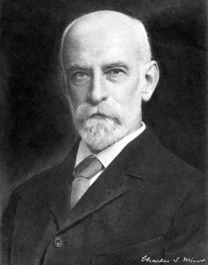
This historic collection of human and other embryos was originally collected by Charles Minot (1852–1914), sometimes referred to as the Minot Collection, now forms part of the larger Carnegie Collection. The collection was described in detail by Minot (1905).[1]
Embryos in the collection are numbered and prefixed in papers by the acronym H.E.C..
Carnegie Collection - HDAC 7 Charles Sedgwick Minot Embryological Collection
- Embryos from the Harvard School of Medicine, as well as drawings and photographs of the embryos.
- A large collection of reprints, printed lectures, class syllabi, and theses on embryology and related topics.
- The reprint collection was started by Charles S. Minot (1852-1914) in the 1800s and added to through the 1960s.
- The reprint collection also includes personal papers and research notes from Charles Wislocki.
- "These considerations have led us to adopt a metal cabinet, which has been specially devised for our needs. It is made of sheet tin in such a manner that the trays are very compact, are absolutely interchangeable, an intake up a minimum amount of room. The construction adopted is such that the tendency to warp is entirely done away with (Fig.3) The trays are all japanned so that they do not rust, and we slip a bit of white paper into each tray to make a background for the sections. Each tray is, moreover, furnished with a litle label holder, and they are put together in cabinets of thirty trays each, the trays themselves being of such a size that they will hold twenty-four of the ordinary slides, three inches by one. Moreover, the cabinets themselves are so devised that they can be stacked one on top of another, taking up a minimum amount of room. We devote a vertical column of these cabinets to a species, and simply interpolate from time to time a new cabinet in the column as the growth of the collection may render necessary. The cabinets are made by Peter Gray & Co., of Union street, Boston, and are now kept in stock by several of the dealers in microscopical supplies in this country. They cost only a trifle more than the wooden cabinets, and are, according to our trial of them, certainly to be preferred to any other form of cabinet which we have tested." (Text fromThe Harvard Embryological Collection (1905)[1])
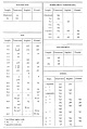
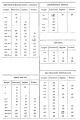
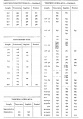
Harvard Collection Catalogue
Human
Many of the collection's human embryos were used by Frederic T. Lewis in his chapter on gastrointestinal tract development in Keibel and Mall's 1912 Human embryology textbook.[2]
- Introduction Frederic Lewis
- Early Development of the Entodermal Tract and the Formation of its Subdivisions Frederic Lewis
- The Development Of The Oesophagus Frederic Lewis
- The Development of the Stomach Frederic Lewis
- The Development of the Small Intestine Frederic Lewis
- The Development of the Large Intestine Frederic Lewis
- The Development of the Liver Frederic Lewis
- Development of the Pancreas Frederic Lewis
|
|
guinea pig, pig and chicken
Garfish, Catfish and Trout
Catfish and Torpedo
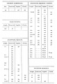
Spiny dogfish and Thornback ray
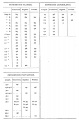
Lamprey and Lancelet


Harvard Collection Embryos
Embryo 55
Harvard Embryo 55
Studied histochemically by Hertig et al. (1958)[3].
- Hysterectomy.
- Presumed age 13 days.
- Chorion, 1.77 x 1.33 x 0.598 mm.
- Chorionic cavity, 0.73 x 0.68 x 0.221 mm. Embryonic disc, 0.296 x 0.196 X 0.044 mm.
Chorionic villi essentially solid, with earliest suggestion of mesoblastic core formation. "Apparently without axial differentiation." Possesses "a very recently formed definitive (secondary) yolk sac." Possible primordial germ cells ("stuffed with glycogen") within endoderm near edge of disc.
Embryo 192
Harvard Embryo 192
Embryo 256
Harvard Embryo 256
Embryo 529
Harvard Embryo 529
Embryo 714
Harvard Embryo 714 published in a paper by Bremer (1906).[4]


Manual of Human Embryology II[2]

|
Fig. 267. — Transverse sections of the epithelial tube of the oesophagus. X 160 diam.
|
Fig. 293. Sections showing the formation of the periportal ducts (D.peri-p.) around a branch of the portal vein (V.p.). A from an embryo of 22.8 mm. (Harvard Collection, Series 871).

Fig. 300 Dorsal views of the hepatic region
Embryo 787
Harvard Embryo 787 Human embryo 22.8 mm[5]
Fig. 10. Photomicrograph x50 H.E.C. Embryo 787, length 22.8 mm., slide 406. Transverse section shows the recess between pubis and ischium to form the acetabular fossa. In it are seen the origins of the ligamentum teres and the Haversian gland. The anteversion of the neck of the femur has developed to produce an angle of 30° to the midline of the embryo. The ligamentum teres develops in situ without formation of a groove in the head of the femur. Short rotator muscles of the hip are well outlined.
Embryo 816
Harvard Embryo 816 human embryo 12.0 mm
Embryo 825
Harvard Embryo 825
Embryo 828
Harvard Embryo 828 human embryo 19.0 mm
Embryo 838
Harvard Embryo 838 human embryo 42 mm
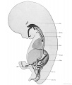
Fig. 246. digestive tract embryo 42 mm
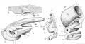
Fig. 247. development of the vermiform process


Embryo 839
Harvard Embryo 839 human embryo 17.8 mm
See Thyng (1914)[6]
- 17.8 mm Embryo, external appearance suggests Carnegie stage 19 embryo (Week 7, 48 - 51 days, 16 - 18 mm).
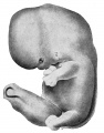
Fig 1
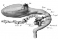
Fig 2
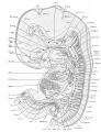
Plate 1a
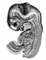
Plate 1b
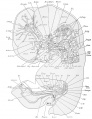
Plate 2a
Plate 2b
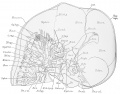
Plate 3a
Plate 3b
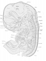
Plate 4a
Plate 4b
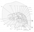
Plate 5a
Plate 5b
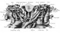
Plate 6


- Links: 1914 Thyng 17.8 mm Embryo
Embryo 871
Harvard Embryo 871
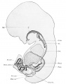
Fig. 243. digestive tract of an embryo 22.8 mm
Fig. 292.

Fig. 292. A section of the gall-bladder of a 29 mm embryo (Harvard Collection, Series 914). X 180 diam. B section of the common bile-duct of a 22.8 mm embryo (Harvard Collection, Series 871). X 180 diam. C epithelium of the gall-bladder, two weeks after birth. X 580 diam.
Embryo 913
Harvard Embryo 913 human embryo 32 mm.
Fig. 293. Sections showing the formation of the periportal ducts around a branch of the portal vein. B from an embryo of 32 mm (Harvard Collection, Series 913). X 185 diam.
Embryo 914
Harvard Embryo 914

Fig. 292. A section of the gall-bladder of a 29 mm embryo (Harvard Collection, Series 914). X 180 diam.
Embryo 918
Harvard Embryo 918 Human embryo 28.8 mm[5]
Fig. 8. Photomicrograph X30 H.E.C. Embryo 918, length 30 mm., slide 1094. Same embryo as Fig. 7. Blastema in the anterior limb of the Y between pubis and ilium is seen. Note location of ossification center in ilium as opposed to nucleus of chondrification near acetabulum in Figs. 3 and 6. Course of ligamentum teres from transverse ligament to fovea is prominent. The glenoid labrum and relationship of the capsule may be seen. Symphysis pubis is approximating.
Embryo 939
Harvard Embryo 939 human embryo 13.6 mm
Embryo 1000
Harvard Embryo 1000 human embryo 10 mm

|
Fig. 274. Sections of the stomach of a 10 mm embryo (Harvard Collection, Series 1000). A, through the cardia. B, through the fundus. C, through the pylorus. A. coel., coeliac artery ; A.g.s., left gastric artery ; A. hep., hepatic artery ; Alien., splenic artery ; Ao., aorta ; B.om., omental bursa ; C.W., Wolffian body ; D.ch., common bile-duct ; D.v., ductus venosus ; F.ep., foramen epiploicum ; N.sym., sympathetic nerve ; O.ma., greater omentum ; O.mi., lesser omentum ; Pul., lung ; Va., vagus nerve ; V.p., portal vein ; V.8., left suprarenal vein. |

|
Fig. 294. A hepatic trabecula containing a lumen. From a 10 mm embryo (Harvard Collection, Series 1000). X 1065 diam. B, bile-capillaries in a 44.3 mm embryo (Harvard Collection, Series 1611). x 1065 diam. Bl., blood-corpuscle; C.hep., hepatic cell; Endo., endothelium ; Lum., lumen of a bile capillary. |
Embryo 1003
Fig. 7 Outline drawing of section #228, 14.5 mm. human embryo (No. 1003, Harvard Collection). X 10.
Embryo 1005
Harvard Embryo 1005
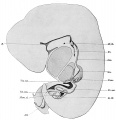
Fig. 242. Digestive tract of an embryo 9.4 mm
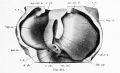
Fig. 301. Dorsal views of the hepatic region


Embryo 1129
Harvard Embryo 1129
Embryo 1322
Harvard Embryo 1322 Human embryo 16 mm
- Keibel Mall 2 311.jpg
Fig. 311. digestive tract of an embryo 9.4 mm
Fig. 311. Section through the stomach, pancreas, and a part of the liver, from an embryo of 16 mm. (Harvard Collection, Series 1322). y>X 40 diam. ; ,A. mes. sup., superior mesenteric artery; tB. oment., omental bursa ;'_Gaster, stomach; Lien, spleen; V. p., portal vein. (Other labels as in preceding figures.)
Embryo 1597
Harvard Embryo 1597 Human embryo 19.3 mm[5]
Fig. 5. Photomicrograph X30 H.E.C. Embryo 1597, length 19.3 mm., slide 807. The center of the femur shows mature fetal cartilage. An angulation of the upper end delineates the neck which forms an angle of 60° to the midline and 160° to the shaft. Note the cell distribution in transverse lines between neck and shaft. The joint region is densely stained.
Embryo 1598
Harvard Embryo 1598 Human embryo 28.8 mm[5]
Fig. 9. Photomicrograph X55 H.E.C. Embryo 1598, length 28.8 mm., slide 218. Sagittal section through the medial wall of the acetabulum shows the relative proportion of ischial and pubic attachments of the ligamentum teres, and the region of the acetabular fossa. At this stage the Y-shaped junction is not present, as fusion in cartilage has already taken place. This cartilage is that which will outline the Y in the child when the centers of ossification of the pelvic bones approximate the acetabulum. Thus we see the Y duplicated in both blastema and cartilage at different stages, although the points of origin of chondrification and ossification are not the same in the pelvic bones as they are in the femur.
Embryo 1707
Fig. 8 Outline drawing of section #457, 16.4 mm. human embryo, (No. 1707, Harvard Collection). X 10.
Embryo 1913
Harvard Embryo 1913 Human embryo 18 mm
(reconstructed by Huntington and McClure in 1915)
Embryo 2046
Harvard Embryo 2046 Human embryo 23 mm[5]
Fig. 6. Photomicrograph x21 H.E.C. Embryo 2046, length 23 mm., slide 1606. The line of the joint is a zone of diminished density. The femur is becoming adducted to an angle of 30-40° with the midline. The neck appears longer and is more angulated on the shaft, 65-70° to the midline and 150-155° to the shaft. The false pelvis with the anterior superior spine of the ilium is shown as a projection of precartilage.
Embryo 2050
Harvard Embryo 2050 Human embryo 36 mm[5]
Fig. 12. Photomicrograph x 140 H.E.C. Embryo Template:HEC2050, length 36 mm., slide 2531. High power of space in Fig. 11. In the spaces float cells possessing normal-appearing nuclei and long fibrils. There are smaller cells with pyknotic nuclei as well as fragmented material suggesting shadows of nuclei. The spaces appear under the ligamentum teres, between it and the head of the femur and within the capsule distal to the glenoid labrum.
Embryo 2051
Harvard Embryo 2051 Human embryo 15 mm[7]
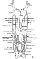
Fig. 9 human embryo 15 mm Harvard Collection no. 2051
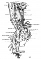
Fig. 10 Ventrolateral aspect.
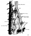
Fig. 11 venous ring of right side


Embryo 2059
Harvard Embryo 2059 Human embryo 36 mm[5]
Fig, 11. Photomicrograph x20 H.E.C. Embryo 2059, length 36 mm., slide 2531. Spaces are shown appearing in the tissue about the head of the femur. All the elements of the joint are defined. The acetabulum, glenoid labrum, and transverse acetabular ligament form 180° of a circle. Blood vessels are present in perichondrium, joint capsule, ligamentum teres, and Haversian gland, but none 1s present in the cartilage or shaft of the bones.
Embryo 2128
Harvard Embryo 2128 Human embryo 45 mm

Fig. 16. Human embryo 45 mm Harvard Collection, no. 2128[7]
Embryo 2155
Harvard Embryo 2155 Human embryo 17.5 mm[5]
Fig. 4. Photomicrograph x30 H.E.C. Embryo 2155, length 17.5 mm., slide 1169. A mass of deeply staining cells marks the joint. Muscle groups are outlined, some converge at a projection of blastema, locating the great trochanter. The femur, covered with perichondrium shows cartilage cells shrinking from matrix at the center and arcuate columns of precartilage cells compose the ends. The elements of the innominate primordia may be identified.
Embryo 2924
Harvard Embryo 2924 Human embryo 25 mm[7]
Harvard Collection Papers
Pharynx
Venous System
Published by McClure (1925).[7]
- No. 2051, 15 mm embryo (reconstructed x 100)
- No. 1913, 18 mm embryo (reconstructed by Huntington and McClure in 1915)
- No. 2924, 25 mm embryo
- No. 2128, 45 mm embryo (reconstructed x 50)
Fig. 9 human embryo 15 mm Harvard Collection, no. 2051
Fig. 10 human embryo 15 mm Harvard Collection, no. 2051 Ventrolateral aspect.
Fig. 11 human embryo 15 mm Harvard Collection, no. 2051 venous ring of right side
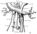
Fig. 16 human embryo 45 mm Harvard Collection, no. 2128
Embryo 2300
Harvard Embryo 2300 Human embryo 6.75 mm[5]
Fig. 1. Photomicrograph x55 H.E.C. Embryo 2300, length 6.75 mm., slide 420. The limb bud is formed. Its relation to the somites, coelom, Wolffian ducts are shown. The cells are indistinguishable from each other, except for the many mitoses. From this period on the limb bud is a distinct entity and displacement caudally and laterally occurs simultaneously with elongation of the embryo as a whole.
References
- ↑ 1.0 1.1 1.2 Minot CS. The Harvard embryological collection. (1905) J Med Res. Aug;13(5):499-522.PMID 19971684 | PDF
- ↑ 2.0 2.1 Keibel F. and Mall FP. Manual of Human Embryology II. (1912) J. B. Lippincott Company, Philadelphia.
- ↑ Hertig AT. Adams EC. Mckay DG. Rock J. Mulligan WJ. and Menkin MF. A thirteen-day human ovum studied histochemically. (1958) Am. J. Obstet. Gynecol., 76(5): 1025-40. PMID 13583048
- ↑ Bremer JL. Description of a 4-mm human embryo. (1906) Amer. J Anat. 5: 459-480.
- ↑ 5.0 5.1 5.2 5.3 5.4 5.5 5.6 5.7 5.8 Strayer MMJr. The embryology of the human hip joint. (1943) Yale J Biol. Med. 16(1): 13–26.6. PMCID: PMC2601352
- ↑ Thyng FW. The anatomy of a 17.8 mm human embryo. (1914) Amer. J Anat. 17: 31-112.
- ↑ 7.0 7.1 7.2 7.3 McClure CFW. and Butler EG. The development of the vena cava inferior in man. (1925) Amer. J Anat. 35(3): 331-383.
External Links
External Links Notice - The dynamic nature of the internet may mean that some of these listed links may no longer function. If the link no longer works search the web with the link text or name. Links to any external commercial sites are provided for information purposes only and should never be considered an endorsement. UNSW Embryology is provided as an educational resource with no clinical information or commercial affiliation.
- National Academy of Sciences Biographical Memoirs Charles Minot (1920)
Glossary Links
- Glossary: A | B | C | D | E | F | G | H | I | J | K | L | M | N | O | P | Q | R | S | T | U | V | W | X | Y | Z | Numbers | Symbols | Term Link
Cite this page: Hill, M.A. (2026, July 12) Embryology Harvard Collection. Retrieved from https://embryology.med.unsw.edu.au/embryology/index.php/Harvard_Collection
- © Dr Mark Hill 2026, UNSW Embryology ISBN: 978 0 7334 2609 4 - UNSW CRICOS Provider Code No. 00098G