BGDA Practical 7 - Week 8
Introduction
Key Events of Human Development during the eighth week (week 8) following fertilization or clinical week 10 (GA week 10).
There are 4 Carnegie stages that show external embryo development during this week. This page will give an overview of limb and neural development and look at other system organization compared with the earlier stages. Use the embryo surface views, histological sections, virtual slides and movies to identify the overall developmental anatomy at the end of the embryonic period.
- Week 8 Stages
Stage 20

Stage 21
Stage 22

Stage 23


|
<html5media height="500" width="300">File:Carnegie stage 23.mp4</html5media> |


Week 8 - Embryo Stage 23
The following MRI movies show details of the external and internal structure of the human embryo at the end of embryonic development.
|
|
|
| ||||||||||||||||||||||
|
|
|
| ||||||||||||||||||||||
| |||||||||||||||||||||||||
| Week 8 - Stage 23 Movies | |||||||||||||||||||||||||||||||
|---|---|---|---|---|---|---|---|---|---|---|---|---|---|---|---|---|---|---|---|---|---|---|---|---|---|---|---|---|---|---|---|
| <html5media height="580" width="612">File:Stage23 MRI 3D01.mp4</html5media> | Embryo Surface Carnegie Stage 23
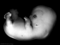
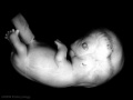
This movie shows an unlabeled MRI 3D volume embryo surface scan around the Kyoto embryo (stage 23, week 8).
| ||||||||||||||||||||||||||||||
| <html5media height="600" width="400">File:Carnegie stage 23.mp4</html5media> | Embryo Surface Carnegie Stage 23
This is a second surface view of the stage 23 embryo from photomicrographs. Compare this to the above MRI view. | ||||||||||||||||||||||||||||||
| <html5media height="600" width="580">File:Stage23 MRI 3D02.mp4</html5media> | Central Nervous System
This movie shows a MRI 3D volume scan inside the Kyoto embryo (stage 23, week 8). The labelled sagittal head section movie show further detail.
| ||||||||||||||||||||||||||||||
| <html5media height="500" width="500">File:Stage23 MRI S04.mp4</html5media> | Midgut Herniation
This movie shows an extract from the whole embryo sagittal MRI scan (left to right) through a Kyoto embryo (stage 23, week 8). The extracted region shows the midgut (intestines) lie outside the ventral body wall (midgut herniation) at the end of the embryonic period. This herniation will not be lost until the early fetal period. Abnormalities can arise from either abnormal body wall development or from gastrointestinal tract development. (More? Ultrasound Gastroschisis | Omphalocele) | ||||||||||||||||||||||||||||||
|
| ||||||||||||||||||||||||||||||
Head
Oral Cavity Region

Sensory
Parts of each sensory system formed from placode origins and each system also has a complex embryonic origin. These images show eye development at the end of the embryonic period.

|
|

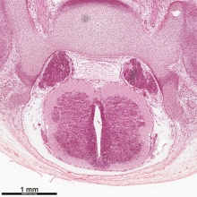
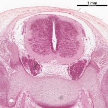
Neural

|
Cortex development commences
choroid plexus
Note
| |||

|
|
Body
Intraembryonic Coelom the pericardioperitoneal canals close, generating the 3 separate body cavities.

|

|
| Lungs, thymus, musculoskeletal. | vertebral column, spinal cord. |
Limbs
| Week 8 Limb Development | ||||||||||||||||||
|---|---|---|---|---|---|---|---|---|---|---|---|---|---|---|---|---|---|---|

Human Embryo (Stage19) limb rotation |
Week 8 (GA week 10) limbs rotate relatively in different directions (Humans Stage 20-23)
| |||||||||||||||||

- Links: Limb Development | Limb Abnormalities | Thalidomide
Gastrointestinal Tract

|

|
|||||||
The gastrointestinal midgut remains herniated outside of the ventral body wall.
|
|
|
Genital
- Testis - mesenchyme, interstitial cells (of Leydig) secrete testosterone, androstenedione.
- Genital 8 to 12 Weeks - hCG stimulates testosterone production.
- required for internal and external genital tract development.
| Human Embryo Male Testis (stage 22) | |
|---|---|

|

|
- Human Stage 22: Testis - labeled overview | Testis - unlabeled overview | Testis - unlabeled detail | Testis - labeled detail | testis | Carnegie stage 22 | Movie - Urogenital stage 22
Genital Development will be covered in detail in BGDB.
Extra-embryonic Coelom
- Chorionic cavity is now lost by fusion with the expanding amniotic cavity.

Covered in more detail in the Placenta practical.
Other Embryo Systems
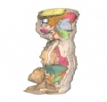
Movies - Embryo Carnegie stage 22 - These are rotating animations based upon reconstruction of a human embryo (stage 22) serial slice images.
|
|
|
|
|
|
Critical Periods
The term "critical period" refers to the specific times during human development when exposure to environmental teratogens can cause congenital abnormalities. In general, the effects for each system are more severe (major anomalies) in the embryonic period during organogenesis in the first trimester. Later teratogen exposure are less severe (minor anomalies) in the fetal period during continued growth and differentiation in the second and third trimester. Different systems have different critical periods.
| Critical Periods of Human Development | ||||||||||||||||||||
|---|---|---|---|---|---|---|---|---|---|---|---|---|---|---|---|---|---|---|---|---|
| Conceptus | Embryonic development (weeks) | Fetal period (weeks) | ||||||||||||||||||

|
||||||||||||||||||||
| Neural | ||||||||||||||||||||
| Heart | ||||||||||||||||||||
| Upper limbs | ||||||||||||||||||||
| Lower limbs | ||||||||||||||||||||
| Ear | ||||||||||||||||||||
| Eye | ||||||||||||||||||||
| Palate | ||||||||||||||||||||
| Teeth | ||||||||||||||||||||
| External genitalia | ||||||||||||||||||||
| Loss | Major abnormalities | Functional and Minor abnormalities | ||||||||||||||||||
| ||||||||||||||||||||
Week 8 Interactive Component
| Attempt the Quiz - Week 8 | |
|---|---|

Here are a few simple Quiz questions that relate to Week 8 (GA week 10) from the lecture and practical. See your Quiz Result - Answer all the questions, then click "submit" to complete. The page will reload and you can then reopen this table to see your result and feedback.
|
Additional Information
| Additional Information - Content shown under this heading is not part of the material covered in this class. It is provided for those students who would like to know about some concepts or current research in topics related to the current class page. |
Timeline
| Week 8 - Human Embryo Stages and Events (GA week 10) | ||
|---|---|---|
| Embryo Week: Week 1 | Week 2 | Week 3 | Week 4 | Week 5 | Week 6 | Week 7 | Week 8 | Week 9 | ||
| Event | ||
| Stage 20 | 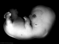
Head scalp vascular plexus visible limb upper limbs begin to rotate ventrally neural - amygdaloid body has at least four individual nuclei[1] oculomotor nerve shows a dorsolateral and a ventromedial portion rhombic lip (rhombencephalon) formation of the cerebellum (intermediate layer) and of the cochlear nuclei cerebellum cell layer (future Purkinje cells) develops choroid plexuses of the fourth and lateral ventricles | |
| Gastrointestinal Tract anal (cloacal) membrane perforates | ||
| Stage 21 | 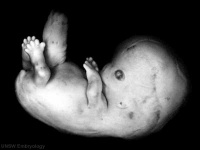
neural - cortical plate appears in the area of future insula[2] limb upper and lower limbs rotate Intraembryonic Coelom pericardioperitoneal canals close | |
| Stage 22 | 
neural - neocortical fibres project to epithalamus, to dorsal thalamus, and to mesencephalon[2] limb fingers and toes lengthen Sense - Smell Stage 22 to early fetal period - migratory streams of neurons from the subventricular zone of the olfactory bulb towards the future claustrum[3] | |
| Genital 8 Weeks Testis - mesenchyme, interstitial cells (of Leydig) secrete testosterone, androstenedione
Genital 8 to 12 Weeks - hCG stimulates testosterone production Tongue Week 8 - nerves penetrate epitheilai basal lamina and synapse with undifferentiated, elongated, epithelial cells (taste bud progenitor cell) | ||
| Stage 23 | 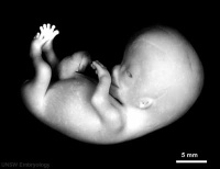 Stage 23 defines the end of the embryonic (organogenesis) period Stage 23 defines the end of the embryonic (organogenesis) period
mesoderm heart prominence, ossification continues Head nose, eye, external acoustic meatus, eyelids, external ears, rounded head Body - straightening of trunk, heart, liver, umbilical cord, intestines herniated at umbilicus limb upper limbs longer and bent at elbow, hands and feet turned inward, foot with separated digits, wrist, hand with separated digits Extraembryonic Coelom chorionic cavity is now lost by fusion with the expanding amniotic cavity neural - rhombencephalon, pyramidal decussation present, nuclei and tracts similar to those present in the newborn cerebellum present as only a plate connected to midbrain and hindbrain through fibre bundles[4] axial skeleton vertebral column 33 or 34 cartilaginous vertebrae (20-33 mm in total length), vertebral pedicles, articular and transverse processes identifiable (no spinous processes)[5] | |
| Week 8 | stomach - Week 8 - Gastrin containing cells in stomach antrum. Somatostatin cells in both the antrum and the fundus. | |
| Note - the day timing of stages is only approximate, system names link to first page of that specific system, and events are based upon the literature cited below. | ||
References
| ||
Week 8 - Stage 22
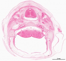
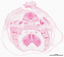
These sections provide a detailed histological overview of the internal structures present within the embryo by the end of the embryonic period.
|
|
|
| ||||||||||||
|
|
| |||||||||||||
|
|
|

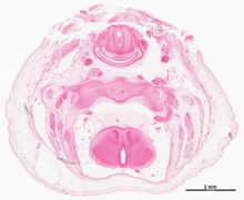

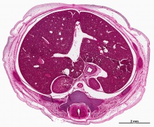
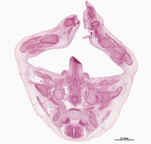
| Selected Embryo Histology - Week 8 (Stage 22) |
|---|
|
| Links: Carnegie stage 22 | Week 8 |


| All Stage 22 Sections | ||||||||||||||||||||||||||||||||||||||||||||||||||||||||||||||||||||||||||||||||||||||||||||||||||||||||||||||||||||||||||||||||||||||||||||||||||||||||||||||||||||||||||||||||||||||||||||||||||||
|---|---|---|---|---|---|---|---|---|---|---|---|---|---|---|---|---|---|---|---|---|---|---|---|---|---|---|---|---|---|---|---|---|---|---|---|---|---|---|---|---|---|---|---|---|---|---|---|---|---|---|---|---|---|---|---|---|---|---|---|---|---|---|---|---|---|---|---|---|---|---|---|---|---|---|---|---|---|---|---|---|---|---|---|---|---|---|---|---|---|---|---|---|---|---|---|---|---|---|---|---|---|---|---|---|---|---|---|---|---|---|---|---|---|---|---|---|---|---|---|---|---|---|---|---|---|---|---|---|---|---|---|---|---|---|---|---|---|---|---|---|---|---|---|---|---|---|---|---|---|---|---|---|---|---|---|---|---|---|---|---|---|---|---|---|---|---|---|---|---|---|---|---|---|---|---|---|---|---|---|---|---|---|---|---|---|---|---|---|---|---|---|---|---|---|---|---|
These are low resolution histology images, the section images start at the head (cranial) end of the embryo and run to the caudal (tail) end. There are available both labelled and unlabelled versions of each section.
|


Week 8 - Stage 23
|
|
|
| ||||||||||||||||||||||
|
|
|
| ||||||||||||||||||||||
| |||||||||||||||||||||||||