|
|
| (147 intermediate revisions by 2 users not shown) |
| Line 1: |
Line 1: |
| | {{Header}}[[File:Paramesonephric ducts.jpg|right]] |
| == Introduction == | | == Introduction == |
| This page introduces the uterus as part of the internal female reproductive tract development. Two paramesonephric ducts form from coelomic epithelium extending beside the mesonephric ducts. In the absence of Mullerian Inhibitory Factor these ducts proliferate and grow extending from the vaginal plate on the wall of the urogenital sinus to lie beside the developing ovary. The paired ducts begin to fuse from the vaginal plate end, forming the primordial body of the uterus and the unfused lateral arms form the uterine tubes. | | [[File:Fetal_uterus_growth.jpg|thumb|Human fetal uterus growth]] |
| | This page introduces the uterus as part of the internal female reproductive tract development. Two paramesonephric ducts form from coelomic epithelium extending beside the mesonephric ducts. In the absence of Mullerian Inhibitory Factor these ducts proliferate and grow extending from the vaginal plate on the wall of the urogenital sinus to lie beside the developing ovary. The paired ducts begin to fuse from the vaginal plate end, forming the primordial body of the uterus and the unfused lateral arms form the {{uterine tube}}s. Recent research points to the paramesonephric ducts also being the entire embryonic origin of the {{vagina}}. For the pregnant uterus see {{Implantation}} and {{Maternal decidua}}. |
|
| |
|
| --[[User:S8600021|Mark Hill]] 17:46, 12 April 2010 (EST)Page transferred from original site, still under construction.
| |
|
| |
|
| Johannes Peter Muller (1801 - 1858) in 1830 was the first to described the duct named after him, the "Mullerian duct" also called the paramesonephric duct.
| | {{Uterus Vignette}} |
|
| |
|
| | {{Genital Links}} |
| | {{Menstrual Links}} |
| | [[:Category:Uterus|Category:Uterus]] |
| | ==Some Recent Findings== |
| | {| |
| | |-bgcolor="F5FAFF" |
| | | |
|
| |
|
| '''Links:''' [[2009_Lecture_16|Lecture - Genital Development]] | [http://embryology.med.unsw.edu.au/Notes/genitalXXuterus.htm original Genital System - Female Uterus page] | | * '''Review - Müllerian duct anomalies coincident with endometriosis'''{{#pmid:32179978|PMID32179978}} "The association between obstructed müllerian duct anomalies and endometriosis has been well established and the pathogenesis is attributed to the theory of retrograde menstruation. However, this relationship with endometriosis is less clear in women with unobstructed müllerian duct anomalies and in those with rudimentary uterine structures that lack functioning endometrial tissue. This article reviews the embryology, genetics, pathophysiology, and American Society for Reproductive Medicine (ASRM) classification for müllerian duct anomalies together with the genetics and pathophysiology of endometriosis to provide a framework for understanding the complex relationship between these two entities. Available published data examining the coexistence of endometriosis in relationship to müllerian duct anomalies, including studies that stratify this relationship according to specific classes of anomalies, are reviewed and organized. Awareness of the increased prevalence of endometriosis among patients with uterine anomalies, particularly those with outflow obstruction, may facilitate early diagnosis of endometriosis and subsequent intervention, with the potential to reverse disease symptoms and arrest disease progression." |
|
| |
|
| == Some Recent Findings ==
| | * '''The histone methyltransferase [https://www.omim.org/entry/601573 EZH2] is required for normal uterine development and function in mice'''{{#pmid:31201420|PMID31201420}} "Enhancer of zeste homolog 2 ([https://www.omim.org/entry/601573 EZH2]) is a rate-limiting catalytic subunit of a histone methyltransferase, polycomb repressive complex, which silences gene activity through the repressive histone mark H3K27me3. EZH2 is critical for epigenetic effects of early estrogen treatment, and may be involved in uterine development and pathologies. We investigated EZH2 expression, regulation and its role in uterine development/function. Uterine epithelial EZH2 expression was associated with proliferation and was high neonatally then declined by weaning. ...In summary, uterine EZH2 expression is developmentally and hormonally regulated, and its loss causes aberrant uterine epithelial proliferation, uterine hypertrophy and cystic endometrial hyperplasia, indicating a critical role in uterine development and function." |
| [http://www.ncbi.nlm.nih.gov:80/entrez/query.fcgi?cmd=Retrieve&db=pubmed&dopt=Abstract&list_uids=17532316 Deutscher E, Hung-Chang Yao H.] Essential roles of mesenchyme-derived beta-catenin in mouse Mullerian duct morphogenesis. Dev Biol. 2007 May 3; | |
|
| |
|
| [http://www.ncbi.nlm.nih.gov:80/entrez/query.fcgi?cmd=Retrieve&db=pubmed&dopt=Abstract&list_uids=17070514 Guioli S, Sekido R, Lovell-Badge R.] The origin of the Mullerian duct in chick and mouse. Dev Biol. 2007 Feb 15;302(2):389-98.
| | * '''Livebirth after uterus transplantation from a deceased donor in a recipient with uterine infertility'''{{#pmid:3052785|PMID3052785}} "Uterus transplantation from live donors became a reality to treat infertility following a successful Swedish 2014 series, inspiring uterus transplantation centres and programmes worldwide. However, no case of livebirth via deceased donor uterus has, to our knowledge, been successfully achieved, raising doubts about its feasibility and viability, including whether the womb remains viable after prolonged ischaemia. ...We describe, to our knowledge, the first case worldwide of livebirth following uterine transplantation from a deceased donor in a patient with congenital uterine absence (Mayer-Rokitansky-Küster-Hauser [MRKH] syndrome). The results establish proof-of-concept for treating uterine infertility by transplantation from a deceased donor, opening a path to healthy pregnancy for all women with uterine factor infertility, without need of living donors or live donor surgery." |
|
| |
|
| "In vertebrates the female reproductive tracts derive from a pair of tubular structures called Mullerian ducts, which are composed of three elements: a canalised epithelial tube, mesenchymal cells surrounding the tube and, most externally, coelomic epithelial cells. ... We show that all Mullerian duct components derive from the coelomic epithelium in both species. Our data support a model of a Mullerian epithelial tube derived from an epithelial anlage at the mesonephros anterior end, which then segregates from the epithelium and extends caudal of its own accord, via a process involving rapid cell proliferation. This tube is surrounded by mesenchymal cells derived from local delamination of coelomic epithelium." | | * '''Review - The cell biology and molecular genetics of Müllerian duct development'''{{#pmid:29350886|PMID29350886}} "The Müllerian ducts are part of the embryonic urogenital system. They give rise to mature structures that serve a critical function in the transport and development of the oocyte and/or embryo. In most vertebrates, both sexes initially develop Müllerian ducts during embryogenesis, but they regress in males under the influence of testis-derived Anti-Müllerian Hormone (AMH)." |
|
| |
|
| | * '''Outcome of assisted reproduction in women with congenital uterine anomalies: a prospective observational study'''{{#pmid:29055072|PMID29055072}} "Consecutive women referred for subfertility between May 2009 and November 2015 who underwent assisted reproduction were included in the study. As part of the initial assessment, each woman underwent three-dimensional transvaginal sonography. Uterine morphology was classified using the modified American Fertility Society (AFS) classification of congenital uterine anomalies proposed by Salim et al. ...Congenital uterine anomalies as a whole, when defined using the modified AFS classification, do not affect clinical pregnancy or live-birth rates in women following assisted reproduction, but do increase the incidence of preterm birth. The presence of uterine abnormalities more severe than arcuate uterus significantly worsens all pregnancy outcomes." |
| | |
| | |} |
| | {| class="wikitable mw-collapsible mw-collapsed" |
| | ! More recent papers |
| | |- |
| | | [[File:Mark_Hill.jpg|90px|left]] {{Most_Recent_Refs}} |
| | |
| | Search term: [http://www.ncbi.nlm.nih.gov/pubmed/?term=Uterus+Development ''Uterus Development''] | [http://www.ncbi.nlm.nih.gov/pubmed/?term=Uterus+Embryology ''Uterus Embryology''] | [http://www.ncbi.nlm.nih.gov/pubmed/?term=Müllerian+Duct ''Müllerian Duct''] | [http://www.ncbi.nlm.nih.gov/pubmed/?term=Uterine+Tube+Development ''Uterine Tube Development''] | [http://www.ncbi.nlm.nih.gov/pubmed/?term=Cervix+Development ''Cervix Development''] | [http://www.ncbi.nlm.nih.gov/pubmed/?term=Broad+Ligament+Development ''Broad Ligament Development''] |
| | |} |
| | {| class="wikitable mw-collapsible mw-collapsed" |
| | ! Older papers |
| | |- |
| | | {{Older papers}} |
| | * '''WNT4 coordinates directional cell migration and extension of the Müllerian duct essential for ontogenesis of the female reproductive tract'''{{#pmid:26721931|PMID26721931}} "The Müllerian duct (MD) is the anlage of the oviduct, uterus and upper part of the vagina, the main parts of the female reproductive tract. Several wingless-type mouse mammary tumor virus (MMTV) integration site family member (Wnt) genes, including Wnt4, Wnt5a and Wnt7a, are involved in the development of MD and its derivatives, with Wnt4 particularly critical, since the MD fails to develop in its absence. We use, here, Wnt4(EGFPCre)-based fate mapping to demonstrate that the MD tip cells and the subsequent MD cells are derived from Wnt4+ lineage cells. Moreover, Wnt4 is required for the initiation of MD-forming cell migration." [[Developmental_Signals_-_Wnt#WNT4|WNT4]] |
| | |
| | * '''LHX1 is required in Müllerian duct epithelium for uterine development'''{{#pmid:24560999|PMID24560999}} "The female reproductive tract organs of mammals, including the oviducts, uterus, cervix and upper vagina, are derived from the Müllerian ducts, a pair of epithelial tubes that form within the mesonephroi. The Müllerian ducts form in a rostral to caudal manner, guided by and dependent on the Wolffian ducts that have already formed. Experimental embryological studies indicate that caudal elongation of the Müllerian duct towards the urogenital sinus occurs in part by proliferation at the ductal tip. The molecular mechanisms that regulate the elongation of the Müllerian duct are currently unclear. Lhx1 encodes a LIM-homeodomain transcription factor that is essential for male and female reproductive tract development. Lhx1 is expressed in both the Wolffian and Müllerian ducts. Wolffian duct-specific knockout of Lhx1 results in degeneration of the Wolffian duct and consequently the non-cell-autonomous loss of the Müllerian duct. To determine the role of Lhx1 specifically in the Müllerian duct epithelium, we performed a Müllerian duct-specific knockout study using Wnt7a-Cre mice. Loss of Lhx1 in the Müllerian duct epithelium led to a block in Müllerian duct elongation and uterine hypoplasia characterized by loss of the entire endometrium (luminal and glandular epithelium and stroma) and inner circular but not the outer longitudinal muscle layer. Time-lapse imaging and molecular analyses indicate that Lhx1 acts cell autonomously to maintain ductal progenitor cells for Müllerian duct elongation. These studies identify LHX1 as the first transcription factor that is essential in the Müllerian duct epithelial progenitor cells for female reproductive tract development." [https://www.genenames.org/cgi-bin/gene_symbol_report?hgnc_id=HGNC:6593 HGNC] |
| | |
| | * '''The origin of the Mullerian duct in chick and mouse'''{{#pmid:17070514|PMID17070514}} "In vertebrates the female reproductive tracts derive from a pair of tubular structures called Mullerian ducts, which are composed of three elements: a canalised epithelial tube, mesenchymal cells surrounding the tube and, most externally, coelomic epithelial cells. ... We show that all Mullerian duct components derive from the coelomic epithelium in both species. Our data support a model of a Mullerian epithelial tube derived from an epithelial anlage at the mesonephros anterior end, which then segregates from the epithelium and extends caudal of its own accord, via a process involving rapid cell proliferation. This tube is surrounded by mesenchymal cells derived from local delamination of coelomic epithelium." |
| | |
| | * '''Essential roles of mesenchyme-derived beta-catenin in mouse Mullerian duct morphogenesis'''{{#pmid:17532316|PMID17532316}} |
| | |
| | |} |
| == Paramesonephric Duct == | | == Paramesonephric Duct == |
| The Mullerian duct (= paramesonephric duct, preferred terminology) paired ducts that form the epithelial lining of female reproductive organs: utererine tube, uterus, upper vaginal canal. The term "paramesonephric" duct means beside the mesonephric (Wolffian) duct, which is its anatomical location in early development. Mullerian refers to Johannes Peter M√ºller (1801-1858) a German scientist who specialised in comparative anatomy. These ducts initially form and then degenerate in the male. | | The Müllerian duct (= paramesonephric duct, preferred terminology) paired ducts that form the epithelial lining of female reproductive organs: utererine tube, uterus, upper vaginal canal. The term "paramesonephric" duct means beside the mesonephric (Wolffian) duct, which is its anatomical location in early development. Mullerian refers to Johannes Peter Müller (1801-1858) a German scientist who specialised in comparative anatomy. These ducts initially form and then degenerate in the male. |
|
| |
|
| A recent study using both chicken and mouse embryos has shown that these initially paired tubular structures derive from the coelomic epithelium. [http://www.ncbi.nlm.nih.gov:80/entrez/query.fcgi?cmd=Retrieve&db=PubMed&list_uids=17070514&dopt=Abstract Guioli S, Sekido R, Lovell-Badge R.] The origin of the Mullerian duct in chick and mouse. Dev Biol. 2006 Oct 3 | | A recent study using both chicken and mouse embryos has shown that these initially paired tubular structures derive from the coelomic epithelium.{{#pmid:17070514|PMID17070514}} |
|
| |
|
| "Mullerian epithelial tube derived from an epithelial anlage at the mesonephros anterior end, which then segregates from the epithelium and extends caudal of its own accord, via a process involving rapid cell proliferation. This tube is surrounded by mesenchymal cells derived from local delamination of coelomic epithelium." | | :"Müllerian epithelial tube derived from an epithelial anlage at the mesonephros anterior end, which then segregates from the epithelium and extends caudal of its own accord, via a process involving rapid cell proliferation. This tube is surrounded by mesenchymal cells derived from local delamination of coelomic epithelium." |
|
| |
|
| Mullerian ducts have three elements: | | Mullerian ducts have three elements: |
| Line 28: |
Line 58: |
| # mesenchymal cells surrounding the tube | | # mesenchymal cells surrounding the tube |
| # coelomic epithelial cells | | # coelomic epithelial cells |
| | |
| | |
| | ==Duct Molecular Development== |
| | |
| | The paired paramesonephic ducts (Müllerian ducts) go through a series of developmental changes recently identified as regulated by a number of molecular factors. |
| | |
| | ===Initiation=== |
| | Coelomic epithelium Lim1 expressing cells are specified to a duct fate.{{#pmid:14695376|PMID14695376}} |
| | |
| | * Lim - proteins named for 'LIN11, ISL1, and MEC3,' are defined by the possession of a highly conserved double zinc finger motif called the LIM domain. |
| | ** LIM domain-binding factors - interact with the LIM domains of nuclear proteins are capable of binding to a variety of transcription factors. |
| | |
| | ===Invagination=== |
| | |
| | * Wnt4 - induces duct invagination to reach the mesonephric (Wolffian) |
| | |
| | ===Elongation=== |
| | |
| | * WNT9b - from mesonephric duct to guide paramesonephric duct elongation. Cysteine-rich secreted glycoprotein. |
| | * Pax2 - also acts in elongation and duct maintenance. Member of the paired box protein family. |
| | |
| | Cells at the leading tip proliferate and form the duct elongating to reach the cloaca (urogenital sinus). |
| | |
| | |
| | |
| | |
| | :'''Links:''' [http://www.ncbi.nlm.nih.gov/omim OMIM - WNT9b] | [http://www.ncbi.nlm.nih.gov/omim/167409 OMIM - Pax2] | [http://www.ncbi.nlm.nih.gov/omim/167410 OMIM - paired box gene] |
| | |
|
| |
|
| == Uterine Development Movie == | | == Uterine Development Movie == |
| Line 39: |
Line 97: |
| The '''urogenital sinus''' (yellow), in contact with the paramesonephric duct, thickens to form the '''sinusal tubercle''' which extends as a solid '''vaginal plate''', then becomes hollow as the '''sinovaginal bulb''', finally forming the '''vagina'''. | | The '''urogenital sinus''' (yellow), in contact with the paramesonephric duct, thickens to form the '''sinusal tubercle''' which extends as a solid '''vaginal plate''', then becomes hollow as the '''sinovaginal bulb''', finally forming the '''vagina'''. |
|
| |
|
| [../Movies/genital/uterus2.mov Female Internal Genitalia] (288 Kb)
| |
|
| |
|
| |} | | |} |
| | |
| == Development Overview == | | == Development Overview == |
| {| class="prettytable" | | {| class="prettytable" |
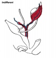
| | [[File:Urogenital_indifferent.jpg]] | | | [[File:Urogenital_indifferent.jpg|300px]] |
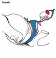
| | [[File:Urogenital_female.jpg]] | | | [[File:Urogenital_female.jpg|300px]] |
|
| |
|
| |- | | |- |
| Line 58: |
Line 116: |
|
| |
|
| |} | | |} |
| The data below gives an overview of the timecourse of embryonic human uterine development. | | The data below gives an overview of the timecourse of embryonic human uterine development.{{#pmid:12740945|PMID12740945}} |
|
| |
|
| stage 18 - Mullerian duct to the coelomic cavity was formed as the result of an invagination of the coelomic epithelium | | :'''Carnegie stage 18''' - Mullerian duct to the coelomic cavity was formed as the result of an invagination of the coelomic epithelium - [[Carnegie_stage_18|stage 18]] |
|
| |
|
| stages 19-23 - duct grows independently from the invagination | | :'''Carnegie stages 19 - 23''' - duct grows independently from the invagination - [[Carnegie_stage_19|stage 19]] |
|
| |
|
| week 20 - uterine horn fimbrial development begins and continues after birth
| | :'''Week 20''' - uterine horn fimbrial development begins and continues after birth - [[Second_Trimester|second trimester]] |
|
| |
|
| Data: [http://www.ncbi.nlm.nih.gov:80/entrez/query.fcgi?cmd=Retrieve&db=PubMed&list_uids=12740945&dopt=Abstract Hashimoto R.] Development of the human Mullerian duct in the sexually undifferentiated stage. Anat Rec A Discov Mol Cell Evol Biol. 2003 Jun;272(2):514-9.
| |
|
| |
|
| |
|
| |
|
| == Fetal Uterus == | | == Fetal Uterus == |
|
| |
|
| {| class="prettytable" | | {| class="prettytable" |
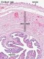
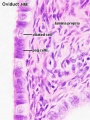
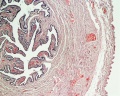
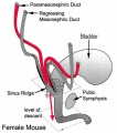
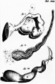
| | [[Image:paramesonephric_ducts.jpg]] | | | [[File:Paramesonephric_ducts.jpg]] |
| | [[Image:paramesonephric2.jpg]] | | | [[File:Paramesonephric_duct.jpg]] |
|
| |
|
| |- | | |- |
| Line 81: |
Line 136: |
|
| |
|
| |} | | |} |
| | |
| == Fetal Uterus Growth == | | == Fetal Uterus Growth == |
|
| |
|
| [[File:Fetal_uterus_growth.jpg|left]] Graph shows the growth during the fetal period of the uterus between week 19 and 38.<ref>Development of the fetal uterus between 19 and 38 weeks of gestation: in-utero ultrasonographic measurements. Soriano D, Lipitz S, Seidman DS, Maymon R, Mashiach S, Achiron R. Hum Reprod. 1999 Jan;14(1):215-8. [http://www.ncbi.nlm.nih.gov/pubmed/10374123 PMID: 10374123]</ref> During this time the uterine circumferunce increases from about 20 mm to just under 60mm and the width increases from less than 10mm to just over 20 mm. | | [[File:Fetal_uterus_growth.jpg|left]] Graph shows the growth during the fetal period of the uterus between week 19 and 38.{{#pmid:10374123|PMID10374123}} During this time the uterine circumferunce increases from about 20 mm to just under 60mm and the width increases from less than 10mm to just over 20 mm. |
|
| |
|
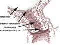
| Uterine horn fimbrial development begins after week 20 and continues after birth. | | Uterine horn fimbrial development begins after week 20 and continues after birth. |
| Line 94: |
Line 150: |
| [[Image:newborn_uterus.jpg]] | | [[Image:newborn_uterus.jpg]] |
|
| |
|
| | {{Collapsible Table Postfetal Uterus Growth}} |
| == Uterine Tubes == | | == Uterine Tubes == |
| | [[File:Human_right_ovary_and_tube_1.jpg|thumb|Adult Human right uterine tube and ovary]] |
| | [[File:Cat_embryo_ovary.jpg|thumb|Developing Uterus (cat) showing relationship to ovary and degenerating mesonephros.]] |
| The unfused portion of the paramesonephric ducts will form the uterine tubes. Note that there are several synonyms used for the paired '''uterine tubes''' or '''Fallopian tubes''' or '''oviducts''' or '''uterine horns'''. | | The unfused portion of the paramesonephric ducts will form the uterine tubes. Note that there are several synonyms used for the paired '''uterine tubes''' or '''Fallopian tubes''' or '''oviducts''' or '''uterine horns'''. |
|
| |
|
| | In the adult, the uterine tube has been described in 4 anatomical regions. |
|
| |
|
| {| class="prettytable" | | {{Uterine Tube Regions table}} |
| | [[Image:uterine_tubes_sm.jpg]]
| |
| | In the adult, the uterine tube has been described in 4 anatomical regions.
| |
|
| |
|
| # '''Infundibulum''' - funnel-shaped open end of the uterine tube with fimbriae (finger-like extensions), which are closely associated with the ovary. Opens into the peritoneal cavity (abdominal ostium, ostium abdominale)
| |
| # '''Ampulla''' - uterine tube with highly folded structure with plicae (mucosal folds) and secondary folds dividing the lumen, usual site for fertilization.
| |
| # '''Isthmus''' - narrow portion of the uterine tube with fewer mucosal folds and a thick muscularis layer.
| |
| # '''Intramural''' - uterine tube which passes through the muscular wall of the uterus. (an alternative interpretation is that it is an extension of the body of the uterus)
| |
|
| |
|
| | ===Mucosa=== |
| | * formed by a ciliated and secretory epithelium resting on a very cellular lamina propria. |
| | * The number of ciliated cells and non-ciliated secretory cells varies along the oviduct. |
| | * Secretory activity varies during the menstrual cycle, and resting secretory cells are also referred to as peg-cells. |
| | * Some of the secreted substances are thought to nourish the oocyte and the very early embryo. |
|
| |
|
| | ===Muscularis=== |
| | * inner circular muscle layer and an outer longitudinal layer. |
| | * An inner longitudinal layer is present in the isthmus and the intramural part of the oviduct. |
| | * Peristaltic muscle action seems to be more important for the transport of sperm and oocyte than the action of the cilia. |
|
| |
|
| |-
| | <gallery> |
| | <center>Peritoneal view of uterus body and tubes </center>
| | File:Uterine tube histology 02.jpg|Uterine tube (monkey) histology overview |
| |
| | File:Uterine tube histology 03.jpg|Uterine tube (monkey) epithelium and underlying histology |
| | | File:Uterine_tube_histology.jpg |
| |-
| | </gallery> |
| | [[Image:cat_embryo_ovary_sm.jpg]]
| |
| | Developing uterine tube (cat) showing relationship to ovary and degenerating mesonephros. | |
| | |
| |-
| |
| |
| |
| |
| |
| | |
| |}
| |
| | |
| {| class="prettytable"
| |
| | [[Image:odu04he.jpg]]
| |
| | [[Image:odu40he.jpg]] | |
| | |
| |-
| |
| | [[Image:uem021he.jpg]]
| |
| | [[Image:uem022he.jpg]]
| |
|
| |
|
| |-
| |
| |
| |
| | (Images: UWA Blue Histology - [http://www.lab.anhb.uwa.edu.au/mb140/CorePages/FemaleRepro/femalerepro.htm#LabOvid Female Reproductive Tract])
| |
|
| |
| |}
| |
| == Uterine Blood Supply == | | == Uterine Blood Supply == |
| | [[File:Gray1170.jpg|600px]] |
|
| |
|
| {| class="prettytable"
| |
| | [[Image:uterine_blood_supply.jpg]]
| |
| |
| |
|
| |
| |}
| |
| == Abnormalities ==
| |
|
| |
| {| class="prettytable"
| |
| | [[Image:uterine_abnormalities.jpg]]
| |
| | A range of uterine and vaginal anatomical anomalies based upon the abnormal development and fusion of the paramesonephric ducts and vaginal plate development.
| |
|
| |
|
| |
|
| |
| |-
| |
| | [[File:Unicornate_uterus.jpg]]
| |
| | '''Unicornate Uterus''' - failure of the paramesonephric ducts to fuse. A single paramesomnephric duct has fused with the vaginal plate and now opens into the vagina, while the other forms a diverticulum.
| |
|
| |
| |}
| |
| '''Uterine Duplication''' (uterus didelphys, double uterus, uterus didelphis) A rare uterine developmental abnormality where the paramesonephric ducts (Mullerian ducts) completely fail to fuse generating two separate uterus parts each connected to the cervix and having an ovary each.
| |
|
| |
| '''Septate Uterus'''
| |
|
| |
|
| '''Cervical:''' cervical agenesis, cervical duplication
| | ==Uterine Glands== |
| | [[File:Uterine_gland_secretory_phase.jpg|thumb|alt=Uterine Gland Secretory Phase histology|Uterine Gland Secretory Phase]] |
| | Uterine adenogenesis is the term used to describe the formation of uterine glands from the epithelial lining of the uterus that begins prenatal in humans. In other species, the overt development occurs postnatally and has been described through a 3 step the sequence: |
| | # differentiation and budding of the glandular epithelium. |
| | # invagination and tubular coiling of the epithelium. |
| | # branching of the glandular elements and their expansion throughout the endometrial stroma toward the myometrium. |
|
| |
|
| '''Vaginal:''' Mayer-Rokitansky syndrome (MRK anomaly, Rokitansky-Küster-Hauser syndrome, RKH syndrome, RKH) congenital absence of the vagina, dyspareunia, vaginal agenesis. | | Epithelial-mesenchymal interaction occurs through Wnt signalling during this process: |
| | * '''Wnt7a''' - expressed in the luminal epithelium |
| | * '''Wnt5a''' - expressed in the mesenchyme |
|
| |
|
| '''Environmental Abnormalities '''
| | In [[Mouse_Timeline_Detailed|mice]], this development sequence occurs between postnatal day (PND) 5 to 7 and involves Wnt up-regulation of Lymphoid Enhancing Factor 1 (Lef1).{{#pmid:22792274|PMID22792274}} |
|
| |
|
| '''DES''' '''D'''i'''e'''thyl'''s'''tilbestrol or diethylstilbetrol, is a drug that was prescribed to women from 1938-1971 to prevent miscarriage in high-risk pregnancies. The drug acted as a potent estrogen (mimics natural hormone) and therefore could also act as a potential endocrine disruptor. This led to a number of developing fetal reproductive tract and other abnormalities. In the female fetus, it increased risk of abnormal reproductive tract and also carcinogenic (cancer forming). In the male fetus, it increased the occurance of abnormal genitalia. The drug was banned by FDA (USA) in 1979 as a teratogen, it had previously also been used as livestock growth promoter and could have potentially entered the human food chain. (More? [endocrine2.htm Endocrine Abnormalities] | [../Defect/drugs.htm Abnormal Development - Drugs])
| | Postnatally both prolactin and estradiol-17 beta (and their receptors) regulate gland development. There are some gland species gestational differences, in both sheep and pigs the glands provide additional histotrophic support by undergoing extensive hyperplasia and hypertrophy.{{#pmid:11673245|PMID11673245}} |
|
| |
|
| '''Links:''' [endocrine2.htm Endocrine Abnormalities] | [../Defect/drugs.htm Abnormal Development - Drugs] | [http://www.childrenshospital.org/clinicalservices/Site2239/mainpageS2239P5sublevel13.html Childrens Hospital Boston - Congenital Anomalies of the Uterus] | [http://www.gfmer.ch/selected_images_v2/detail_list.php?cat1=4&cat3=502&stype=d Medical Education Image Link - Cervical agenesis] | [http://www.ncbi.nlm.nih.gov/entrez/dispomim.cgi?id=277000 OMIM - Rokitansky-Küster-Hauser syndrome] | | | :'''Links:''' [[Developmental_Signals_-_Wnt|Wnt]] | [[Developmental_Mechanism_-_Epithelial_Mesenchymal_Interaction|Epithelial Mesenchymal Interaction]] |
|
| |
|
| == Vagina Development == | | ==Postnatal Growth== |
| The embryonic origin of the vagina has been a historically hotly debated issue with several different contributions and origins described.
| |
|
| |
|
| One description shows the vagina arising by downward growth of Wolffian and Mullerian ducts. The sinovaginal bulbs are the caudal ends of the Wolffian ducts. Vaginal development is also under negative control of androgens.
| | {{Table Postfetal Uterus Growth}} |
|
| |
|
| An earlier understanding was that the upper part of the vagina derived from Müllerian ducts and the lower part from the sinovaginal bulbs (formed by fusion form the vaginal plate) all derived from the urogenital sinus. The terms sinovaginal bulbs and vaginal plate were first coined by Koff in 1933.
| | ==Uterus Histology== |
|
| |
|
| '''References:''' Koff AK. Development of the vagina in the human fetus. Contributions to Embryology No. 140, Carnegie Inst. 1933; 24:61–90.
| | See also [[Menstrual Cycle - Histology]] |
|
| |
|
| == Broad Ligament ==
| |
|
| |
| {| class="prettytable"
| |
| | The broad ligament is found associated with the internal human female genital tract. It forms a mesentery consisting of a double fold of the peritoneum that connects the uterus to the peritoneal floor and walls.
| |
|
| |
| Anatomically it has three parts:
| |
|
| |
| # mesometrium - surrounding the uterus
| |
| # mesosalpinx - surrounding the uterine tube
| |
| # mesovarium - surrounding the ovary
| |
|
| |
| Abnormalities include peritoneal endometriosis.
| |
| | [[Image:image1161.gif]]
| |
|
| |
| |}
| |
| == Molecular ==
| |
| '''Wnt genes''' - Wnt4, Wnt5a, and Wnt7a implicated in the formation and morphogenesis of the Müllerian duct.
| |
|
| |
| '''Wnt7a''' - mediates the patterning of the oviduct and differentiation of the uterus.
| |
|
| |
| '''beta-catenin''' - manufactured in the mesenchyme is a downstream effector of Wnt7a.
| |
|
| |
| '''Bmp2''' - decidualization regulator of gene expression and function (shown in mouse uterus).
| |
|
| |
| Lim1, Lhx9, Emx, Pax-2, Hox-A9, Hox-A10, Hox-A11, Hox-A13, WT1, SF-1, GATA-4. TGF-beta
| |
|
| |
| == References ==
| |
| <references/>
| |
|
| |
|
| |
| ===Reviews===
| |
|
| |
| [http://www.ncbi.nlm.nih.gov:80/entrez/query.fcgi?cmd=Retrieve&db=PubMed&list_uids=16208476&dopt=Abstract Farage M, Maibach H.] Lifetime changes in the vulva and vagina. Arch Gynecol Obstet. 2006 Jan;273(4):195-202.
| |
|
| |
| [http://www.ncbi.nlm.nih.gov:80/entrez/query.fcgi?cmd=Retrieve&db=PubMed&list_uids=15467266&dopt=Abstract Kavlock R, Cummings A] [http://www.ncbi.nlm.nih.gov/entrez/query.fcgi?db=pubmed&cmd=Display&dopt=pubmed_pubmed&from_uid=15467266&tool=ExternalSearch [See Related Articles]] Function of sexual glands and mechanism of sex differentiation. J Toxicol Sci. 2004 Aug;29(3):167-78. Review.
| |
|
| |
| ===Articles===
| |
|
| |
| * Essential roles of mesenchyme-derived beta-catenin in mouse Mullerian duct morphogenesis. Deutscher E, Hung-Chang Yao H. Dev Biol. 2007 May 3; [http://www.ncbi.nlm.nih.gov:80/entrez/query.fcgi?cmd=Retrieve&db=pubmed&dopt=Abstract&list_uids=17532316 PMID: 17532316]
| |
|
| |
| * [http://www.ncbi.nlm.nih.gov:80/entrez/query.fcgi?cmd=Retrieve&db=pubmed&dopt=Abstract&list_uids=17070514 Guioli S, Sekido R, Lovell-Badge R.] The origin of the Mullerian duct in chick and mouse. Dev Biol. 2007 Feb 15;302(2):389-98.
| |
|
| |
| * [http://www.ncbi.nlm.nih.gov:80/entrez/query.fcgi?cmd=Retrieve&db=PubMed&list_uids=12740945&dopt=Abstract Hashimoto R.] Development of the human Mullerian duct in the sexually undifferentiated stage. Anat Rec A Discov Mol Cell Evol Biol. 2003 Jun;272(2):514-9.
| |
|
| |
| ===Search PubMed===
| |
|
| |
| Search May 2007 "embryonic uterine development" '''3,025''' reference articles of which '''491''' were reviews.
| |
|
| |
| '''Search PubMed:''' term = [http://www.ncbi.nlm.nih.gov/entrez/query.fcgi?db=pubmed&cmd=search&term=embryonic%20uterine%20development embryonic uterine development] | [http://www.ncbi.nlm.nih.gov/entrez/query.fcgi?db=pubmed&cmd=search&term=Uterine%20Development Uterine Development] | [http://www.ncbi.nlm.nih.gov/entrez/query.fcgi?db=pubmed&cmd=search&term=Paramesonephric+Duct Paramesonephric Duct] | [http://www.ncbi.nlm.nih.gov/entrez/query.fcgi?db=pubmed&cmd=search&term=Mullerian+Duct Mullerian Duct] | [http://www.ncbi.nlm.nih.gov/entrez/query.fcgi?db=pubmed&cmd=search&term=Endocrine+Disruptors Endocrine Disruptors]
| |
|
| |
| ==Additional Images==
| |
| <gallery> | | <gallery> |
| File:Urogenital_indifferent.jpg|Urogenital indifferent | | File:Uterine tube histology 02.jpg|Uterine tube histology overview showing epithelium and underlying muscular layers |
| File:Urogenital_female.jpg|Urogenital female | | File:Uterine tube histology 03.jpg|Uterine tube epithelium histology showing secretory and ciliated cells |
| File:Paramesonephric duct.jpg|Mouse - paramesonephric duct | | File:Uterus_proliferative_phase.jpg|Uterine body endometrium and myometrium during the proliferative phase of the menstrual cycle overview |
| File:Unicornate_uterus.jpg|Unicornate uterus | | File:Uterine_gland_proliferative_phase.jpg|Uterine body endometrium during the proliferative phase of the menstrual cycle |
| | File:Uterus_secretory_phase.jpg|Uterine body endometrium during the secretory phase of the menstrual cycle overview |
| | File:Uterine_gland_secretory_phase.jpg|Uterine body endometrium during the secretory phase of the menstrual cycle |
| </gallery> | | </gallery> |
|
| |
|
| == Terms == | | == Abnormalities == |
| '''abdomen''' - trunk between diaphragm and pelvis.
| | [[File:Female genital and ureter abnormality 02.jpg|thumb|Uterine didelphys, obstructed hemivagina, and ectopic ureter on MR imaging in a 17-year-old girl.{{#pmid:19924410|PMID19924410}}]] |
|
| |
|
| '''abdominal circumference''' - An ultrasound measurement of Abdominal Circumference (AC) is used to determine fetal age and normal development (small/large/abnormal) parameters. Measured at the outer edge of the circumference of the body plane in which the portal vein or stomach can be visualized in a tangential section. It is one of the four typical ultrasound assessments of fetal size and age: Biparietal Diameter (BPD), Head Circumference (HC), Abdominal Circumference (AC), and Femur Length] (FL). Abdominal Circumference of less than 31 cm at 36 to 40 weeks gestation is a predictor of intrauterine growth retardation (IUGR).
| |
|
| |
|
| '''AC''' - Acronym for Abdominal Circumference.
| | There are at least two clinical society classifications for female genital tract abnormalities: |
| | # European Society of Human Reproduction and Embryology—European Society for Gynaecological Endoscopy (ESHRE-ESGE){{#pmid:23894234|PMID23894234}} |
| | # American Society for Reproductive Medicine (ASRM) {{#pmid:3371491|PMID3371491}} |
|
| |
|
| '''adenohypophysis''' - (anterior pituitary, pars distalis) The anterior part of the pituitary, which develops in the early embryo from a transient region on the roof of the pharynx called Rathke's pouch.
| | ===ESHRE-ESGE Classification=== |
| | European Society for Gynaecological Endoscopy (ESHRE-ESGE){{#pmid:23894234|PMID23894234}} |
|
| |
|
| '''adnexa''' - (Latin, ''adnexae'' = appendages) Term used to describe any anatomical appendage (accessory structure, extension or outgrowth from the body). In reproductive anatomy used to describe appendages of the [U.htm#uterus uterus] "body"; ovaries, uterine tubes and uterus supporting ligaments. | | Uterine anatomical deviations deriving from the same embryological origin: |
| | * '''U0''' - normal uterus |
| | * '''U1''' - dysmorphic uterus |
| | * '''U2''' - septet uterus |
| | * '''U3''' - bicorporeal uterus |
| | * '''U4''' - hemi-uterus |
| | * '''U5''' - aplastic uterus |
| | * '''U6''' - for still unclassified cases |
|
| |
|
| '''adrenal gland''' - (suprarenal gland) The endocrine organ that anatomically sits on top of the kidneys (renal). It has two different embryonic origins, neurat crest (aderenal medulla) and mesoderm (adrenal cortex).
| | Main classes have been divided into sub-classes expressing anatomical varieties with clinical significance. Cervical and vaginal anomalies are classified independently into sub-classes having clinical significance. |
|
| |
|
| '''adventitia''' - Anatomical term describing the outermost connective tissue covering of any organ, vessel, or other structure not covered by a serosa. The covering is from the surrounding connective tissue and does not form an integral part of such organ or structure.
| |
|
| |
|
| '''amnion''' - An extraembryonic membrane ectoderm and extraembryonic mesoderm in origin and forms the innermost fetal membrane, produces amniotic fluid. This fluid-filled sac initially lies above the trilaminar embryonic disc and with embryoic disc folding this sac is drawn ventrally to enclose (cover) the entire embryo, then fetus. The presence of this membrane led to the description of reptiles, bird, and mammals as amniotes.
| | {{ESHRE/ESGE Uterine Anomalies table}} |
|
| |
|
| '''amniotic fluid''' - The fluid that fills amniotic cavity totally encloses and cushions the embryo. Amniotic fluid enters both the gastrointestinal and respiratory tract following rupture of the buccopharyngeal membrane. The late fetus swallows amniotic fluid.
| | [[File:Uterine anomalies ESHRE-ESGE classification.jpg|600px|alt=Uterine anomalies ESHRE-ESGE classification]] |
|
| |
|
| '''ampulla''' - Term used to describe an anatomical dilation of a tube or canal lumen. Anatomical description of the opening end of the uterine tube lying above the ovary and the enlarged initial segmeny of the semicircular canals of the inner ear vestibular system.
| | ===Uterine Duplication=== |
|
| |
|
| '''anastomosis''' - Term used to describe the connection between two tubes. Applied to describe the connection between peripheral blood vessels without an intervening capillary bed.
| | {| class="prettytable" |
| | | [[Image:uterine_abnormalities.jpg]] |
| | | A range of uterine and vaginal anatomical anomalies based upon the abnormal development and fusion of the paramesonephric ducts and vaginal plate development. |
|
| |
|
| '''androgens''' - The male sex hormones, eg testosterone.
| | |
|
| |
|
| '''anterior''' - Anatomical term used to describe the front or ventral surface. | | |- |
| | | [[File:Unicornate_uterus.jpg]] |
| | | '''Unicornate Uterus''' - failure of the paramesonephric ducts to fuse. A single paramesomnephric duct has fused with the vaginal plate and now opens into the vagina, while the other forms a diverticulum. |
|
| |
|
| '''Anti-Mullerian Hormone''' - (AMH, Mullerian Inhibiting Substance, MIS) A secreted factor (transforming growth factor-beta, TGF-beta superfamily) that regulates gonadal and genital tract development. Inhibits paramesonephric (Mullerian) duct development in males. (More? [http://www.ncbi.nlm.nih.gov/entrez/dispomim.cgi?id=600957 OMIM - AMH])
| | {{Ultrasound Bicornuate Ectopic}} |
| | |} |
| | [[File:Bicornuate uterus01.jpg|600px]] |
|
| |
|
| '''antral follicle''' - (secondary follicle) Term used to describe the developmental stage of ovarian follicle development following preantral (primary) in describing the sequence (primordial, preantral, antral) of follicle development within the ovary. In humans, a number of primordial follicles will begin to develop into primary follicles, some of which will then form antral follicles (secondary), with only a single antral follicle developing into the ovulating follicle (Graafian) each menstrual cycle.
| | Bicornuate uterus containing conceptus chorionic sac with placental cord on one side. |
| | ===Septate Uterus=== |
|
| |
|
| '''antrum''' - (Latin from Greek, ''antron'' = a cave, cavity; a nearly-closed cavity or bulge). Identified anatomically in many structures (ovarian follicle, bone, cardiac, gastric). In the ovary this refers to the follicular fluid-filled space within the follicle.
| | Uterine residual septum classification: |
| | # American Society for Reproductive Medicine (ASRM) criterion with an internal fundal indentation length equal or greater than 1 cm{{#pmid:20052665|PMID20052665}} |
| | # European Society of Human Reproduction and Embryology—European Society for Gynaecological Endoscopy (ESHRE-ESGE) classification of female genital tract congenital anomalies with an internal indentation at the fundal midline greater than 50% myometrial thickness.{{#pmid:23894234|PMID23894234}} |
|
| |
|
| '''atresia''' - (Greek, ''a'' = without + ''tresis'' = perforation) Term used for anatomical closing or absence of a cavity or opening that should exist. Used as an antomical, pathological and clinical term: esophageal atresia, biliary atresia, duodenal atresia, jejunal atresia, choanal atresia, vaginal atresia, urethral atresia, pulmonary atresia, bronchial atresia, tricuspid atresia.
| | [[File:Septate uterus ultrasound 01.jpg|alt=Septate uterus ultrasound|800px]] |
|
| |
|
| '''autosomal''' - The term decribing all the chromosomes that contribute to a cell's genetic material, except for the sex chromosomes X, Y. (More? [../DNA/DNA.htm DNA Notes])
| | Septate Uterus Ultrasound{{#pmid:24838703|PMID24838703}} |
|
| |
|
| '''autosomal inheritance''' - Some hereditary diseases are described as autosomal which means that the disease is due to a DNA error in one of the 22 pairs that are not sex chromosomes. Both boys and girls can then inherit this error. If the error is in a sex chromosome, the inheritance is said to be sex-linked. (More? [../DNA/DNA.htm DNA Notes])
| | ===Uterine Duplication=== |
|
| |
|
| '''birth''' - (parturition) Term describing the pysiological process of offspring (child) being born. (More? [../Child/birth1.htm Normal Development - Birth])
| | (uterus didelphys, double uterus, uterus didelphis) A rare uterine developmental abnormality where the paramesonephric ducts (Mullerian ducts) completely fail to fuse generating two separate uterus parts each connected to the cervix and having an ovary each. |
|
| |
|
| '''bladder exstrophy''' - (Greek, ''ekstriphein'' = "turn inside out") A congenital malformation with bladder open to ventral wall of abdomen (between umbilicus and pubic symphysis) and may have other anomolies associated with failure of closure of abdominal wall and bladder (epispadias, pubic bone anomolies). (More? [urogenital2.htm Urogenital Abnormalities])
| |
|
| |
|
| '''Bulbourethral Gland''' - (= Cowper's Gland) A male genital tract gland which secretes a small amount of a thick clear mucous fluid prior to ejaculation, the alkaline content apparently buffers acidity of the urethra. The equivalent female gland are Bartholin's glands.
| | ===Uterus/Vaginal=== |
| | Mayer-Rokitansky-Kuster-Hauser syndrome (MRKH, MRK anomaly, Rokitansky-Kuster-Hauser syndrome, RKH syndrome, RKH) consists of congenital aplasia of the uterus and the upper part of vagina due to anomalous development of Müllerian ducts, either isolated or associated with other congenital malformations, including renal, skeletal, hearing and heart defects. Has an incidence of approximately 1 in 4500 newborn girls and has been associated with a microdeletion at 17q12.{{#pmid:19889212|PMID19889212}} |
|
| |
|
| '''caudal''' - (Latin, ''caudal'' = tail) Anatomical term referring to structures that are more towards the tail.
| | There has been recently a single report of a MRKH syndrome woman giving a live-birth after uterus transplantation from a deceased donor.{{#pmid:3052785|PMID3052785}} |
|
| |
|
| '''chryptochid testes''' - A male genital abnormality where the testes remain undescended in the abdominopelvic cavity. | | '''Cervical:''' cervical agenesis, cervical duplication |
|
| |
|
| '''ciliated epithelium''' - (Latin, ''cilium'' = eyelid) An epithelium named on the basis of the cells having surface hair-like appearance of a cilium; singular, cilium. In many tissues, cilia are found as epithelial cell apical surface motile specializations. In the uterine tube epithelium, after ovulation used to move the unfertilized egg, then the fertilized zygote, then blastocyst during the first week of development.
| |
|
| |
|
| '''cloacal membrane''' - Forms the external lower membrane limit (caudal end) of the early gastrointestinal tract (GIT). This membrane is formed during gastrulation by ectoderm and endoderm without a middle (intervening) layer of mesoderm. The membrane breaks down to form the initial "anal opening" of the gastrointestinal tract.
| | ===Environmental Abnormalities=== |
|
| |
|
| '''coelom''' - Term used to describe a space. There are extraembryonic and intraembryonic coeloms that form during vertebrate development. The single intraembryonic coelom will form the 3 major body cavities: pleural, pericardial and peritoneal. (More? [../coelom.htm Coelom Notes]) | | '''DES''' '''D'''i'''e'''thyl'''s'''tilbestrol or diethylstilbetrol, is a drug that was prescribed to women from 1938-1971 to prevent miscarriage in high-risk pregnancies. The drug acted as a potent estrogen (mimics natural hormone) and therefore could also act as a potential endocrine disruptor. This led to a number of developing fetal reproductive tract and other abnormalities. In the female fetus, it increased risk of abnormal reproductive tract and also carcinogenic (cancer forming). In the male fetus, it increased the occurance of abnormal genitalia. The drug was banned by FDA (USA) in 1979 as a teratogen, it had previously also been used as livestock growth promoter and could have potentially entered the human food chain. (More? {{endocrine abnormalities}} | {{chemicals}} | {{drugs}}) |
|
| |
|
| '''congenital''' - Already present at birth, often used to describe defects present at birth, congenital defects. (More? [../Defect/page1.htm Abnormal Development])
| |
|
| |
|
| '''congenital adrenal hyperplasia''' - (CAH, adrenal virilism''') '''Abnormality of the fetal adrenal cortex, alters cortisol and androgens with different effects dependent upon sex: in females masculization of the external genitalia; in males, disorder often unnoticed until postnatally. In both sexes, accelerated skeletal growth and sexual maturation is seen in late childhood. | | :'''Links:''' [http://www.childrenshospital.org/clinicalservices/Site2239/mainpageS2239P5sublevel13.html Childrens Hospital Boston - Congenital Anomalies of the Uterus] | [http://www.gfmer.ch/selected_images_v2/detail_list.php?cat1=4&cat3=502&stype=d Medical Education Image Link - Cervical agenesis] | [http://www.ncbi.nlm.nih.gov/entrez/dispomim.cgi?id=277000 OMIM - Rokitansky-Kuster-Hauser syndrome] | |
|
| |
|
| '''corticosteroid''' - A steroidal hormone produced by the adrenal cortex.
| | ===Cervical Cancer=== |
| | {{ICD-11 Cervical Cancer table}} |
|
| |
|
| '''critical period''' - (critical period of development) The term used to describe a developmental time when exposure to a teratogen can lead to a developmental abnormality, which can be further divided into an early major and later minor developmental abnormality. The defined critical period will differ in timing and length for different systems.
| | In Australia, the "Pap Smear" test was replaced in 2017 (1 December) by a new "National Cervical Screening Program". This new program will use new technologies to detect HPV DNA rather than pathological screening for abnormal cells from a "Pap Smear". See the last report Cervical screening in Australia 2019<ref name=AIHW-2019_Cervical_Screening>{{Ref-AIHW-2019_Cervical_Screening}}</ref>{{Ref-AIHW-2011–2012 Physical activity pregnancy2019}}</ref>, that used Pap tests as the screening tool (data for women screened between 1 January 2016 and 30 June 2017) |
|
| |
|
| '''CRL''' - Acronym for '''C'''rown-'''R'''ump''' L'''ength. Used in embryology to accurately stage the early embryo. Used in clinical ultrasound as a measurement between the periods of 7 to 13 weeks as an accurate estimation of the gestational age.
| |
|
| |
|
| '''dihydrotestosterone''' - The hormonally active form of testosterone (male sex hormone) produced by enzyme (5-alpha reductase) conversion. In the male embryo, this can occur in the genital skin which then supports external genital development. In the adult, this conversion occurs in a number of different tissues. A known treatment for prostate cancer include 5-alpha reductase inhibitors.
| | For more information see the external links below. |
|
| |
|
| '''ectoderm''' - (Greek, ''ecto'' = outside + ''derma'' = skin) One of the initial 3 germ cell layers, which will form the nervous system from the neural tube and neural crest and also generates the entire epithelial layer of the skin covering the embryo. (More? [week3.htm Week 3 Notes])
| | {| class="wikitable mw-collapsible mw-collapsed" |
| | ! DOH Information Video |
| | |- |
| | | |
| | <html5media width="480" height="360">https://www.youtube.com/embed/a22VIXp3cxc</html5media> |
|
| |
|
| '''embryology''' - (Greek, ''en'' = in + ''bryein'' = to be full of) The science of studying embryo development, usually applied to all development before birth (in humans, included both the embryonic and fetal period).
| | Australian Department of Health (Published on Nov 1, 2017) |
| | |} |
|
| |
|
| '''endocrine''' - (Greek, ''endon'' = within) Glands which release hormones into the blood stream. There are many specialized organs and tissues that release hormones into the bloodstream. | | :"''The two yearly Pap test for women aged 18 to 69 will change to a five yearly human papillomavirus (HPV) test for women aged 25 to 74. Women will be due for the first Cervical Screening Test two years after their last Pap test.''" |
|
| |
|
| '''endocrine gland''' - (Greek, ''endon'' = within) A gland (organ, tissue) that is specialized for secretion of a hormone into the bloodstream for general circulation.
| |
|
| |
|
| '''endoderm''' - (Greek, ''endo'' = inside + ''derma'' = skin) One of the initial 3 germ cell layers, formed by the process of gastrulation. The endoderm forms as a cuboidal epithelium and contributes not only to the trilaminar embryo, but also lines the yolk sac. It will form the entire epithelial lining of the gastrointestinal tract (GIT), contribute to the accessory organs of GIT and also forms the epithelial lining of the respiratory tract. Note that in the GIT it contributes both epithelium and the associated epithelial glands. In humans, endoderm forms during week 3 of development.
| | :"The Cervical Screening Test detects infection with human papillomavirus (HPV). Partial genotyping is used to determine the type of HPV into one of two groups: oncogenic HPV 16/18 or oncogenic HPV types other than 16/18 as a pooled result." ([http://www.cancerscreening.gov.au/internet/screening/publishing.nsf/Content/1-december-changes-fact-sheet NCSP Factsheet]) |
|
| |
|
| '''epiblast''' - (Greek, ''epi'' = above, upon) the layer (of the bilaminar embryo) that generates endoderm and mesoderm by migration of cells through the primitive streak. The remaing cells form ectoderm.
| |
|
| |
|
| '''epithelium''' - (Greek, ''epi'' = upon + ''thele'' = nipple) Cells tightly linked together to form a sheet with little extracellular matrix. Most epithelia (plural) in the body are embryonically derived from ectoderm or endoderm germ layers. Note: not "skin" which is the epithelium and includes the underlying connective tissue layers (mesoderm) and melanocytes (neural crest) forming a complex tissue.
| |
|
| |
|
| '''epoophoron''' - (rete ovarii, broad ligament cyst) A group of epithelial tubules that can be located in the mesosalpinx possibly mesonephric in origin. Occurs when a segment of the mesonephric duct remains in the female, associated with either the ovary and broad ligament. This "male remnant" will appear as a cyst (broad ligament cyst, adnexal papillary cystadenoma of probable mesonephric origin, APMO) with an appearance that differs depending upon the state of differentiation when the original abnormality occurred. | | :'''Links:''' [http://www.cancerscreening.gov.au/internet/screening/publishing.nsf/Content/cervical-screening-1 National Cervical Screening Program] | [http://www.cancerscreening.gov.au/internet/screening/publishing.nsf/Content/1-december-changes-fact-sheet Factsheet] | [http://www.compasstrial.org.au Compass Trial] | |
| | [http://www.abc.net.au/radionational/programs/lifematters/pap-smear-tests/8388756 ABC radio program Monday 27 March 2017 - Death of the pap smear?] | [http://mpegmedia.abc.net.au/rn/podcast/2017/03/lms_20170327_0906.mp3 ABC Audio - Death of the pap smear?] |
|
| |
|
| '''estrogens''' - Sex hormone found in both male and female. In the female, this hormone is produced by the ovaries and is responsible for development of secondary feminine sex characteristics. Together with progesterone these hormones also regulate changes that occur each menstral cycle. In the male, Leydig cells produce estrogen into the rete testis fluid at variable levels in different species. During male embryonic development exposure to high levels of estrogen can lead to genital abnormalities.
| | ====History of the Pap Smear==== |
| | The information below relates to the original "Pap Smear" (Papanicolaou smear, pap test, cervical smear) The text below is from the ABC - Great Moments In Science. |
|
| |
|
| '''exstrophy '''- (Greek, ''ekstriphein'' = "turn inside out", bladder exstrophy, cloacal exstrophy) Term used to describe developmental abnormalities where the structure has been anatomically inverted. For example, bladder exstrophy, a congenital malformation with bladder open to ventral wall of abdomen (between umbilicus and pubic symphysis) and may have other anomolies associated with failure of closure of abdominal wall and bladder (epispadias, pubic bone anomolies).
| | :"Luckily, we have the famous Pap Smear - an excellent way to find cancer of the cervix before it digs in locally and/or spreads throughout the body. The Pap Smear is named after a certain Dr. Papanicolaou - who did a Pap Smear on his wife virtually every day for 20 years. |
|
| |
|
| '''exstrophy of the bladder''' - See bladder exstrophy
| | :George Nikolas Papanicolaou was born in 1883 in Kymi, a small town overlooking the Aegean Sea on the Island of Euboea in Greece. His father, Nikolas Papanicolaou was both the Major of Kymi and a medical doctor. His older brother, Naso, had studied law, so his father convinced George to continue in the family medical tradition. So George studied medicine, and did well, graduating with a degree in honours in 1904............" |
|
| |
|
| '''extraembryonic membrane '''- Term used to describe each of the amnion, yolk sac, allantois and chorion membranes. [index/A.htm#amnion Amniotic membrane], ectoderm origin innermost membrane, produces amniotic fluid (reptiles, bird, and mammals are amniotes). [index/A.htm#yolk_sac Yolk sac], endoderm origin, associated with nutrition in reptiles and birds (mammals source of primordial germ cells and blood cells). [index/A.htm#allantois Allantois], endoderm origin, in reptiles and birds acts as a reservoir for wastes and mediates gas exchange; in mammals is associated/incorporated with connecting stalk/placental cord fetal-maternal interface. [index/C.htm#chorion Chorioic membrane], mesoderm origin, outermost layer in reptiles and birds acts in gas exchange; in mammals incorporated into the placenta and its functions.
| |
|
| |
|
| '''extraembryonic mesoderm '''- Cells from the conceptus that contribute to placenta and fetal membranes. Described as "extraembryonic" because it is tissue lying outside the embryonic trilaminar disc (ectoderm, mesoderm and endoderm) and "mesoderm", because of the connective tissue cellular organization. | | :'''Links:''' [[Menstrual_Cycle_-_Histology|Menstrual Cycle - Histology]] | [http://www.betterhealth.vic.gov.au/bhcv2/bhcarticles.nsf/pages/Dilatation_and_curettage?open Dilatation and curettage (D&C)] | [http://www.abc.net.au/science/articles/2003/05/26/855235.htm?site=science/greatmomentsinscience ABC - Great Moments In Science] |
|
| |
|
| '''fallopian tube '''- (see [#uterine_tube uterine tube], uterine horn, oviducts) A pair of tubular structures designed to transport the oocyte (egg) from the ovary to the [U.htm#uterus uterus] body, named after Gabriel Fallopius (1523-1562), an anatomists and physician.
| |
|
| |
|
| '''fetal period''' - (foetal period) In humans, the development week 9 to 36 is the fetal stage (second and third trimester) and during this time organs formed in the embryonic period continue to develop and the fetus grows in size and weight. The first 8 weeks of development is considered the embryonic period and is divided into 23 Carnegie stages based upon developmental milestones. Note when searching an alternate spelling "foetal".
| | == Broad Ligament == |
|
| |
|
| '''fetus''' - (foetus) In mammals, term describes the period of development following the embryonic period. In humans, the development week 9 to 36 is the fetal stage (second and third trimester).
| | {| class="prettytable" |
| | | The broad ligament is found associated with the internal human female genital tract. It forms a mesentery consisting of a double fold of the peritoneum that connects the uterus to the peritoneal floor and walls. |
|
| |
|
| '''fimbriae''' - (Latin, ''fimbria'' = a fringe) The finger-like projections at the ovarian end of uterine tube. At ovulation they sit over the ovary to aid egg movement into the uterine tube.
| | Anatomically it has three parts: |
|
| |
|
| '''Finasteride''' - A chemical used to prevent male pattern baldness and enlargement of prostate glands. An anti-androgen (blocks synthesis of dihydrotestosterone) and therefore a potential endocrine disruptor, exposed pregnant women can impact on male fetus genetial development.
| | # mesometrium - surrounding the uterus |
| | # mesosalpinx - surrounding the uterine tube |
| | # mesovarium - surrounding the ovary |
|
| |
|
| '''first trimester''' - Clinical term used to describe and divide human pregnancy period (9 months) into three equal parts of approximately three calendar months. The first trimester corresponds approximately to embryonic development (week 1 to 8) of organogenesis and early fetal. The second and third trimester correspond to the fetal period of growth in size (second trimester) and weight (third trimester), as well as continued differentiation of existing organs and tissues.
| | Abnormalities include peritoneal endometriosis. |
| | | [[File:Gray1161.jpg|600px]] |
|
| |
|
| '''follicle''' - (Latin, ''folliculus'' = little bag, dim. of Latin ''follis'') The functional unit within the ovary that includes the developing oocyte (egg) and the surrounding layers of cells that support that oocyte. Some cells within the follicle are released along with the ooctye at ovulation, while other cells are involved with female sex hormone secretion into the maternal bloodstream. | | |} |
| | == Molecular == |
| | '''Wnt genes''' - Wnt4, Wnt5a, and Wnt7a implicated in the formation and morphogenesis of the Müllerian duct. |
|
| |
|
| '''follicle stimulating hormone''' - (FSH, gonadotropin) Glycoprotein hormone secreted by anterior pituitary and acts on gametogenesis and other systems in both males and females. In females, FSH acts on the ovary to stimulate follicle development. Negative feedback by inhibin from the developing follicle decreases FSH secretion. In males, acts on the testis Sertoli cells to increase androgen-binding protein (ABP) that binds androgens and has a role in spermatogenesis. FSH-deficientcy in females results in infertile (block in folliculogenesis prior to antral follicle formation) and in males does not affect fertility (have small testes but are fertile). FSH protein has a molecular weight 30 kDa and a 3-4 hour half-life in circulation. Gonadotrophins have been used clinically in humans for the treatment of infertility. Other glycoproetin hormones include luteinizing hormone (LH), thyroid stimulating hormone (TSH), and chorionic gonadotropin. (More? [http://www.ncbi.nlm.nih.gov:80/entrez/query.fcgi?cmd=Retrieve&db=pubmed&dopt=Abstract&list_uids=9020850 Kumar TR, Wang Y, Lu N, Matzuk MM.] Follicle stimulating hormone is required for ovarian follicle maturation but not male fertility. Nat Genet. 1997 Feb;15(2):201-4.) | | '''Wnt7a''' - mediates the patterning of the oviduct and differentiation of the uterus. |
|
| |
|
| '''folliculogenesis''' - The term used to describe the process of [#follicle follicle] development within the ovary. The follicle is the structure developing within the ovary that includes the oocyte (egg) and surrounding support cells. | | '''beta-catenin''' - manufactured in the mesenchyme is a downstream effector of Wnt7a. |
|
| |
|
| '''gametes''' - (Greek, ''gamos'' = marriage) A specialized reproductive cell through which sexually reproducing parents pass chromosomes to their offspring; a sperm or an egg. | | '''Bmp2''' - decidualization regulator of gene expression and function (shown in mouse uterus). |
|
| |
|
| '''gameteogenesis''' - The production of either the haploid germ cells of spermatazoa (male) or eggs (female).
| | Lim1, Lhx9, Emx, Pax-2, Hox-A9, Hox-A10, Hox-A11, Hox-A13, WT1, SF-1, GATA-4. TGF-beta |
|
| |
|
| '''Gartner's duct''' - a female developmental abnormality caused by the persistance of the mesonephric duct (normally lost in females) when the ureteric bud fails to separate from the mesonephric duct. Can generate a broad ligament or vaginal cyst. Named after Hermann Treschow Gartner (1785-1827) a Danish surgeon and anatomist.
| |
|
| |
|
| '''gene''' - A DNA sequence that is transcribed as a single unit and encodes a single polypeptide (protein) or a set of closely related polypeptides. There are approximately 20,000-25,000 protein encoding genes in the human genome. In each cell, DNA is found within the nucleus and also within mitochondria.
| | == References == |
| | <references/> |
|
| |
|
| '''genitalia''' - (Latin, ''genitalia'' = ) The term used to describe either the external or internal male and female sexual and reproductive organs. (More? [urogen.htm Urogenital Notes])
| |
|
| |
|
| '''genital tubercle''' - A prominence or rounded protuberance extending ventrally at the inferior end of the body of the embryo. It has initially a sexually indifferent external genitalia structure and contributes to either male (glans penis) and female (clitoris) external genitalia.
| | ===Reviews=== |
| | {{#pmid:32127135}} |
|
| |
|
| '''genome''' - The collection of all the DNA in an organism.
| | {{#pmid:16208476}} |
|
| |
|
| '''germ layers''' - The first three cellular layers (ectoderm, mesoderm, and endoderm) that will form all tissues of the embryo. In humans, these layers begin to form during week 3 of development. Term should not be confused with germ cells, which are the oocyte and spermatazoa forming cells. Named originally by Robert Remak (1815 - 1865) a German scientist and embryologist.
| | {{#pmid:15467266}} |
| | ===Articles=== |
|
| |
|
| '''germinal epithelium''' - cellular component covering surface of ovary, it is continuous with mesothelium covering mesovarium. Note that it is a historical misnomer, as it is not the actual site of germ cell formation.
| | {{#pmid:17532316}} |
|
| |
|
| '''GHRH''' - Arconym for '''G'''rowth '''H'''ormone '''R'''eleasing '''H'''ormone, secreted by the Hypothalamus it is a protein that activates Growth Hormone synthesis and release from the pituitary.
| | {{#pmid:17070514}} |
|
| |
|
| '''gonad''' - (Greek, ''gonos'' = seed) A gamete-producing (germ cell) organ. A non-sexual term which is used to describe both the female ovary and male testis.
| | {{#pmid:12740945}} |
|
| |
|
| '''gonadotrophin releasing hormone''' - (Greek, ''gonos'' = seed) (GnRH) Hormone released from hypothalamus that stimulates pituitary gonadotropin synthesis and secretion ([L.htm#luteinizing_hormone luteinizing hormone, LH] and [F.htm#follicle_stimulating_hormone follicle stimulating hormone, FSH]). The cyclic release of GnRH has been shown to differentially affect gonadotropin release (rapid frequency, more than 1 pulse / hour LH; slower frequencies FSH secretion). (More?[http://www.ncbi.nlm.nih.gov:80/entrez/query.fcgi?cmd=Retrieve&db=pubmed&dopt=Abstract&list_uids=11604221 Marshall JC, Eagleson CA, McCartney CR.] Hypothalamic dysfunction. Mol Cell Endocrinol. 2001 Oct 25;183(1-2):29-32. Review.)
| | ===Search PubMed=== |
|
| |
|
| '''granulosa cell''' - A specific cell type that proliferates in association with the oocyte within the developing follicles of the ovary. These cells form the follicle stratum granulosa and are also given specific names based upon their position within the follicle. In the antral follicle, [index/M.htm#membrana_granulosa membrana granulosa] sits on the [index/F.htm#follicular_basal_lamina follicular basal lamina] and lines the antrum as a stratified epithelium. The [index/C.htm#cumulus_oophorus cumulus oophorus] isindex/ a column of granulosa cells that attaches the oocyte to the follicle wall. The [C.htm#corona_radiata corona radiata] are the granulosa cells that directly surround the oocyte, and are released along with it at ovulation. Following ovulation the corona radiata provide physical protection to the oocyte and granulosa cells within the ovulating follicle contribute to corpus luteum. | | Search May 2007 "embryonic uterine development" '''3,025''' reference articles of which '''491''' were reviews. |
|
| |
|
| '''growth hormone''' - (GH) A peptide hormone, made in the anterior pituitary, that stimulates tissue and skeletal growth. | | '''Search Pubmed:''' [http://www.ncbi.nlm.nih.gov/sites/entrez?db=pubmed&cmd=search&term=uterus+development Uterus Development] | [http://www.ncbi.nlm.nih.gov/sites/entrez?db=pubmed&cmd=search&term=embryonic+uterine+development embryonic uterine development] | [http://www.ncbi.nlm.nih.gov/sites/entrez?db=pubmed&cmd=search&term=Paramesonephric+Duct Paramesonephric Duct] | [http://www.ncbi.nlm.nih.gov/entrez/query.fcgi?db=pubmed&cmd=search&term=Mullerian+Duct Mullerian Duct] | [http://www.ncbi.nlm.nih.gov/entrez/query.fcgi?db=pubmed&cmd=search&term=Endocrine+Disruptors Endocrine Disruptors] | [http://www.ncbi.nlm.nih.gov/sites/entrez?db=pubmed&cmd=search&term=uterine+adenogenesis uterine+adenogenesis] |
|
| |
|
| '''growth hormone releasing hormone''' - (GHRH) secreted by the hypothalamus it is a protein that activates Growth Hormone synthesis and release from the pituitary.
| | ==Additional Images== |
| | | <gallery> |
| '''hCG''' - An acronym for the hormone human Chorionic Gonadotrophin.
| | File:Human_uterine_tube_ciliated_epithelium_SEM.jpg|Human uterine tube ciliated epithelium SEM |
| | | File:Urogenital_indifferent.jpg|Urogenital indifferent |
| '''hernia''' - A general discription of protrusion of an organ through a weak spot in the surrounding tissue. In normal development, herniated midgut, describes the gastrointestinal tract growth outside the abdominal wall prior to body wall growth. In abnormal development, abnormal protrusion of organs in the diaphragm, abdominal or groin areas (hiatal hernias or inguinal hernias).
| | File:Urogenital_female.jpg|Urogenital female |
| | | File:Paramesonephric duct.jpg|Mouse - paramesonephric duct |
| '''herniated''' - The discription of the process of protrusion of an organ through a weak spot in the surrounding tissue. In normal development, herniated midgut, describes the gastrointestinal tract growth outside the abdominal wall prior to body wall growth. In abnormal development, abnormal protrusion of organs in the diaphragm, abdominal or groin areas (hiatal hernias or inguinal hernias). Occurs normally in the development of the gastrointestinal tract when the midgut is initially herniated at the umbilicus during embryonic development.
| | File:Ultrasound uterine and ovarian vascularity.jpg|Ultrasound uterine and ovarian vascularity |
| | | File:Unicornate_uterus.jpg|Unicornate uterus |
| '''hilum''' - Term used to describe an anatomical depression in an organ where vessels and nerves enter or leave.
| | File:Reinier_De_Graaf_-_Plate_XIX_uterine_tube_drawings.jpg|Historic drawing of the uterine tube (Reinier De Graaf) |
| | | File:Cervical_mucus_plug.jpg|Cervical mucus plug |
| '''hindgut''' - The last of the three part/division ([F.htm#foregut foregut] - [M.htm#midgut midgut] - '''hindgut''') of the early forming gastrointestinal tract. The hindgut forms all the tract from the distral transverse colon to the cloacal membrane and extends into the connecting stalk (placental cord) as the allantois. In addition, a ventral of the hindgut will also form the urinary tract (bladder, urethra) epithelium. (More? [git.htm Gastrointestinal Tract Notes] | [urogenital.htm Urogenital Notes])
| | File:Corner001.jpg |
| | | </gallery> |
| '''hormone''' - A substance, made and released by cells in a specific organ or structure, that moves throughout the organism and exerts specific effects on specific cells in other organs or structures. (More? [endocrine.htm Endocrine Notes])
| |
| | |
| '''human chorionic gonadotrophin''' - (hCG) Placental hormone initially secreted by cells (syncitiotrophoblasts) from the implanting conceptus during week two, supporting the ovarian corpus luteum, which in turn supports the endometrial lining and therefore maintains pregnancy. Hormone can be detected in maternal blood and urine and is teh basis of many pregnancy tests. Hormone also stimulates the onset of fetal gonadal steroidogenesis, high levels are teratogenic to fetal gonadal tissues.
| |
| | |
| '''hyperplasia''' - An abnormal increase in organ due to cell proliferation.
| |
| | |
| '''hypospadia''' - A male external genital abnormality, which is the most common penis abnormality (1 in 300) resulting from a failure of male urogenital folds to fuse in various regions and are therefore classified by the location of the opening (meatus).
| |
| | |
| '''ICSH''' - acronym for [#interstitial_cell_stimulating_hormone Interstitial Cell Stimulating Hormone] an anterior pituitary hormone.
| |
| | |
| '''inferior''' - Anatomical term meaning below, beneath or lying below, a relative anatomical term.
| |
| | |
| '''interstitial cell stimulating hormone''' - (ICSH, gonadotropin, lutropin, Interstitial Cell Stimulating Hormone, ICSH) Glycoprotein hormone releasd from anterior pituitary hormone that acts on the gonad and has a role in male and female reproduction. In male, stimulates testis interstital cell (Leydig cell) production of testosterone. In female, increase in concentration during the menstrual cycle triggers ovulation (release of the oocyte).
| |
| | |
| '''intraembryonic coelom''' - The "horseshoe-shaped" space (cavity) that forms initially in the third week of development in the lateral plate mesoderm that will eventually form the 3 main body cavities: pericardial, pleural, peritoneal. The intraembryonic coelom communicates transiently with the extraembryonic coelom.
| |
| | |
| '''intrauterine''' - Term means lying within the uterus.
| |
| | |
| '''karyotype''' - (Greek, ''karyon'' = kernel or nucleus + ''typos'' = stamp) Term used to describe the chromosomal (genetic) makeup (complement) of a cell.
| |
| | |
| '''kidney''' - In humans the metanephros forms the final adult kidney. An excretory organ which also has endocrine functions.
| |
| | |
| '''Leydig cells''' - (interstitial cells) Testis (male gonad) cell which secrete testosterone, beginning in the fetus. These cells are named after Franz von Leydig (1821 - 1908) a German scientist who histologically described these cells.
| |
| | |
| '''ligamentum teres''' - (ligamentum teres uteri, Hunter's ligament) The round ligament of uterus which maintains the ventral uterine position.
| |
| | |
| '''lobule''' - Term used to describe a small lobe. Can be used to describe part of a gland, ear, organ structure.
| |
| | |
| '''luteinizing hormone''' - (LH, gonadotropin, lutropin, Interstitial Cell Stimulating Hormone, ICSH) Glycoprotein hormone releasd from anterior pituitary hormone that acts on the gonad and has a role in male and female reproduction. In female, increase in concentration during the menstrual cycle triggers ovulation (release of the oocyte). In male, stimulates testis interstital cell (Leydig cell) production of testosterone. Gonadotrophins have been used clinically in humans for the treatment of female infertility.
| |
| | |
| '''lutenizing hormone''' - alternative spelling, (LH, gonadotropin) - (LH, gonadotropin, lutropin, Interstitial Cell Stimulating Hormone, ICSH) Glycoprotein hormone releasd from anterior pituitary hormone that acts on the gonad and has a role in male and female reproduction. In female, increase in concentration during the menstrual cycle triggers ovulation (release of the oocyte). In male, stimulates testis interstital cell (Leydig cell) production of testosterone.
| |
| | |
| '''luteinizing hormone/chorionic gonadotropin receptor''' - (LHCGR) a G protein-coupled receptor expressed in male testis (Leydig cells) and female ovary (granulosa-lutein and theca cells).
| |
| | |
| '''medial''' - (Latin, ''medialis'' = middle) Anatomically towards the midline of the body or structure. The opposite term is lateral.
| |
| | |
| '''mesenchyme''' - Term used to describe the cellular organisation of undifferentiated embryonic connective tissue . Mesenchymal tissue is mainly derived from mesoderm and neural crest, which will form most of the adult connective tissues. This connective tissue organization contrasts with the other main form of cellular organization, epithelial tissue.
| |
| | |
| '''mesonephros''' - The second temporary stage of kidney development (pro-, meso-, meta-). The intermediate mesonephros develops and disappears with the exception of its duct, the '''mesonephric duct''', which will form the male reproductive duct system. In males, the mesonephric tubules go on to form the ducts of the testis. In females, these degenerate. A few mesonephric tubules remain as efferent ductules in the male and vestigial remnants in the female. (More? [urogen.htm Urogenital Notes])
| |
| | |
| '''mesonephric duct''' - (Wollfian duct) An early developing urogenital paired duct system that initially runs the length of the embryo, that will differentiate and form the male reproductive duct system (ductus deferens). In females, this duct degenerates occasionally some remnants may remain associated in broad ligament. (More? [genital.htm Genital Notes])
| |
| | |
| '''mesovarium''' - The mesentry of the ovary formed from a fold of the broad ligament that attaches the ovary.
| |
| | |
| '''metanephric mesenchyme''' - Metanephric mesenchyme caudal part of intermediate mesoderm that will develop into nephrons within the kidney. The intermediate mesoderm forms as an unsegmented strip running rostro-caudally between the somite and lateral plate mesoderm. The very caudal (tail) end of this mesoderm strip where the uteric bud forms is the metanephric mesenchyme, which induces the formation of, and surrounds the end of, the ureteric bud.
| |
| | |
| '''midgut''' - The middle of the three part/division ([index/F.htm#foregut foregut] - '''midgut''' - [index/H.htm#hindgut hindgut]) of the early forming gastrointestinal tract. The midgut is initially connected on the ventral embryo surface to the external yolk sac by a yolk stalk, a narrow tubular connection. The midgut forms all the tract from beneath the stomach (duodenum, small intestine and large intestine) to the distral transverse colon. The midgut develops as an external loop "herniated" ventrally, until early fetal growth of the body wall recaptures this external loop, which also undergoes a rotation about the superior mesenteric artery to establish the adult anatomical position.
| |
| | |
| '''Mullerian Duct''' - (paramesonephric duct) An embryonic paired duct system that will form the epithelial lining of female reproductive organs: utererine tube, [U.htm#uterus uterus], upper vaginal canal. This duct system degenerate in male gonadal development. Named after Johannes Peter Muller (1801-1858) a German scientist.
| |
| | |
| '''Johannes Peter Müllerian''' - Johannes Peter Muller (1801 - 1858) in 1830 was the first to describe the duct named after him, the "Mullerian duct" also called the paramesonephric duct.
| |
| | |
| '''Mullerian Inhibiting Substance''' - (MIS, Anti-Mullerian Hormone, AMH, Mullerian inhibiting hormone, MIH). A sertoli cell secreted glycoprotein (transforming growth factor-beta, TGF-beta superfamily) that regulates gonadal and genital tract development. The main role is to inhibit paramesonephric (Mullerian) duct development in males. Postnatally, after puberty it is also expressed in females by ovarian granulosa cells and has a role in follicle development. (More? [http://www.ncbi.nlm.nih.gov/entrez/dispomim.cgi?id=600957 OMIM - AMH])
| |
| | |
| '''oviduct''' - (uterine horn, fallopian tube, oviduct, salpinx) see uterine tube. A pair of tubular structures designed to transport the oocyte (egg) from the ovary to the uterus body.
| |
| | |
| '''paramesonephric duct''' - (Mullerian duct) (Greek, ''para'' = "beside") The paired ducts that lie beside the mesonephric ducts, that will differentiate in the female embryo to form the female internal genital tract (uterine tubes, uterus, upper vaginal canal).
| |
| | |
| '''parietal pleura''' - Serous membrane which forms the outer lining of pleural cavity. Mesoderm of the thoracic cavity body wall and derived from epithelia of pericardioperitoneal canals from intraembryonic coelom. The inner pleural layer, visceral pleura, is splanchnic mesoderm in origin.
| |
| | |
| '''peritoneal cavity''' - The anatomical body cavity in which the lower body organs lie: intestines, liver, bladder, uterus, ovary. The peritoneal cavity forms initially from two separate regions of the early intraembryonic coelom (formed in the lateral plate mesoderm), which with embryo folding, fuse to form a single cavity. Note the single intraembryonic coelom forms all three major body cavities: pericardial, pleural, peritoneal.
| |
| | |
| '''Pouch of Douglas '''- (rectouterine pouch or rectovaginal) A female anatomical region describing the portion of the peritoneal cavity lying between the back wall of the uterus and rectum.
| |
| | |
| '''preantral follicle''' - (primary follicle]) Term used to describe the developmental stage of ovarian follicle development following primordial in describing the sequence (primordial, preantral, antral) of follicle development within the ovary. In humans, a number of primordial follicles will begin to develop into preantral follicles (primary), some of which will then form antral follicles (secondary), with only a single antral follicle developing into the ovulating follicle (Graafian) each menstrual cycle.
| |
| | |
| '''processus vaginalis''' - A transient communicating channel in testes development between tunica vaginalis and peritoneal cavity.
| |
| | |
| '''progesterone''' - A steroidal hormone of the progestogens class, which has many roles in the female. Functions include regulation of the menstrual cycle, uterine changes, maintaining pregnancy and effects on systems throughout the body. Biological sources include: adrenal glands, gonads (corpus luteum), brain, and placenta. Male progesterone has a suggested role in neural development. Progesterone is also used clinically as a part of hormone replacement therapy (HRT) in women. The human progesterone receptor has two isoforms (PRA and PRB). (More? [[Menstrual Cycle]])
| |
| | |
| '''progestins''' - these compounds are synthetically produced progestogens used clinically and experimentally. (More? (More? [[Menstrual Cycle]])
| |
| | |
| '''prolactin''' - (PRL) anterior pituitary hormone which stimulates breast development and milk production in pregnancy. Also has a role in regulating follicle stimulating hormone (FSH) effect on the ovary. Protein hormone is similar in structure to both growth hormone (anterior pituitary) and chorionic somatomammotropin (placenta). Anterior pituitary release of prolactin is in turn regulated by the hypothalamus [#prolactin-releasing_hormone prolactin-releasing hormone] (PRLH, prolactin-releasing peptide). Recently been shown to to mimic in pregnancy effects of increased maternal myelination processes (oligodendrocyte precursor proliferation). (More? [http://www.ncbi.nlm.nih.gov/entrez/dispomim.cgi?id=176760 OMIM - PRL])
| |
| | |
| '''prolactin-releasing hormone''' - (PRLH, prolactin-releasing peptide, PRRP) an 87 amino acid peptide hypothalamus hormone which regulates anterior pituitary release of prolactin. (More? [http://www.ncbi.nlm.nih.gov/entrez/dispomim.cgi?id=602663 OMIM - PRLH])
| |
| | |
| '''puberty''' - (Latin, ''pubertas'' = adulthood) process involving maturation of the reproductive system. A complex process, initiated by an unknown mechanism, but involving the brain driving the hormonal axis.
| |
| | |
| '''rectouterine pouch''' - (Pouch of Douglas or rectovaginal) Anatomical description of the female peritoneal cavity lying between the back wall of the [U.htm#uterus uterus] and rectum.
| |
| | |
| '''renal''' - (Latin, ''renes'' = kidney) Term used in relation to the kidney and associated structures (renal pelvis, renal artery) (More?[urogen.htm Urogenital Notes])
| |
| | |
| '''rete ovarii''' - A group of epithelial tubules located at the hilum of the ovary possibly mesonephric origin. (More? [urogen.htm Urogenital Notes])
| |
| | |
| '''second trimester''' - Clinical term used to describe and divide human pregnancy period (9 months) into three equal parts of approximately three calendar months. The first trimester corresponds approximately to embryonic development (week 1 to 8) of organogenesis and early fetal. The second and third trimester correspond to the fetal period of growth in size (second trimester) and weight (third trimester), as well as continued differentiation of existing organs and tissues.
| |
| | |
| '''sry''' - (Sry, human; Testis-Determining Factor, TDF; Testis-Determining Factor on Y, TDY ) Gene name '''s'''ex-determining '''r'''egion of '''Y''', the gene locus on the Y chromosome encoding the male "testis determining factor", a protein transcription factor and a member of the high mobility group (HMG)-box family of DNA binding proteins. See also the transcription factor SRY-related protein, SOX9 (SRY-related high-mobility group (HMG) box 9) (More? [../MolDev/MolDev.htm Molecular Notes] | [week1.htm Week 1 Notes] | [http://www.ncbi.nlm.nih.gov/entrez/dispomim.cgi?id=480000 OMIM])
| |
| | |
| '''stromal cells''' - (Greek, ''stroma'' = "a cover, table-cloth, bedding") Descriptive term in the ovary, for cells surrounding the developing follicle that form a connective tissue sheath (theca folliculi). This layer then differentiates into 2 layers (theca interna, theca externa). This region is vascularized and involved in hormone secretion.
| |
| | |
| '''testis-determining factor''' - (TDF, Sry, Testis-Determining Factor on Y, TDY ) Protein name for the protein transcription factor product of the Sry gene on the Y chromosome responsible for maleness. This protein is a member of the high mobility group (HMG)-box family of DNA binding proteins. See also the transcription factor SRY-related protein, SOX9 (SRY-related high-mobility group (HMG) box 9) (More? [../MolDev/MolDev.htm Molecular Notes] | [week1.htm Week 1 Notes] | [http://www.ncbi.nlm.nih.gov/entrez/dispomim.cgi?id=480000 OMIM])
| |
| | |
| '''theca externa''' - (Greek, ''thek '' = box) The ovarian follicle stromal cells forming the outer layer of the theca folliculi surrounding the developing follicle within the ovary. Consisting of connective tissue cells, smooth muscle and collagen fibers.
| |
| | |
| '''theca interna''' - (Greek, ''thek '' >= box) The ovarian follicle endocrine cells forming the inner layer of the theca folliculi surrounding the developing follicle within the ovary. This vascularized layer of cells respond to leutenizing hormone (LH) synthesizing and secreting androgens (androstendione) transported to glomerulosa cells which process initially into testosterone and then by aromatase into estrogen (estradiol). Theca cells do not begin hormonal functions until puberty.
| |
| | |
| '''third trimester''' - Clinical term used to describe and divide human pregnancy period (9 months) into three equal parts of approximately three calendar months. The third trimester corresponds to the fetal period of growth in weight, as well as continued differentiation of existing organs and tissues. The respiratory system matures late in teh third trimester. The first trimester corresponds approximately to embryonic development (week 1 to 8) of organogenesis and early fetal period, the second trimester is the fetal period of growth in size.
| |
| | |
| '''trimegestone''' - A synthetic progesterone potentially used in postmenopausal women (with an intact [index/U.htm#uterus uterus]) in combination with estrogen as hormone-replacement therapy (HRT). (Other Progestins: levonorgestrel, 3-keto-desogestrel, dienogest, drospirenone, Nestorone and nomegestrol acetate ) Note that Trimegestone and Nestorone are currently the most potent fourth-generation progestins with no androgenic or estrogenic actions.
| |
| | |
| '''trimester''' - Clinical term used to describe and divide human pregnancy period (9 months) into three equal parts of approximately three calendar months. The first trimester corresponds approximately to embryonic development (week 1 to 8) of organogenesis and early fetal. The second and third trimester correspond to the fetal period of growth in size (second trimester) and weight (third trimester), as well as continued differentiation of existing organs and tissues.
| |
| | |
| '''tubulogenesis''' - Term used to describe the development of branched tubes from an initially unbranched epithelial bud. A fundamental process in the development of many organ systems (pancreas, mammary gland, lung, and kidney).
| |
| | |
| '''tunica albuginea''' - A dense connective tissue layer lying between germinal epithelium and cortical region of female ovary, or the equivilaent capsule of the male testis.
| |
|
| |
|
| '''ultrasound''' - A noninvasive technique for visualizing and prenatal diagnosis of several features of development including: follicles in the ovaries, the gestational sac, fetus in the [U.htm#uterus uterus], fetal parameters, and the placenta. Uses high-frequency sound waves that are reflected off internal structures. These reflections can be analysed and displayed by computer.
| | ==Terms== |
| | Note some of these terms relate to the adult or the maternal uterus during pregnancy. |
|
| |
|
| '''ureter''' - The ureters are hollow tubes that link and carry urine from each kidney to the bladder. The tubes have a muscular wall lined with transitional epithelium. | | * '''cervical cerclage''' - A clinical birth procedure involving circumferential banding or suture of the cervix early (between 12 -14 weeks) or when required to prevent or treat passive dilation prior to completion of pregnancy (37 weeks), described as [[C#cervical insufficiency|cervical insufficiency]]. |
|
| |
|
| '''urethra''' - The single muscular tube that links and carries urine from the bladder to the exterior. In humans, the urethral length differs between the sexes (male longer, female shorter). | | * '''cervical insufficiency''' - (CI) A clinical term describing a painless and progressive dilatation and effacement of the [[C#cervix|cervix]] that may lead to second trimester abortions or preterm delivery. It has also been described as inability of the uterine [[C#cervix|cervix]] to retain a pregnancy in the absence of uterine contractions. The condition may in some instances treated clinically by [[C#cervical cerclage|cervical cerclage]]. The biological basis is currently undetermined with some evidence showing a genetic relationship. |
|
| |
|
| '''urinary''' - Term used to describe all components of the kidney system including the bladder, ureters and urethra. | | * '''cervical length''' - There is some data that shows the risk of spontaneous preterm labour and delivery increases in women who have a short cervix PMID 8569824. |
|
| |
|
| '''urine''' - Term used to describe the liquid waste produced by the kidney, stored in the bladder and excreted from the body through the urethra. | | * '''cervical mucus plug''' - (CMP) During early pregnancy, maternal glands located at the cervical junction between vagina and [[U#uterus|uterus]] secrete mucus that forms a plug or barrier between these two structures. |
|
| |
|
| '''urorectal septum''' - (URS) The structure which develops to separate the cloaca (common urogenital sinus) into an anterior urinary part and a posterior rectal part. (More? [urogen.htm Urogenital Notes]) | | * '''cervical pregnancy''' - A rare type of {{ectopic pregnancy}} with implantation at the cervical canal, occurring with an incidence ranging between 1:1,000 and 1:18,000 pregnancies. Clinically, when an associated haemorrhage occurs a hysterectomy is usually performed. |
|
| |
|
| '''URSMS''' - An acronym for urorectal septum malformation sequence, clinically describing abnormalities of the urorectal septum (URS) and urogenital organs. | | * '''cervical ripening''' - Clinical birth term describing the hormonal softening of the [[C#cervix|cervix]] to allow expansion in preparation for birth. |
|
| |
|
| '''urinary bladder''' - muscular sac for the storage of urine. | | * '''cervix''' - (Latin, ''cervix'' = neck) The female anatomical region of the [[U#uterus|uterus]] forming a canal that opens and connects to the {{vagina}}. |
|
| |
|
| '''uterine horn''' - (fallopian tube, oviduct, salpinx) see uterine tube. | | * '''fallopian tube obstruction''' - (tubal occlusion) A blockage of the uterine tube that can affect fertility due to a pathologic occlusion, spasm or plugging and also be either unilateral (single tube) or bilateral (both tubes). Described anatomically as in the proximal, the mid or the distal part of the tube. |
|
| |
|
| '''uterine peristalsis''' - rhythmic muscular contraction of the [#uterus uterus] which occurs during the menstrual cycle, maximally just before ovulation, in the non-pregnant [#uterus uterus]. | | * '''fundus''' - (Latin, ''fundus'' = "bottom") Top part of the uterus body lying between the two uterine tubes and a common implantation site. |
|
| |
|
| '''uterine tube''' - (uterine horn, oviduct, fallopian tube, salpinx) A pair of tubular structures that transport the oocyte (egg) from the ovary to the [#uterus uterus] body. They are located laterally on the upper uterus and consist medial to lateral of three main parts: isthmus (medial constricted third), ampulla (intermediate dilated portion) and infundibulum (containing the abdominal opening/ostium, surrounded by finger-like fimbriæ). The tube has structurally several layers: a lining mucosa (mix of ciliated and secretory epithelium), a middle muscularis layer (inner circular muscle layer and an outer longitudinal layer) and outer serous layer (peritoneal). | | * '''hysterosalpingography''' - A clinical diagnostic technique used to visualise the uterine cavity by X-ray. |
|
| |
|
| '''uterus''' - The female internal genital (reproductive) tract forming a hollow muscular walled organ, embryonically derived from the [P.htm#paramesonephric paramesonephric ducts]. The human uterus has two uterine tubes (fallopian tubes, oviducts) where the first week of development occurs and a single hollow body where implantation of the blastocyst normally occurs. Following puberty, the non-pregnant uterus (epithelium and underlying stroma) undergoes cyclic changes under the influence of hormones, the menstrual cycle. This cycle of uterine changes ceases during pregnancy. In other species females of non-primate vertebrates (eg rats, mice, horses, pig) have a reproductive cycle called the estrous cycle (oestrous, British spelling). In pregnancy, the uterus contributes the maternal component of the placenta. | | * '''hysteroscopy''' - A clinical diagnostic technique used to visualise the uterine cavity by a camera or video. |
|
| |
|
| '''villi''' - Plural of villus, which is a thin projection from a surface. | | * '''peg-cell''' - (resting secretory cell) A histological term for the non-ciliated secretory epithelial cells located within the uterus. |
|
| |
|
| '''vitelline duct''' - (yolk stalk) Is a narrow endodermal channel between the yolk sac and the developing mid-gut. An abnormality associated with this duct failing to regress is called Meckel's diverticulum. | | * '''Pouch of Douglas''' - (rectouterine pouch or rectovaginal) Anatomical description of the female peritoneal cavity lying between the back wall of the uterus and rectum. |
|
| |
|
| '''Wolffian duct''' - (= mesonephric duct, preferred terminology), A developmental duct that runs from the mesonephros to cloaca. The duct in male differentiates to form the vas deferens and in female regresses. Named after Caspar Friedrich Wolff (1733-1794), a German scientist and early embryology researcher and is said to have established the doctrine of germ layers. | | * '''rectouterine pouch''' - (Pouch of Douglas or rectovaginal) Anatomical description of the female peritoneal cavity lying between the back wall of the uterus and rectum. |
|
| |
|
| '''X chromosome''' - The female sex chromosome, which following sexual reproduction is inherited from each parent in females, and inherited from the mother in males. This inheritence pattern impacts upon the pattern of genetic disease. (More? [genitalX.htm X Chromosome]) | | * '''sonohysterography''' - A clinical diagnostic technique used to visualise the uterine cavity by {{ultrasound}}. Firstly, fluid is injected through the cervix into the uterus, then ultrasound is carried out to image the uterine cavity. |
|
| |
|
| '''Xist''' - The name for a non-translated RNA (18 Kb) that is associated with the inactivated X chromosome in female cells to correct for the double gene dosage, 2 copies of teh X chromosome.
| |
|
| |
|
| '''X inactivation''' - Process that occurs in all cells within females, each cell has 2 copies of the X chromosome (one from father and one from mother) one of copy of which is randomly inactivated throughout the entire body in order to maintain gene dosage.
| | ==External Links== |
| | {{External Links}} |
|
| |
|
| '''X linked''' - Term used to refer to genes, and genetic diseases, located on the X chromosome. Therefore more likely to be expressed in males, where there is only a single maternal X chromosome.
| | * Blue Histology [http://www.lab.anhb.uwa.edu.au/mb140/CorePages/FemaleRepro/femalerepro.htm#LabOvid Female Reproductive Tract] |
|
| |
|
| '''yolk sac''' - An [index/E.htm#extraembryonic_membrane extraembryonic membrane]which is endoderm origin and covered with extraembryonic mesoderm. Yolk sac lies outside the embryo connected initially by a yolk stalk to the midgut with which it is continuous with. The endodermal lining is continuous with the endoderm of the gastrointestinal tract. In reptiles and birds, the yolk sac has a function associated with nutrition. In mammals the yolk sac acts as a source of primordial germ cells and blood cells. (More? [week2.htm Week 2 Notes] | [git.htm Gastrointestinal Tract Notes] | [heart20.htm Cardiovascular System - Blood])[http://www.ncbi.nlm.nih.gov:80/books/bv.fcgi?call=bv.View..ShowTOC&rid=dbio.TOC&depth=2 2010-04-12T17:17:11Search Google]
| |
|
| |
|
| {{Template:Glossary}} | | {{Glossary}} |
|
| |
|
| {{Template:Footer}}
| |
|
| |
|
| [[Category:Uterus]] [[Category:Genital]] [[Category:Menstrual Cycle]] [[Category:Reproductive Cycle]] [[Category:Ovary]] [[Category:Endocrine]] | | {{Footer}} |
| | [[Category:Uterus]][[Category:Female]] |
Introduction
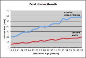
Human fetal uterus growth
This page introduces the uterus as part of the internal female reproductive tract development. Two paramesonephric ducts form from coelomic epithelium extending beside the mesonephric ducts. In the absence of Mullerian Inhibitory Factor these ducts proliferate and grow extending from the vaginal plate on the wall of the urogenital sinus to lie beside the developing ovary. The paired ducts begin to fuse from the vaginal plate end, forming the primordial body of the uterus and the unfused lateral arms form the uterine tubes. Recent research points to the paramesonephric ducts also being the entire embryonic origin of the vagina. For the pregnant uterus see implantation and maternal decidua.
| Historic Embryology
|
| Johannes Peter Müller (1801 - 1858) in 1830 was the first to describe the female genital duct that develops as the uterus and vagina, historically named after him as the "Müllerian duct". The current terminology is the "paramesonephric duct".
|
Category:Uterus
Some Recent Findings
- Review - Müllerian duct anomalies coincident with endometriosis[1] "The association between obstructed müllerian duct anomalies and endometriosis has been well established and the pathogenesis is attributed to the theory of retrograde menstruation. However, this relationship with endometriosis is less clear in women with unobstructed müllerian duct anomalies and in those with rudimentary uterine structures that lack functioning endometrial tissue. This article reviews the embryology, genetics, pathophysiology, and American Society for Reproductive Medicine (ASRM) classification for müllerian duct anomalies together with the genetics and pathophysiology of endometriosis to provide a framework for understanding the complex relationship between these two entities. Available published data examining the coexistence of endometriosis in relationship to müllerian duct anomalies, including studies that stratify this relationship according to specific classes of anomalies, are reviewed and organized. Awareness of the increased prevalence of endometriosis among patients with uterine anomalies, particularly those with outflow obstruction, may facilitate early diagnosis of endometriosis and subsequent intervention, with the potential to reverse disease symptoms and arrest disease progression."
- The histone methyltransferase EZH2 is required for normal uterine development and function in mice[2] "Enhancer of zeste homolog 2 (EZH2) is a rate-limiting catalytic subunit of a histone methyltransferase, polycomb repressive complex, which silences gene activity through the repressive histone mark H3K27me3. EZH2 is critical for epigenetic effects of early estrogen treatment, and may be involved in uterine development and pathologies. We investigated EZH2 expression, regulation and its role in uterine development/function. Uterine epithelial EZH2 expression was associated with proliferation and was high neonatally then declined by weaning. ...In summary, uterine EZH2 expression is developmentally and hormonally regulated, and its loss causes aberrant uterine epithelial proliferation, uterine hypertrophy and cystic endometrial hyperplasia, indicating a critical role in uterine development and function."
- Livebirth after uterus transplantation from a deceased donor in a recipient with uterine infertility[3] "Uterus transplantation from live donors became a reality to treat infertility following a successful Swedish 2014 series, inspiring uterus transplantation centres and programmes worldwide. However, no case of livebirth via deceased donor uterus has, to our knowledge, been successfully achieved, raising doubts about its feasibility and viability, including whether the womb remains viable after prolonged ischaemia. ...We describe, to our knowledge, the first case worldwide of livebirth following uterine transplantation from a deceased donor in a patient with congenital uterine absence (Mayer-Rokitansky-Küster-Hauser [MRKH] syndrome). The results establish proof-of-concept for treating uterine infertility by transplantation from a deceased donor, opening a path to healthy pregnancy for all women with uterine factor infertility, without need of living donors or live donor surgery."
- Review - The cell biology and molecular genetics of Müllerian duct development[4] "The Müllerian ducts are part of the embryonic urogenital system. They give rise to mature structures that serve a critical function in the transport and development of the oocyte and/or embryo. In most vertebrates, both sexes initially develop Müllerian ducts during embryogenesis, but they regress in males under the influence of testis-derived Anti-Müllerian Hormone (AMH)."
- Outcome of assisted reproduction in women with congenital uterine anomalies: a prospective observational study[5] "Consecutive women referred for subfertility between May 2009 and November 2015 who underwent assisted reproduction were included in the study. As part of the initial assessment, each woman underwent three-dimensional transvaginal sonography. Uterine morphology was classified using the modified American Fertility Society (AFS) classification of congenital uterine anomalies proposed by Salim et al. ...Congenital uterine anomalies as a whole, when defined using the modified AFS classification, do not affect clinical pregnancy or live-birth rates in women following assisted reproduction, but do increase the incidence of preterm birth. The presence of uterine abnormalities more severe than arcuate uterus significantly worsens all pregnancy outcomes."
|
| Older papers
|
| These papers originally appeared in the Some Recent Findings table, but as that list grew in length have now been shuffled down to this collapsible table.
See also the Discussion Page for other references listed by year and References on this current page.
- WNT4 coordinates directional cell migration and extension of the Müllerian duct essential for ontogenesis of the female reproductive tract[6] "The Müllerian duct (MD) is the anlage of the oviduct, uterus and upper part of the vagina, the main parts of the female reproductive tract. Several wingless-type mouse mammary tumor virus (MMTV) integration site family member (Wnt) genes, including Wnt4, Wnt5a and Wnt7a, are involved in the development of MD and its derivatives, with Wnt4 particularly critical, since the MD fails to develop in its absence. We use, here, Wnt4(EGFPCre)-based fate mapping to demonstrate that the MD tip cells and the subsequent MD cells are derived from Wnt4+ lineage cells. Moreover, Wnt4 is required for the initiation of MD-forming cell migration." WNT4
- LHX1 is required in Müllerian duct epithelium for uterine development[7] "The female reproductive tract organs of mammals, including the oviducts, uterus, cervix and upper vagina, are derived from the Müllerian ducts, a pair of epithelial tubes that form within the mesonephroi. The Müllerian ducts form in a rostral to caudal manner, guided by and dependent on the Wolffian ducts that have already formed. Experimental embryological studies indicate that caudal elongation of the Müllerian duct towards the urogenital sinus occurs in part by proliferation at the ductal tip. The molecular mechanisms that regulate the elongation of the Müllerian duct are currently unclear. Lhx1 encodes a LIM-homeodomain transcription factor that is essential for male and female reproductive tract development. Lhx1 is expressed in both the Wolffian and Müllerian ducts. Wolffian duct-specific knockout of Lhx1 results in degeneration of the Wolffian duct and consequently the non-cell-autonomous loss of the Müllerian duct. To determine the role of Lhx1 specifically in the Müllerian duct epithelium, we performed a Müllerian duct-specific knockout study using Wnt7a-Cre mice. Loss of Lhx1 in the Müllerian duct epithelium led to a block in Müllerian duct elongation and uterine hypoplasia characterized by loss of the entire endometrium (luminal and glandular epithelium and stroma) and inner circular but not the outer longitudinal muscle layer. Time-lapse imaging and molecular analyses indicate that Lhx1 acts cell autonomously to maintain ductal progenitor cells for Müllerian duct elongation. These studies identify LHX1 as the first transcription factor that is essential in the Müllerian duct epithelial progenitor cells for female reproductive tract development." HGNC
- The origin of the Mullerian duct in chick and mouse[8] "In vertebrates the female reproductive tracts derive from a pair of tubular structures called Mullerian ducts, which are composed of three elements: a canalised epithelial tube, mesenchymal cells surrounding the tube and, most externally, coelomic epithelial cells. ... We show that all Mullerian duct components derive from the coelomic epithelium in both species. Our data support a model of a Mullerian epithelial tube derived from an epithelial anlage at the mesonephros anterior end, which then segregates from the epithelium and extends caudal of its own accord, via a process involving rapid cell proliferation. This tube is surrounded by mesenchymal cells derived from local delamination of coelomic epithelium."
- Essential roles of mesenchyme-derived beta-catenin in mouse Mullerian duct morphogenesis[9]
|
Paramesonephric Duct
The Müllerian duct (= paramesonephric duct, preferred terminology) paired ducts that form the epithelial lining of female reproductive organs: utererine tube, uterus, upper vaginal canal. The term "paramesonephric" duct means beside the mesonephric (Wolffian) duct, which is its anatomical location in early development. Mullerian refers to Johannes Peter Müller (1801-1858) a German scientist who specialised in comparative anatomy. These ducts initially form and then degenerate in the male.
A recent study using both chicken and mouse embryos has shown that these initially paired tubular structures derive from the coelomic epithelium.[8]
- "Müllerian epithelial tube derived from an epithelial anlage at the mesonephros anterior end, which then segregates from the epithelium and extends caudal of its own accord, via a process involving rapid cell proliferation. This tube is surrounded by mesenchymal cells derived from local delamination of coelomic epithelium."
Mullerian ducts have three elements:
- a canalised epithelial tube
- mesenchymal cells surrounding the tube
- coelomic epithelial cells
Duct Molecular Development
The paired paramesonephic ducts (Müllerian ducts) go through a series of developmental changes recently identified as regulated by a number of molecular factors.
Initiation
Coelomic epithelium Lim1 expressing cells are specified to a duct fate.[10]
- Lim - proteins named for 'LIN11, ISL1, and MEC3,' are defined by the possession of a highly conserved double zinc finger motif called the LIM domain.
- LIM domain-binding factors - interact with the LIM domains of nuclear proteins are capable of binding to a variety of transcription factors.
Invagination
- Wnt4 - induces duct invagination to reach the mesonephric (Wolffian)
Elongation
- WNT9b - from mesonephric duct to guide paramesonephric duct elongation. Cysteine-rich secreted glycoprotein.
- Pax2 - also acts in elongation and duct maintenance. Member of the paired box protein family.
Cells at the leading tip proliferate and form the duct elongating to reach the cloaca (urogenital sinus).
- Links: OMIM - WNT9b | OMIM - Pax2 | OMIM - paired box gene
Uterine Development Movie
|
|
Anterior view of development of the female uterus and vagina between Week 9 and 20.
The paramesonephric ducts (red) fuse in the midline to form the genital canal.
The urogenital sinus (yellow), in contact with the paramesonephric duct, thickens to form the sinusal tubercle which extends as a solid vaginal plate, then becomes hollow as the sinovaginal bulb, finally forming the vagina.
|
Development Overview

|
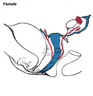
|
| Common
|
Female
|
| Internal Genital Tract Differentiation
|
Mullerian Duct (paramesonephric) - blue
(This historic image mislabels the vaginal origin)
|
The data below gives an overview of the timecourse of embryonic human uterine development.[11]
- Carnegie stage 18 - Mullerian duct to the coelomic cavity was formed as the result of an invagination of the coelomic epithelium - stage 18
- Carnegie stages 19 - 23 - duct grows independently from the invagination - stage 19
- Week 20 - uterine horn fimbrial development begins and continues after birth - second trimester
Fetal Uterus
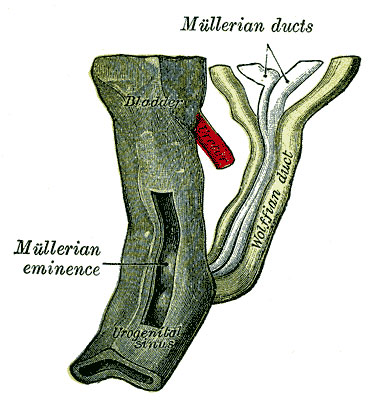
|

|
| Urogenital sinus of female human embryo of eight and a half to nine weeks old (From model by Keibel) (Image: Gray's Anatomy)
|
(Image modified from: Drews U, Sulak O, Schenck PA. Androgens and the development of the vagina.Biol Reprod. 2002 Oct;67(4):1353-9. PMID: 12297555)
|
Fetal Uterus Growth
Graph shows the growth during the fetal period of the uterus between week 19 and 38.[12] During this time the uterine circumferunce increases from about 20 mm to just under 60mm and the width increases from less than 10mm to just over 20 mm.
Uterine horn fimbrial development begins after week 20 and continues after birth.
Uterine growth continues postnatally, increasing outer muscle thickness and cyclic changes in the lining with puberty.
Adult external uterine orifice to the fundus is approximately 6.25 cm.
Newborn Uterus

| Growth of the Uterus in the Postfetal Period
|
| Age
|
Length of corpus (mm)
|
Length of isthmus (mm)
|
Length of cervix (mm)
|
Total length (mm)
|
| Fetus of 7 months
|
|
|
|
22
|
| Child of 5 weeks
|
|
|
|
27
|
| 1 year
|
10
|
|
|
23
|
| 14 months
|
10
|
5
|
12
|
27
|
| 2.5 years
|
8
|
6
|
12
|
26
|
| 3 years
|
9-10
|
5-6
|
10
|
25
|
| 3.5 years
|
6
|
5
|
16
|
27
|
| 9 years
|
9
|
4.5
|
13
|
27
|
| 11 years
|
12
|
6
|
19
|
37
|
| 13 years
|
|
|
27
|
56
|
| 15 years
|
|
|
|
59
|
| 16 years
|
41
|
12
|
25
|
78
|
| 17 years
|
27
|
6
|
22
|
55
|
| 17 years
|
20
|
4
|
16
|
40
|
| 18 years
|
36
|
5
|
31
|
72
|
| 19 years
|
27
|
5
|
28
|
60
|
| 19 years
|
28
|
6
|
27
|
61
|
| 19 years
|
24
|
8
|
21
|
53
|
| 20 years
|
30
|
6
|
16
|
52
|
| 20 years
|
30
|
7
|
21
|
58
|
| 22 years
|
35
|
5
|
29
|
69
|
| 28 years
|
40
|
10
|
28
|
78
|
| 29 years (nulliparous wife)
|
34
|
10
|
34
|
78
|
| 30 years (virgin)
|
38
|
7
|
29
|
74
|
| Data compiled from Hegar (1908) | Uterus Growth Table | Collapsible Table | Uterus Development
|
Uterine Tubes
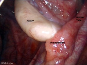
Adult Human right uterine tube and ovary
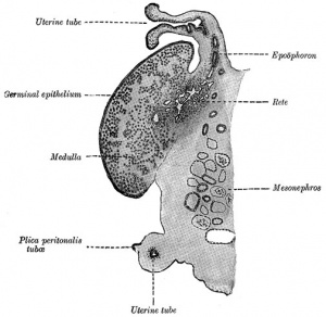
Developing Uterus (cat) showing relationship to ovary and degenerating mesonephros.
The unfused portion of the paramesonephric ducts will form the uterine tubes. Note that there are several synonyms used for the paired uterine tubes or Fallopian tubes or oviducts or uterine horns.
In the adult, the uterine tube has been described in 4 anatomical regions.
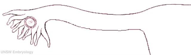
|
| infundibulum
|
ampulla
|
isthmus
|
intramural
|
| funnel-shaped (up to 10 mm in diameter) end of the oviduct. Finger-like extensions of its margins, the fimbriae, are closely applied to the ovary. Ciliated cells are frequent.
|
mucosal folds, or plicae, and secondary folds which arise from the plicae divide the lumen of the ampulla into a very complex shape. Fertilization usually takes place in the ampulla.
|
narrowest portion (2-3 mm in diameter) of the tube located in the peritoneal cavity. Mucosal folds are less complex and the muscularis is thick. An inner, longitudinal layer of muscle is present in the isthmus.
|
penetrates the wall of the uterus. The mucosa is smooth, and the inner diameter of the duct is very small.
|
Mucosa
- formed by a ciliated and secretory epithelium resting on a very cellular lamina propria.
- The number of ciliated cells and non-ciliated secretory cells varies along the oviduct.
- Secretory activity varies during the menstrual cycle, and resting secretory cells are also referred to as peg-cells.
- Some of the secreted substances are thought to nourish the oocyte and the very early embryo.
Muscularis
- inner circular muscle layer and an outer longitudinal layer.
- An inner longitudinal layer is present in the isthmus and the intramural part of the oviduct.
- Peristaltic muscle action seems to be more important for the transport of sperm and oocyte than the action of the cilia.
Uterine tube (monkey) histology overview
Uterine tube (monkey) epithelium and underlying histology
Uterine Blood Supply
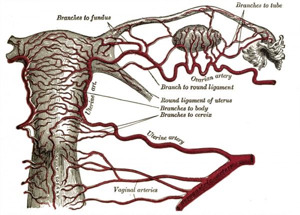
Uterine Glands
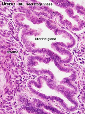
Uterine Gland Secretory Phase
Uterine adenogenesis is the term used to describe the formation of uterine glands from the epithelial lining of the uterus that begins prenatal in humans. In other species, the overt development occurs postnatally and has been described through a 3 step the sequence:
- differentiation and budding of the glandular epithelium.
- invagination and tubular coiling of the epithelium.
- branching of the glandular elements and their expansion throughout the endometrial stroma toward the myometrium.
Epithelial-mesenchymal interaction occurs through Wnt signalling during this process:
- Wnt7a - expressed in the luminal epithelium
- Wnt5a - expressed in the mesenchyme
In mice, this development sequence occurs between postnatal day (PND) 5 to 7 and involves Wnt up-regulation of Lymphoid Enhancing Factor 1 (Lef1).[13]
Postnatally both prolactin and estradiol-17 beta (and their receptors) regulate gland development. There are some gland species gestational differences, in both sheep and pigs the glands provide additional histotrophic support by undergoing extensive hyperplasia and hypertrophy.[14]
- Links: Wnt | Epithelial Mesenchymal Interaction
Postnatal Growth
| Growth of the Uterus in the Postfetal Period
|
| Age
|
Length of corpus (mm)
|
Length of isthmus (mm)
|
Length of cervix (mm)
|
Total length (mm)
|
| Fetus of 7 months
|
|
|
|
22
|
| Child of 5 weeks
|
|
|
|
27
|
| 1 year
|
10
|
|
|
23
|
| 14 months
|
10
|
5
|
12
|
27
|
| 2.5 years
|
8
|
6
|
12
|
26
|
| 3 years
|
9-10
|
5-6
|
10
|
25
|
| 3.5 years
|
6
|
5
|
16
|
27
|
| 9 years
|
9
|
4.5
|
13
|
27
|
| 11 years
|
12
|
6
|
19
|
37
|
| 13 years
|
|
|
27
|
56
|
| 15 years
|
|
|
|
59
|
| 16 years
|
41
|
12
|
25
|
78
|
| 17 years
|
27
|
6
|
22
|
55
|
| 17 years
|
20
|
4
|
16
|
40
|
| 18 years
|
36
|
5
|
31
|
72
|
| 19 years
|
27
|
5
|
28
|
60
|
| 19 years
|
28
|
6
|
27
|
61
|
| 19 years
|
24
|
8
|
21
|
53
|
| 20 years
|
30
|
6
|
16
|
52
|
| 20 years
|
30
|
7
|
21
|
58
|
| 22 years
|
35
|
5
|
29
|
69
|
| 28 years
|
40
|
10
|
28
|
78
|
| 29 years (nulliparous wife)
|
34
|
10
|
34
|
78
|
| 30 years (virgin)
|
38
|
7
|
29
|
74
|
| Data compiled from: Hegar K. Anatomical investigations on the nullipara uterus with special consideration of isthmus (Anatomische Untersuchungen am nullipara Uterus mit besonderer Berücksichtigung der Isthmus). (1908) Beitraae zur Geburtsh. u. Gynak., vol. 13, 1908. reference list | Uterus Growth Table | Collapsible Table | Uterus Development
|
Uterus Histology
See also Menstrual Cycle - Histology
Uterine tube histology overview showing epithelium and underlying muscular layers
Uterine tube epithelium histology showing secretory and ciliated cells
Uterine body endometrium and myometrium during the proliferative phase of the menstrual cycle overview
Uterine body endometrium during the proliferative phase of the menstrual cycle
Uterine body endometrium during the secretory phase of the menstrual cycle overview
Uterine body endometrium during the secretory phase of the menstrual cycle
Abnormalities
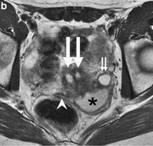
Uterine didelphys, obstructed hemivagina, and ectopic ureter on MR imaging in a 17-year-old girl.
[15]
There are at least two clinical society classifications for female genital tract abnormalities:
- European Society of Human Reproduction and Embryology—European Society for Gynaecological Endoscopy (ESHRE-ESGE)[16]
- American Society for Reproductive Medicine (ASRM) [17]
ESHRE-ESGE Classification
European Society for Gynaecological Endoscopy (ESHRE-ESGE)[16]
Uterine anatomical deviations deriving from the same embryological origin:
- U0 - normal uterus
- U1 - dysmorphic uterus
- U2 - septet uterus
- U3 - bicorporeal uterus
- U4 - hemi-uterus
- U5 - aplastic uterus
- U6 - for still unclassified cases
Main classes have been divided into sub-classes expressing anatomical varieties with clinical significance. Cervical and vaginal anomalies are classified independently into sub-classes having clinical significance.
| ESHRE/ESGE Classification of Uterine Anomalies
|
European Society of Human Reproduction and Embryology (ESHRE) and the European Society for Gynaecological Endoscopy (ESGE)
- Class U0 - incorporates all cases with normal uterus.
- Class U1 - (Dysmorphic uterus) incorporates all cases with normal uterine outline but with an abnormal shape of the uterine cavity excluding septa. Class I is further subdivided into three categories
- Class U1a - (T-shaped uterus) characterized by a narrow uterine cavity due to thickened lateral walls with a correlation 2/3 uterine corpus and 1/3 cervix,
- Class U1b - (uterus infantilis) characterized also by a narrow uterine cavity without lateral wall thickening and an inverse correlation of 1/3 uterine body and 2/3 cervix
- Class U1c - (others) which is added to include all minor deformities of the uterine cavity including those with an inner indentation at the fundal midline level of less than 50 % of the uterine wall thickness.
- Class U2 - (septate uterus) internal indentation >50 % of the uterine wall thickness & external contour straight or with indentation <50 %
- Class U2a - (partial septate uterus) characterized by the existence of a septum dividing partly the uterine cavity above the level of the internal cervical os
- Class U2b - (complete septate uterus) characterized by the existence of a septum fully dividing the uterine cavity up to the level of the internal cervical os.
- Class U3 - (bicorporeal uterus) external indentation >50 % of the uterine wall thickness
- Class U3a - (partial bicorporeal uterus) characterized by an external fundal indentation partly dividing the uterine corpus above the level of the cervix
- Class U3b - (complete bicorporeal uterus) width of the fundal indentation at the midline >150 % of the uterine wall thickness) completely dividing the uterine corpus up to the level of the cervix
- Class U3c - (bicorporeal septate uterus) characterized by the presence of an absorption defect in addition to the main fusion defect.
- Class U4 - (hemi-uterus) incorporates all cases of unilateral formed uterus. Hemi-uterus is defined as the unilateral uterine development; the contralateral part could be either incompletely formed or absent.
- Class U4a - (hemi-uterus with a rudimentary (functional) cavity) characterized by the presence of a communicating or non-communicating functional contralateral horn
- Class U4b - (hemi-uterus without rudimentary (functional) cavity) characterized either by the presence of non-functional contralateral uterine horn or by aplasia of the contralateral part.
- Class U5 - (aplastic uterus) incorporates all cases of uterine aplasia, formation defect characterized by the absence of any fully or unilaterally developed uterine cavity.
- Class U5a - (aplastic uterus with rudimentary (functional) cavity) characterized by the presence of bi- or unilateral functional horn
- Class U5b - (aplastic uterus without rudimentary (functional) cavity) characterized either by the presence of uterine remnants or by full uterine aplasia.
- Class U6 - is kept for still unclassified cases.
|
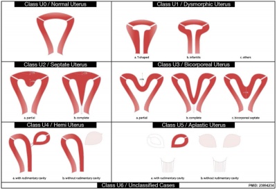
|
| <pubmed>23894234</pubmed>
See also ICD10 Congenital malformations of genital organs (Q50-Q56)
- Links: Genital System - Abnormalities | Uterus Development | image - simplified cartoon
|
Uterine Duplication
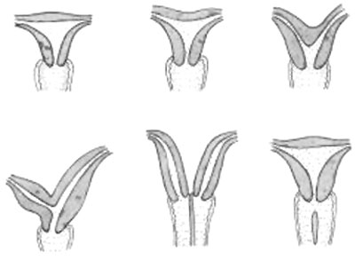
|
A range of uterine and vaginal anatomical anomalies based upon the abnormal development and fusion of the paramesonephric ducts and vaginal plate development.
|
|
Unicornate Uterus - failure of the paramesonephric ducts to fuse. A single paramesomnephric duct has fused with the vaginal plate and now opens into the vagina, while the other forms a diverticulum.
|
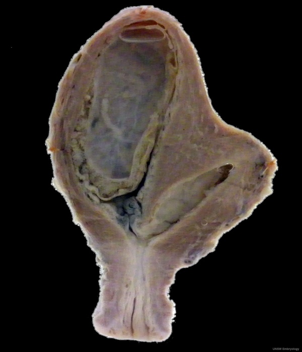
Bicornuate uterus containing conceptus chorionic sac with placental cord on one side.
Septate Uterus
Uterine residual septum classification:
- American Society for Reproductive Medicine (ASRM) criterion with an internal fundal indentation length equal or greater than 1 cm[18]
- European Society of Human Reproduction and Embryology—European Society for Gynaecological Endoscopy (ESHRE-ESGE) classification of female genital tract congenital anomalies with an internal indentation at the fundal midline greater than 50% myometrial thickness.[16]
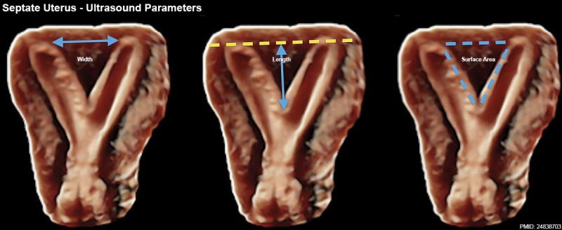
Septate Uterus Ultrasound[19]
Uterine Duplication
(uterus didelphys, double uterus, uterus didelphis) A rare uterine developmental abnormality where the paramesonephric ducts (Mullerian ducts) completely fail to fuse generating two separate uterus parts each connected to the cervix and having an ovary each.
Uterus/Vaginal
Mayer-Rokitansky-Kuster-Hauser syndrome (MRKH, MRK anomaly, Rokitansky-Kuster-Hauser syndrome, RKH syndrome, RKH) consists of congenital aplasia of the uterus and the upper part of vagina due to anomalous development of Müllerian ducts, either isolated or associated with other congenital malformations, including renal, skeletal, hearing and heart defects. Has an incidence of approximately 1 in 4500 newborn girls and has been associated with a microdeletion at 17q12.[20]
There has been recently a single report of a MRKH syndrome woman giving a live-birth after uterus transplantation from a deceased donor.[3]
Cervical: cervical agenesis, cervical duplication
Environmental Abnormalities
DES Diethylstilbestrol or diethylstilbetrol, is a drug that was prescribed to women from 1938-1971 to prevent miscarriage in high-risk pregnancies. The drug acted as a potent estrogen (mimics natural hormone) and therefore could also act as a potential endocrine disruptor. This led to a number of developing fetal reproductive tract and other abnormalities. In the female fetus, it increased risk of abnormal reproductive tract and also carcinogenic (cancer forming). In the male fetus, it increased the occurance of abnormal genitalia. The drug was banned by FDA (USA) in 1979 as a teratogen, it had previously also been used as livestock growth promoter and could have potentially entered the human food chain. (More? endocrine abnormalities | chemicals | drugs)
- Links: Childrens Hospital Boston - Congenital Anomalies of the Uterus | Medical Education Image Link - Cervical agenesis | OMIM - Rokitansky-Kuster-Hauser syndrome |
Cervical Cancer
| ICD-11 2C77 Malignant neoplasms of cervix uteri - Primary or metastatic malignant neoplasm involving the cervix.
|
- 2C77.0 Squamous cell carcinoma of cervix uteri - A squamous cell carcinoma arising from the cervical epithelium. It usually evolves from a precancerous cervical lesion. Increased numbers of sexual partners and human papillomavirus (HPV) infection are risk factors for cervical squamous cell carcinoma. The following histologic patterns have been described: Conventional squamous cell carcinoma, papillary squamous cell carcinoma, transitional cell carcinoma, lymphoepithelioma-like carcinoma, verrucous carcinoma, condylomatous carcinoma and spindle cell carcinoma. Survival is most closely related to the stage of disease at the time of diagnosis.
- 2C77.1 Adenocarcinoma of cervix uteri - An adenocarcinoma arising from the cervical epithelium. It accounts for approximately 15% of invasive cervical carcinomas. Increased numbers of sexual partners and human papillomavirus (HPV) infection are risk factors. Grossly, advanced cervical adenocarcinoma may present as an exophytic mass, an ulcerated lesion, or diffuse cervical enlargement. Microscopically, the majority of cervical adenocarcinomas are of the endocervical (mucinous) type.
- 2C77.2 Adenosquamous carcinoma of cervix uteri
- 2C77.3 Neuroendocrine carcinoma of cervix uteri
|
| Links: cervical cancer | human papillomavirus | viral infection | uterus
|
In Australia, the "Pap Smear" test was replaced in 2017 (1 December) by a new "National Cervical Screening Program". This new program will use new technologies to detect HPV DNA rather than pathological screening for abnormal cells from a "Pap Smear". See the last report Cervical screening in Australia 2019[21]Australian Institute of Health and Welfare 2019. Physical activity during pregnancy 2011–12. Cat. no. PHE 243. Canberra: AIHW. PDF</ref>, that used Pap tests as the screening tool (data for women screened between 1 January 2016 and 30 June 2017)
For more information see the external links below.
- "The two yearly Pap test for women aged 18 to 69 will change to a five yearly human papillomavirus (HPV) test for women aged 25 to 74. Women will be due for the first Cervical Screening Test two years after their last Pap test."
- "The Cervical Screening Test detects infection with human papillomavirus (HPV). Partial genotyping is used to determine the type of HPV into one of two groups: oncogenic HPV 16/18 or oncogenic HPV types other than 16/18 as a pooled result." (NCSP Factsheet)
- Links: National Cervical Screening Program | Factsheet | Compass Trial |
ABC radio program Monday 27 March 2017 - Death of the pap smear? | ABC Audio - Death of the pap smear?
History of the Pap Smear
The information below relates to the original "Pap Smear" (Papanicolaou smear, pap test, cervical smear) The text below is from the ABC - Great Moments In Science.
- "Luckily, we have the famous Pap Smear - an excellent way to find cancer of the cervix before it digs in locally and/or spreads throughout the body. The Pap Smear is named after a certain Dr. Papanicolaou - who did a Pap Smear on his wife virtually every day for 20 years.
- George Nikolas Papanicolaou was born in 1883 in Kymi, a small town overlooking the Aegean Sea on the Island of Euboea in Greece. His father, Nikolas Papanicolaou was both the Major of Kymi and a medical doctor. His older brother, Naso, had studied law, so his father convinced George to continue in the family medical tradition. So George studied medicine, and did well, graduating with a degree in honours in 1904............"
- Links: Menstrual Cycle - Histology | Dilatation and curettage (D&C) | ABC - Great Moments In Science
Broad Ligament
| The broad ligament is found associated with the internal human female genital tract. It forms a mesentery consisting of a double fold of the peritoneum that connects the uterus to the peritoneal floor and walls.
Anatomically it has three parts:
- mesometrium - surrounding the uterus
- mesosalpinx - surrounding the uterine tube
- mesovarium - surrounding the ovary
Abnormalities include peritoneal endometriosis.
|
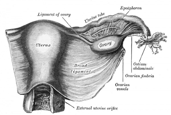
|
Molecular
Wnt genes - Wnt4, Wnt5a, and Wnt7a implicated in the formation and morphogenesis of the Müllerian duct.
Wnt7a - mediates the patterning of the oviduct and differentiation of the uterus.
beta-catenin - manufactured in the mesenchyme is a downstream effector of Wnt7a.
Bmp2 - decidualization regulator of gene expression and function (shown in mouse uterus).
Lim1, Lhx9, Emx, Pax-2, Hox-A9, Hox-A10, Hox-A11, Hox-A13, WT1, SF-1, GATA-4. TGF-beta
References
- ↑ Pitot MA, Bookwalter CA & Dudiak KM. (2020). Müllerian duct anomalies coincident with endometriosis: a review. Abdom Radiol (NY) , , . PMID: 32179978 DOI.
- ↑ Nanjappa MK, Mesa AM, Medrano TI, Jefferson WN, DeMayo FJ, Williams CJ, Lydon JP, Levin ER & Cooke PS. (2019). The histone methyltransferase EZH2 is required for normal uterine development and function in mice. Biol. Reprod. , , . PMID: 31201420 DOI.
- ↑ 3.0 3.1 Parikh PM, Charak BS, Banavali SD, Koppikar SB, Giri N, Nadkarni P, Saikia TK, Gopal R & Advani SH. (1988). A prospective, randomized double-blind trial comparing metoclopramide alone with metoclopramide plus dexamethasone in preventing emesis induced by high-dose cisplatin. Cancer , 62, 2263-6. PMID: 3052785
- ↑ Roly ZY, Backhouse B, Cutting A, Tan TY, Sinclair AH, Ayers KL, Major AT & Smith CA. (2018). The cell biology and molecular genetics of Müllerian duct development. Wiley Interdiscip Rev Dev Biol , , . PMID: 29350886 DOI.
- ↑ Prior M, Richardson A, Asif S, Polanski L, Parris-Larkin M, Chandler J, Fogg L, Jassal P, Thornton JG & Raine-Fenning NJ. (2018). Outcome of assisted reproduction in women with congenital uterine anomalies: a prospective observational study. Ultrasound Obstet Gynecol , 51, 110-117. PMID: 29055072 DOI.
- ↑ Prunskaite-Hyyryläinen R, Skovorodkin I, Xu Q, Miinalainen I, Shan J & Vainio SJ. (2016). Wnt4 coordinates directional cell migration and extension of the Müllerian duct essential for ontogenesis of the female reproductive tract. Hum. Mol. Genet. , 25, 1059-73. PMID: 26721931 DOI.
- ↑ Huang CC, Orvis GD, Kwan KM & Behringer RR. (2014). Lhx1 is required in Müllerian duct epithelium for uterine development. Dev. Biol. , 389, 124-36. PMID: 24560999 DOI.
- ↑ 8.0 8.1 Guioli S, Sekido R & Lovell-Badge R. (2007). The origin of the Mullerian duct in chick and mouse. Dev. Biol. , 302, 389-98. PMID: 17070514 DOI.
- ↑ Deutscher E & Hung-Chang Yao H. (2007). Essential roles of mesenchyme-derived beta-catenin in mouse Müllerian duct morphogenesis. Dev. Biol. , 307, 227-36. PMID: 17532316 DOI.
- ↑ Kobayashi A, Shawlot W, Kania A & Behringer RR. (2004). Requirement of Lim1 for female reproductive tract development. Development , 131, 539-49. PMID: 14695376 DOI.
- ↑ Hashimoto R. (2003). Development of the human Müllerian duct in the sexually undifferentiated stage. Anat Rec A Discov Mol Cell Evol Biol , 272, 514-9. PMID: 12740945 DOI.
- ↑ Soriano D, Lipitz S, Seidman DS, Maymon R, Mashiach S & Achiron R. (1999). Development of the fetal uterus between 19 and 38 weeks of gestation: in-utero ultrasonographic measurements. Hum. Reprod. , 14, 215-8. PMID: 10374123
- ↑ Shelton DN, Fornalik H, Neff T, Park SY, Bender D, DeGeest K, Liu X, Xie W, Meyerholz DK, Engelhardt JF & Goodheart MJ. (2012). The role of LEF1 in endometrial gland formation and carcinogenesis. PLoS ONE , 7, e40312. PMID: 22792274 DOI.
- ↑ Gray CA, Bartol FF, Tarleton BJ, Wiley AA, Johnson GA, Bazer FW & Spencer TE. (2001). Developmental biology of uterine glands. Biol. Reprod. , 65, 1311-23. PMID: 11673245
- ↑ Wang ZJ, Daldrup-Link H, Coakley FV & Yeh BM. (2010). Ectopic ureter associated with uterine didelphys and obstructed hemivagina: preoperative diagnosis by MRI. Pediatr Radiol , 40, 358-60. PMID: 19924410 DOI.
- ↑ 16.0 16.1 16.2 Grimbizis GF, Gordts S, Di Spiezio Sardo A, Brucker S, De Angelis C, Gergolet M, Li TC, Tanos V, Brölmann H, Gianaroli L & Campo R. (2013). The ESHRE-ESGE consensus on the classification of female genital tract congenital anomalies. Gynecol Surg , 10, 199-212. PMID: 23894234 DOI.
- ↑ . (1988). The American Fertility Society classifications of adnexal adhesions, distal tubal occlusion, tubal occlusion secondary to tubal ligation, tubal pregnancies, müllerian anomalies and intrauterine adhesions. Fertil. Steril. , 49, 944-55. PMID: 3371491
- ↑ Bermejo C, Martínez Ten P, Cantarero R, Diaz D, Pérez Pedregosa J, Barrón E, Labrador E & Ruiz López L. (2010). Three-dimensional ultrasound in the diagnosis of Müllerian duct anomalies and concordance with magnetic resonance imaging. Ultrasound Obstet Gynecol , 35, 593-601. PMID: 20052665 DOI.
- ↑ Ludwin A, Ludwin I, Pityński K, Banas T & Jach R. (2014). Role of morphologic characteristics of the uterine septum in the prediction and prevention of abnormal healing outcomes after hysteroscopic metroplasty. Hum. Reprod. , 29, 1420-31. PMID: 24838703 DOI.
- ↑ Bernardini L, Gimelli S, Gervasini C, Carella M, Baban A, Frontino G, Barbano G, Divizia MT, Fedele L, Novelli A, Béna F, Lalatta F, Miozzo M & Dallapiccola B. (2009). Recurrent microdeletion at 17q12 as a cause of Mayer-Rokitansky-Kuster-Hauser (MRKH) syndrome: two case reports. Orphanet J Rare Dis , 4, 25. PMID: 19889212 DOI.
- ↑ Australian Institute of Health and Welfare 2019. Cervical screening in Australia 2019. Cancer series no. 123. Cat. no. CAN 124. Canberra: AIHW. PDF
Reviews
Passos IMPE & Britto RL. (2020). Diagnosis and treatment of müllerian malformations. Taiwan J Obstet Gynecol , 59, 183-188. PMID: 32127135 DOI.
Farage M & Maibach H. (2006). Lifetime changes in the vulva and vagina. Arch. Gynecol. Obstet. , 273, 195-202. PMID: 16208476 DOI.
Cummings AM & Kavlock RJ. (2004). Function of sexual glands and mechanism of sex differentiation. J Toxicol Sci , 29, 167-78. PMID: 15467266
Articles
Deutscher E & Hung-Chang Yao H. (2007). Essential roles of mesenchyme-derived beta-catenin in mouse Müllerian duct morphogenesis. Dev. Biol. , 307, 227-36. PMID: 17532316 DOI.
Guioli S, Sekido R & Lovell-Badge R. (2007). The origin of the Mullerian duct in chick and mouse. Dev. Biol. , 302, 389-98. PMID: 17070514 DOI.
Hashimoto R. (2003). Development of the human Müllerian duct in the sexually undifferentiated stage. Anat Rec A Discov Mol Cell Evol Biol , 272, 514-9. PMID: 12740945 DOI.
Search PubMed
Search May 2007 "embryonic uterine development" 3,025 reference articles of which 491 were reviews.
Search Pubmed: Uterus Development | embryonic uterine development | Paramesonephric Duct | Mullerian Duct | Endocrine Disruptors | uterine+adenogenesis
Additional Images
Human uterine tube ciliated epithelium SEM
Mouse - paramesonephric duct
Ultrasound uterine and ovarian vascularity
Historic drawing of the uterine tube (Reinier De Graaf)
Terms
Note some of these terms relate to the adult or the maternal uterus during pregnancy.
- cervical cerclage - A clinical birth procedure involving circumferential banding or suture of the cervix early (between 12 -14 weeks) or when required to prevent or treat passive dilation prior to completion of pregnancy (37 weeks), described as cervical insufficiency.
- cervical insufficiency - (CI) A clinical term describing a painless and progressive dilatation and effacement of the cervix that may lead to second trimester abortions or preterm delivery. It has also been described as inability of the uterine cervix to retain a pregnancy in the absence of uterine contractions. The condition may in some instances treated clinically by cervical cerclage. The biological basis is currently undetermined with some evidence showing a genetic relationship.
- cervical length - There is some data that shows the risk of spontaneous preterm labour and delivery increases in women who have a short cervix PMID 8569824.
- cervical mucus plug - (CMP) During early pregnancy, maternal glands located at the cervical junction between vagina and uterus secrete mucus that forms a plug or barrier between these two structures.
- cervical pregnancy - A rare type of ectopic pregnancy with implantation at the cervical canal, occurring with an incidence ranging between 1:1,000 and 1:18,000 pregnancies. Clinically, when an associated haemorrhage occurs a hysterectomy is usually performed.
- cervical ripening - Clinical birth term describing the hormonal softening of the cervix to allow expansion in preparation for birth.
- cervix - (Latin, cervix = neck) The female anatomical region of the uterus forming a canal that opens and connects to the vagina.
- fallopian tube obstruction - (tubal occlusion) A blockage of the uterine tube that can affect fertility due to a pathologic occlusion, spasm or plugging and also be either unilateral (single tube) or bilateral (both tubes). Described anatomically as in the proximal, the mid or the distal part of the tube.
- fundus - (Latin, fundus = "bottom") Top part of the uterus body lying between the two uterine tubes and a common implantation site.
- hysterosalpingography - A clinical diagnostic technique used to visualise the uterine cavity by X-ray.
- hysteroscopy - A clinical diagnostic technique used to visualise the uterine cavity by a camera or video.
- peg-cell - (resting secretory cell) A histological term for the non-ciliated secretory epithelial cells located within the uterus.
- Pouch of Douglas - (rectouterine pouch or rectovaginal) Anatomical description of the female peritoneal cavity lying between the back wall of the uterus and rectum.
- rectouterine pouch - (Pouch of Douglas or rectovaginal) Anatomical description of the female peritoneal cavity lying between the back wall of the uterus and rectum.
- sonohysterography - A clinical diagnostic technique used to visualise the uterine cavity by ultrasound. Firstly, fluid is injected through the cervix into the uterus, then ultrasound is carried out to image the uterine cavity.
External Links
External Links Notice - The dynamic nature of the internet may mean that some of these listed links may no longer function. If the link no longer works search the web with the link text or name. Links to any external commercial sites are provided for information purposes only and should never be considered an endorsement. UNSW Embryology is provided as an educational resource with no clinical information or commercial affiliation.
Glossary Links
- Glossary: A | B | C | D | E | F | G | H | I | J | K | L | M | N | O | P | Q | R | S | T | U | V | W | X | Y | Z | Numbers | Symbols | Term Link
Cite this page: Hill, M.A. (2024, May 1) Embryology Uterus Development. Retrieved from https://embryology.med.unsw.edu.au/embryology/index.php/Uterus_Development
- What Links Here?
- © Dr Mark Hill 2024, UNSW Embryology ISBN: 978 0 7334 2609 4 - UNSW CRICOS Provider Code No. 00098G