Placenta - Membranes: Difference between revisions
mNo edit summary |
mNo edit summary |
||
| Line 119: | Line 119: | ||
This swallowed amniotic fluid moves through the GIT from esophagus, to stomach, to small intestine, stopping at the large bowel. In the large bowel the majority of fluid (water) is absorbed, along with electrolytes, glucose, urea and hormones. This process may contribute to fetal nutrition and prepare the GIT for its postnatal function. The process of swallowing amniotic fluid has been suggested to also help regulate fluid volume. | This swallowed amniotic fluid moves through the GIT from esophagus, to stomach, to small intestine, stopping at the large bowel. In the large bowel the majority of fluid (water) is absorbed, along with electrolytes, glucose, urea and hormones. This process may contribute to fetal nutrition and prepare the GIT for its postnatal function. The process of swallowing amniotic fluid has been suggested to also help regulate fluid volume. | ||
[[File:Yolk sac and amniotic cavity volume graph.jpg|400px]] | |||
Yolk sac and amniotic cavity volume week 2 to 3 (stage [[Carnegie stage 5|5]], [[Carnegie stage 6|6]], [[Carnegie stage 7|7]] and [[Carnegie stage 8|8]]). | |||
:'''Links:''' [[Amniocentesis]] | :'''Links:''' [[Amniocentesis]] | ||
| Line 124: | Line 128: | ||
==Chorionic Sac== | ==Chorionic Sac== | ||
[[File:Gray0039.jpg|thumb]] | [[File:Gray0039.jpg|thumb]] | ||
The chorionic sac (cavity) is lined by extra-embryonic mesoderm and covered in trophoblast cells forming villi. In the human embryo during week 3 this space forms outside the yolk sac and surrounding the amniotic sac. | The chorionic sac (cavity) is lined by extra-embryonic mesoderm and covered in trophoblast cells forming villi. In the human embryo during week 3 this space forms outside the yolk sac and surrounding the amniotic sac. | ||
Revision as of 10:45, 7 March 2017
| Embryology - 19 May 2024 |
|---|
| Google Translate - select your language from the list shown below (this will open a new external page) |
|
العربية | català | 中文 | 中國傳統的 | français | Deutsche | עִברִית | हिंदी | bahasa Indonesia | italiano | 日本語 | 한국어 | မြန်မာ | Pilipino | Polskie | português | ਪੰਜਾਬੀ ਦੇ | Română | русский | Español | Swahili | Svensk | ไทย | Türkçe | اردو | ייִדיש | Tiếng Việt These external translations are automated and may not be accurate. (More? About Translations) |
Introduction


The placental membranes is a term often used to describe the all the fetal components of the placenta (Greek, plakuos = flat cake).
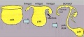
This page will not cover the whole placenta, just the development of the extra-embryonic membranes that form the extra-embryonic coeloms (cavities or spaces); amnionic sac, chorionic sac, yolk sac and allantois.
In monozygotic twinning, depending upon when the twinning event occurred, each embryo will either share or have completely separate set of placental membranes.
|
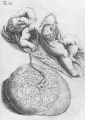
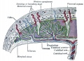
Connecting stalk (body stalk) shown to the right linking to the developing placenta. |
Some Recent Findings
|
| More recent papers |
|---|
 This table allows an automated computer search of the external PubMed database using the listed "Search term" text link.
More? References | Discussion Page | Journal Searches | 2019 References | 2020 References Search term: Placental Membrane Development <pubmed limit=5>Placental Membrane Development</pubmed> |
Reading

- Human Embryology (2nd ed.) Larson Chapter 7 p151-188 Heart, Chapter 8 p189-228 Vasculature
- The Developing Human: Clinically Oriented Embryology (6th ed.) Moore and Persaud Chapter 14: p304-349
- Before we Are Born (5th ed.) Moore and Persaud Chapter 12; p241-254
- Essentials of Human Embryology Larson Chapter 7 p97-122 Heart, Chapter 8 p123-146 Vasculature
- Human Embryology Fitzgerald and Fitzgerald Chapter 13-17: p77-111

Day 8 to 9 early (Week 2) extra-embryonic coeloms (cavities)
Movies
|
|
|
|
- Links: Movies
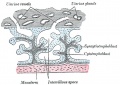
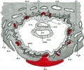
Initial Conceptus Cavities
| <mediaplayer width='220' height='260' image="http://php.med.unsw.edu.au/embryology/images/0/08/Chorion_001_icon.jpg">File:Chorion 001.mp4</mediaplayer> | blue - epiblast layer yellow - hypoblast layer red cells - extraembryonic mesoderm layer green - trophoblast layer red spaces - blood-filled spaces, maternal lacunae white cells - (left) endometrial gland (right) endometrial epithelium |
Amnionic Sac

The amniotic sac (cavity) is initially lined by ectoderm and covered by extra-embryonic mesoderm. In the human embryo during week 3 to 4, folding of the embryonic disc draws the amniotic membrane ventrally over the embryo leading to the enclosing of the embryo within the amniotic sac. Embryonic and fetal development from this time onward occurs fully enclosed within the amniotic sac floating in the amniotic fluid.
Amniotic Fluid
The amniotic fluid has been described as having several functions:
- cushions the fetus against physical trauma.
- allows fetal growth free from restriction or distortion by the adjacent structures.
- provides a thermally stable environment.
- contributes to respiratory and gastrointestinal development.
- helps prevent infection.
- can provide a short-term source of nutrients and fluid to the embryo.
Amniotic fluid is often assessed for both quality and quantity. The volume increases as the fetus grows and rate of change varies during the pregnancy.
- up to 8 weeks - increases at the rate of 10 ml/week
- 8 to 13 weeks - increases at the rate of 25 ml/week
- 13 to 21 weeks - increases at the rate of 60 ml/week
- 21 to 33 weeks - amniotic volume increase starts decreasing and eventually plateaus.
- 34 weeks (GA) - peaks at about 800 mL.
- 40 weeks (GA) - about 600 mL at term.
Fluid Facts
- Circulated by fetal inhaling and swallowing.
- Replaced by fetal exhalation and urination.
- Magnesium low levels associated with preeclampsia and diabetes.
- normal magnesium value at 16 weeks (GA) is 1.65 ± 0.16 mg/dL in amniotic fluid and 1.97 ± 0.23 mg/dL in serum.[3]
Amniotic Fluid Swallowing
In early embryonic development both the buccopharyngeal and cloacal membranes degenerated, allowing the GIT to be filled with amniotic fluid. Towards the end of the fetal period the fetus is now swallowing approximately 500 ml of amniotic fluid / day.
This swallowed amniotic fluid moves through the GIT from esophagus, to stomach, to small intestine, stopping at the large bowel. In the large bowel the majority of fluid (water) is absorbed, along with electrolytes, glucose, urea and hormones. This process may contribute to fetal nutrition and prepare the GIT for its postnatal function. The process of swallowing amniotic fluid has been suggested to also help regulate fluid volume.

Yolk sac and amniotic cavity volume week 2 to 3 (stage 5, 6, 7 and 8).
- Links: Amniocentesis
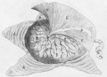
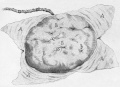
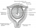
Chorionic Sac

The chorionic sac (cavity) is lined by extra-embryonic mesoderm and covered in trophoblast cells forming villi. In the human embryo during week 3 this space forms outside the yolk sac and surrounding the amniotic sac.
This forms a transient fluid-filled space that is lost by expansion of the amniotic sac, which eventually fuses to the chorionic membrane.
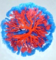
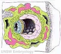
Yolk Sac

The ventral endoderm lines and extra-embryonic mesoderm covers the space called the yolk sac (yellow). Folding of the embryonic disc "pinches off" part of this yolk sac forming the first primitive gastrointestinal tract.

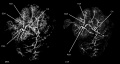
Notch and yolk sac blood vessels model[4]
Allantois
An extra-embryonic membrane, endoderm in origin as an extension from the early hindgut, then cloaca into the connecting stalk of placental animals, connected to the superior end of developing bladder.
In reptiles and birds, acts as a reservoir for wastes and mediates gas exchange. In mammals is associated/incorporated with connecting stalk/placental cord fetal-maternal interface.
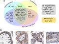
A recent comparative study looking at allantois structure in mouse, pig, rabbit and man[5] found the following:
- tissue interaction between endoderm and mesoderm required for allantoic development and vascular differentiation in species with a rudimentary allantoic diverticulum
- allantoic mesothelium plays a role in chorioallantoic attachment, allantoic differentiation and vascularization
- a pronounced diversity in the extraembryonic migratory pathways of primordial germ cells among mammals
Allantois from hindgut

Placental cord cross-section

Placental allantois


Term Membranes
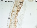
Cannabinoid Receptor 1 (CB1)
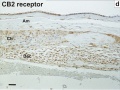
Cannabinoid Receptor 2 (CB2)
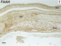
Fatty acid amide hydrolase (FAAH)


Abnormalities
Monoygotic Twinning
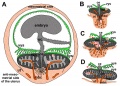
Monoygotic twins (identical) produced from a single fertilization event (one fertilised egg and a single spermatazoa, form a single zygote), these twins therefore share the same genetic makeup. Occurs in approximately 3-5 per 1000 pregnancies, more commonly with aged mothers.
The later the twinning event occurs, the less common are the initially separate placental membranes (diamniotic, dichorionic) and finally resulting in conjoined twins.
| Week | Week 1 | Week 2 | |||||||||||||
| Day | 0 | 1 | 2 | 3 | 4 | 5 | 6 | 7 | 8 | 9 | 10 | 11 | 12 | 13 | 14 |
| Cell Number | 1 | 1 | 2 | 16 | 32 | 128 | bilaminar | ||||||||
| Event | Ovulation | fertilization | First cell division | Morula | Early blastocyst | Late blastocyst
Hatching |
Implantation starts | X inactivation | |||||||

|

|

|
|||||||||||||
| Monoygotic
Twin Type |
Diamniotic
Dichorionic |
Diamniotic
Monochorionic |
Monoamniotic
Monochorionic |
Conjoined | |||||||||||
Table based upon[6] =
Chorioamnionitis
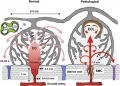
(CA) An intraamniotic puerperal infection described as having 3 forms: histologic, clinical (clinical chorioamnionitis, IAI), and subclinical. Intraamniotic infection is a common (2-4%) event in labor and the systemic inflammatory response can also lead to preterm birth and neonatal complications.
- Links: Placenta - Abnormalities
References
Reviews
<pubmed>20711983</pubmed> <pubmed>11312634</pubmed> <pubmed>8671414</pubmed>
Articles
Search PubMed
Search April 2010
- Placental Membranes - All (10083) Review (748) Free Full Text (1728)
Search Pubmed: Placental Membranes | amniotic sac development | chorionic sac development | yolk sac development | allantois development
Additional Images
see all online Placental materials
Placenta and Fetus
Placenta Fetal Side
Placenta Maternal Side

Fetal membrane and placenta cartoon
Placenta spiral artery conversion
Uterine and placental vasculature
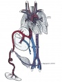
Fetal circulation overview
Placental trophospongium

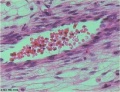
Placenta anchoring villi
Fetal blood
Placental cord cross-section
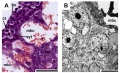
Placenta_abnormalities
Mouse placenta E16.5

Mouse placenta E16.5
Human placenta viewed from the fetal side
Cord with one artery and one vein
Placenta gene expression


Terms
| Placenta Terms (expand to view) |
|---|
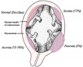
with an incidence of about 2.8 per 1,000 pregnancies, there is also a rarer form of extra-abdominal varices.PMID 24883288
with an incidence of about 2.8 per 1,000 pregnancies, there is also a rarer form of extra-abdominal varices. PMID 24883288
|
| Other Terms Lists |
|---|
| Terms Lists: ART | Birth | Bone | Cardiovascular | Cell Division | Endocrine | Gastrointestinal | Genital | Genetic | Head | Hearing | Heart | Immune | Integumentary | Neonatal | Neural | Oocyte | Palate | Placenta | Radiation | Renal | Respiratory | Spermatozoa | Statistics | Tooth | Ultrasound | Vision | Historic | Drugs | Glossary |
External Links
External Links Notice - The dynamic nature of the internet may mean that some of these listed links may no longer function. If the link no longer works search the web with the link text or name. Links to any external commercial sites are provided for information purposes only and should never be considered an endorsement. UNSW Embryology is provided as an educational resource with no clinical information or commercial affiliation.
Glossary Links
- Glossary: A | B | C | D | E | F | G | H | I | J | K | L | M | N | O | P | Q | R | S | T | U | V | W | X | Y | Z | Numbers | Symbols | Term Link
Cite this page: Hill, M.A. (2024, May 19) Embryology Placenta - Membranes. Retrieved from https://embryology.med.unsw.edu.au/embryology/index.php/Placenta_-_Membranes
- © Dr Mark Hill 2024, UNSW Embryology ISBN: 978 0 7334 2609 4 - UNSW CRICOS Provider Code No. 00098G